Morphological characteristics of eosinophilic neuronal death after transient unilateral forebrain ischemia in Mongolian gerbils
Abstract
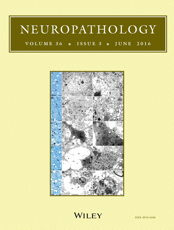
Various types of eosinophilic neurons (ENs) are found in the post-ischemic brain. The aim of the present study was to elucidate the temporal and spatial profile of ENs, the expression of TUNEL staining and ultrastructural characteristics in the core and peripheral regions of the cortex post-ischemia. Unilateral forebrain ischemia was induced in Mongolian gerbils by transient common carotid artery occlusions, and the brains from 3 h to 2 weeks post-ischemia were prepared for morphometric, electron microscopy (EM) and TUNEL staining of the ENs. Light microscopy showed that ENs with minimally abnormal nuclei and swollen cell bodies appeared at 3 h in the ischemic core and at 12 h in the periphery. Thereafter, ENs with pyknosis and irregular atrophic cytoplasm peaked at 12 h, pyknosis with scant cytoplasm peaked at 4 days, and TUNEL-positive staining was observed in the ischemic core. In the ischemic periphery, ENs had slightly atrophic cytoplasm and sequentially developed pyknosis, karyorrhexis and karyolysis over 1 week. These cells were also positive for TUNEL. In EM, severe organelle dilation and vacuolization preceded chromatin fragmentation in the ischemic core, while chromatin fragmentation and homogenization were the vital characteristics in the ischemic periphery. There might be two region-dependent pathways for EN changes in the post-ischemic brain: pyknosis with cytoplasmic shrinkage in the core and nuclear disintegration with slightly atrophic cytoplasm in the periphery. These pathways were comparable to necrosis and proceeded from non-classical apoptosis to necrosis, respectively.