To what extent does smoking affect gingival bleeding response to supragingival plaque? Site-specific analyses in a population-based study
Abstract
Background and objective
The aim of this study was to investigate the influence of smoking on the site-specific association between bleeding on gingival probing and supragingival plaque and to assess whether this differs in different regions of the dentition.
Methods
Data from a representative sample of 1911 adults (20-79 years old) in Northern Norway were analyzed. Periodontal examinations consisted of full-mouth recordings of periodontal probing depth (PD), bleeding on probing (BOP), and presence of supragingival plaque. Smoking status and background characteristics were self-reported by questionnaire. The association between plaque and BOP was assessed in several three-level (subject, tooth, and site) random intercept logistic regression models adjusted for PD, smoking status, socioeconomic factors, and body mass index. In a further model, it was assessed whether the association between supragingival plaque and BOP differed in different parts of the dentition.
Results
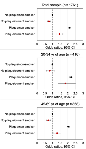
For plaque-free sites, bleeding tendency was lower in smokers, the odds ratio (OR) was 0.773 with a 95% confidence interval of 0.678-0.881 as compared to non-smokers (OR: 1; ref., P < .001). The odds of BOP at plaque-covered sites in non-smokers were increased twofold (OR: 2.117; 2.059-2.177). Albeit bleeding tendency was slightly increased in plaque-covered sites in smokers, it was considerably lower as compared to plaque-covered sites in non-smokers (OR: 1.459; 1.282-1.662, P < .001). Smoking ≥ 20 pack-years further attenuated the association. In smokers, the odds of BOP were reduced in all parts of the dentition, lower and upper anterior and posterior teeth ( = 32.043, P < .001). When restricting the data to younger adults (20-34 year old), smoking had only a slight effect on the association between plaque and BOP. For plaque-free and plaque-covered sites, differences in ORs were not statistically noticeable (P = .221 and P = .235, respectively).
Conclusions
Smoking considerably attenuates the site-specific association between plaque and BOP with a dose-dependent effect. The effect of smoking did not differ across tooth types.
1 INTRODUCTION
Smoking increases susceptibility to periodontitis and is associated with higher levels of periodontal destruction,1 but also reduces the inflammatory response to dental plaque in the gingiva.2 Gingival inflammation is considered a key risk factor for the development and progression of periodontitis.3, 4 Therefore, it is important to investigate the extent to which smoking affects the gingival bleeding response to dental plaque.
In studies of experimental gingivitis, it has been reported that smokers and non-smokers presented similar levels of dental plaque, while the severity of gingival inflammation was less pronounced in smokers as compared to non-smokers.5-9 This was also demonstrated in observational studies where smokers had similar, or even higher, levels of plaque than non-smokers but less gingival bleeding after probing.10-12 What the above-mentioned studies have in common is that the relationship between gingival bleeding and plaque has been studied using subjects’ mean values. Respective associations have been designated as ecological correlations.13 In ecological studies, data are analyzed at a higher level, for example, at the population or group level, rather than at the individual level. When data are analyzed in aggregate form, associations found at the population or group level cannot be inferred to the individual.14 The same applies for the association between subjects’ mean gingival bleeding and mean plaque levels. The gingival inflammatory response to plaque occurs locally at the tooth site, so the (causal) relationship between plaque and gingival inflammation is preferably studied at the site level in order to avoid bias and confounding, the so-called ecological fallacy.15
Site-level analyses of the effects of smoking on gingival bleeding have been assessed in some studies. A large study of a representative sample of the US population showed that smoking had a strong and dose-dependent suppressive effect on gingival bleeding after probing at the site level.16 In a study of Italian dental patients, the odds for a site to bleed on probing were lower in smokers as compared to non-smokers.17 Plaque was not considered in these studies, and consequently, the possible site-specific effects of smoking on the (causal) association between plaque and gingival bleeding were not studied. Possible effects of smoking were, however, explicitly addressed in a six-month longitudinal experiment conducted in a cohort of young adults with mild gingivitis.18 In a steady state, where participants were asked not to alter oral hygiene habits, heavy smokers consistently presented with higher plaque and calculus scores. In this study, site-specific analyses did not reveal evidence for an enhanced or attenuated association between plaque and bleeding on probing in smokers.
Compared to non-smokers, more periodontal destruction has been reported in smokers in all parts of the dentition, which is consistent with a systemic effect of tobacco smoking.19, 20 Anterior teeth seem to be more affected,20, 21 which indicates a possible local effect, as well. As regards the suppressive effect on gingival bleeding in smokers, no differences in maxillary and mandibular molars, premolars, and incisors had been found in a previous study.16
So far, possible effects of smoking on the association between plaque and gingival inflammation have not been studied in a representative sample. Therefore, the aim of the present study was to investigate the influence of smoking on the site-specific association between bleeding on gingival probing and supragingival plaque in a general adult population. A second aim was to assess local effects of smoking by examining whether smoking affects respective associations differently in different parts of the dentition.
2 MATERIAL AND METHODS
2.1 Study population
This is a secondary analysis of data from a dental health survey in Northern Norway (Tromstannen—Oral Health in Northern Norway, TOHNN). The TOHNN study was a cross-sectional study of adults, 20 to 79 years old, living in Troms County, Norway. The sampling and invitation procedures have been described in detail elsewhere.22, 23 In brief, 3000 individuals were randomly selected from the population registry by the National Statistical Institute of Norway. The sample size was based on a hypothesized 10% prevalence of severe periodontitis with a 95% confidence level and margin of error of 1.5%, accounting for a response rate of about 50%. A letter of invitation was sent by mail to 2909 individuals (the remaining 91 individuals had moved out of the county or died). Data were collected between October 2013 and November 2014, with 1986 participants completing the clinical examination and questionnaire. The Regional Committee for Medical and Health Research Ethics North, Norway, approved the study (2013/348/REC North). All participants provided written informed consent.
2.2 Inclusion and exclusion criteria
All subjects with two or more natural teeth were included in the analysis (n = 1933). Individuals with incomplete periodontal recordings (n = 4) were excluded. This resulted in 1929 individuals (946 males and 983 females, aged 20-79 years; mean age ± standard deviation: 47.5 ± 15.3 years).
2.3 Clinical examinations
Examinations were performed in dental offices by 11 dentists (employed by the Public Dental Health Service in Troms County) assisted by dental nurses. All examiners were trained by an experienced periodontist (NO) prior to the study regarding periodontal measurements. Measurements were made for all teeth; however, third molars and implants were excluded from analyses. The presence of supragingival plaque, bleeding on probing (BOP), and periodontal probing depth (PD) were originally assessed at six sites per tooth. PD was measured to the nearest millimeter with a periodontal probe with single millimeter gradations (PUNC 15, American Eagle Instruments, Inc, Missoula, MT, United States). BOP was registered within about 20 s after probing to the bottom of the gingival sulcus or periodontal pocket as present or absent. A modification of the Plaque Control Record24 was applied in order to assess supragingival plaque as present or not using a mouth mirror and periodontal probe. No disclosing agent was used. Data were entered in T4 software (Carestream Dental LLC, Atlanta, GA, USA) with recordings of six sites per tooth for PD and BOP but only 4 sites (mesial, buccal, distal, and palatal/lingual) for the presence of plaque due to restrictions of the software. Hence, plaque was considered present at mesial surfaces when it could either be ascertained mesiobuccally or mesiopalatally/mesiolingually (or at both surfaces). For distal surfaces, the respective procedure was applied. For site-level analysis of PD and BOP, we had to collapse 6-site recordings to 4: distal, buccal, mesial, and palatal/lingual. In order to match the presence of plaque with the presence of BOP, we applied the same procedure: BOP was considered present at mesial surfaces when it had occurred either mesiobuccally or mesiopalatally/mesiolingually (or at both surfaces). For distal surfaces, the respective procedure was applied. In order to match PD with plaque and BOP, the deeper measurement (either mesiobuccally or mesiopalatally/mesiolingually) was selected. Again, for distal surfaces the respective procedure was applied. Height (m) and weight (kg) were measured at time of examination, and body mass index (BMI, kg/m2) was calculated. Inter-examiner reliability of PD measurements was assessed between each of the examiners and the experienced periodontist at the site level (median intraclass correlation coefficient: 0.81, range: 0.43-0.94), described in detail elsewhere.23 Inter-examiner reliability was not assessed for registration of BOP and dental plaque.
2.4 Questionnaire
Information about demographics, socioeconomic factors, behaviors, and health was collected by self-reported questionnaire. Age was stratified in categories 20-34, 35-44, 45-69, and 70-79 years. Education was categorized as less than high school, high school, and university level. Annual household income was analyzed in three categories (high, intermediate, and low) according to national tertiles of household income in 2013.25 Smoking was assessed by smoking status (daily smoker: yes/no) and categorized as non-smoker and smoker. Smoking level was categorized as non-smoker, smoker with < 20 pack-years, and smoker with ≥ 20 pack-years. Number of pack-years was estimated from two questions: number of cigarettes per day and number of years smoking. Number of pack-years was calculated as (number of cigarettes per day/20) × number of years smoked. A cutoff value of 20 pack-years has been used in previous studies to define heavy smoking.26 Former smokers (n = 42) were excluded from analyses because of unclear reporting of former smoking status. Missing data in other covariates also resulted in exclusion from analysis. See Table 1 for number of excluded participants in each category.
| Individual-related variables (level 3) | N = 1929 |
|---|---|
| Age, years [mean (SD)] | 47.5 (15.3) |
| Age-group [n (%)] | |
| 20-34 years | 462 (24.0) |
| 35-44 years | 386 (20.0) |
| 45-69 years | 926 (48.0) |
| 70-79 years | 155 (8.0) |
| Gender [n (%)] | |
| Female | 983 (51.0) |
| Male | 946 (49.0) |
| Education [n (%)] | |
| University level | 796 (41.3) |
| High school | 835 (43.3) |
| Less than high school | 280 (14.5) |
| Missing | 18 (0.9) |
| Income [n (%)] | |
| High | 371 (19.2) |
| Intermediate | 917 (47.5) |
| Low | 566 (29.3) |
| Missing | 75 (3.9) |
| Smoking [n (%)] | |
| Smoker | 284 (14.7) |
| Non-smoker | 1590 (82.4) |
| Missing | 55 (2.9) |
| Smoking level [n (%)] | |
| ≥20 pack-years | 74 (3.8) |
| <20 pack-years | 180 (9.3) |
| Non-smoker | 1590 (82.4) |
| Missing | 85 (4.4) |
| Diabetes [n (%)] | 71 (3.7) |
| BMI (kg/m2) [n (%)] | |
| Normal weight (<25) | 656 (34.0) |
| Overweight (25-29.9) | 773 (40.1) |
| Obese (≥30) | 472 (24.5) |
| Missing | 28 (1.4) |
| BOP scorea [mean (SD)] | 37.1 (19.9) |
| Plaque scorea [mean (SD)] | 44.6 (22.7) |
| Tooth-related variables (level 2) | N = 48 043 |
|---|---|
| Tooth type [n (%)] | |
| Upper anterior teeth | 10 734 (22.3) |
| Lower anterior teeth | 11 374 (23.7) |
| Upper posterior teeth | 12 790 (26.6) |
| Lower posterior teeth | 13 145 (27.4) |
| Site-related variables (level 1) | N = 192 172 |
|---|---|
| PD, mm [mean (SD)] | 2.1 (1.0) |
| BOP, % [mean (SD)] | 36.6 (48.2) |
| Plaque, % [mean (SD)] | 43.6 (49.6) |
- Abbreviations: BMI, body mass index; BOP, bleeding on probing; PD, probing depth; SD, standard deviation.
- a Subjects' averages.
2.5 Statistical analysis
Descriptive data are presented as means with standard deviations (SD) or numbers with proportions in parentheses. Three-level (subject, tooth, and site), random intercept, logistic regression models were built, with BOP as the outcome. A detailed description of the models can be found in Appendix S1.
Plaque, PD, smoking status (non-smoker and smoker), age-group, gender, education, income, BMI, and tooth type were entered as covariates. In order to assess how much smoking status modifies the association between plaque and bleeding on probing, interaction terms of “plaque × smoking status” were included as well. Bleeding tendency was also assessed at different tooth types, that is, upper anterior, lower anterior, upper posterior, and lower posterior teeth. In further analyses, the association between plaque and BOP was assessed in young adults (20-34 years old) and middle-aged adults (45-69 years old). Results are reported as regression coefficients, odds ratios (OR), and respective 95% confidence intervals (CI). If considered necessary, p-values were derived from Wald tests. However, any inferential statistics (P-values, CIs) were intended to be exploratory, not confirmatory. No correction for multiple testing was done. P-values < 0.05 were considered as statistically noticeable.
Data were analyzed using special software (MLwiN, version 3.02, Centre for Multilevel Modelling, University of Bristol, Bristol, UK). For details, see Appendix S1.
3 RESULTS
There were 1929 dentate individuals with 192 172 sites with complete records of BOP, plaque, and PD. Because of missing values in education, income, smoking status, and BMI, the final model included 1761 individuals with 176 220 sites. Mean percent BOP for excluded participants was 39.5%, and mean percent plaque was 46.9%, compared to 36.9% and 44.4%, respectively, for included participants (BOP: t(1927) = −1.48, P = .141; plaque: t(1927) = −1.39, P = .165). Characteristics of the study population are presented in Table 1.
Estimates of three-level random intercept models of BOP are listed in Table 2. According to the null model (without covariates), on average 34.3% gingival units bled upon probing (ALOGit of −0.649). The reason for the discrepancy with the respective figure in Table 1 (37.1%) might be explained by the fact that the latter was calculated based on aggregate data. In the null model, the variance partition coefficient (VPC) was 0.236, meaning 23.6% of the total variance was attributable to differences between subjects. In the model with main effects, plaque, PD, and smoking, the OR of BOP when plaque was present at a site was (exponential of 0.733) 2.08 (95% CI: 2.03; 2.14). PD had an even stronger influence on the odds of BOP. With every millimeter increase in PD, the odds for BOP increased by a factor of 2.82 (2.78; 2.87). On the other hand, being a smoker drastically decreased the odds of BOP. The OR was 0.744 (0.659; 0.840).
| Null model (1) | Main effects (2) | Full model (3) | |
|---|---|---|---|
| Estimate (SE) | Estimate (SE) | Estimate (SE) | |
| Fixed effects | |||
| β0jk (intercept) | −0.649 (0.024) | −1.020 (0.025) | −0.823 (0.092) |
| Plaque vs no plaque | 0.733 (0.013) | 0.750 (0.014) | |
| PD (centered on mean) | 1.038 (0.008) | 1.039 (0.009) | |
| Smoker vs non-smoker | −0.296 (0.062) | −0.258 (0.067) | |
| Plaque × smoker | −0.114 (0.039) | ||
| Female vs male | 0.041 (0.046) | ||
| Age-group (reference: 20-34 years) | |||
| 35-44 years | −0.309 (0.069) | ||
| 45-69 years | −0.388 (0.058) | ||
| 70-79 years | −0.357 (0.098) | ||
| Education (reference: less than high school) | |||
| High school | −0.084 (0.072) | ||
| University level | −0.219 (0.076) | ||
| Income (reference: low income) | |||
| Intermediate income | 0.063 (0.054) | ||
| High income | 0.010 (0.071) | ||
| BMI (reference: normal weight) | |||
| Overweight | 0.149 (0.053) | ||
| Obese | 0.306 (0.060) | ||
| Random effects | |||
| v0k (subject-level variance) | 1.022 (0.035) | 0.831 (0.030) | 0.773 (0.029) |
| u0jk (tooth-level variance) | 0.026 (0.008) | 0.144 (0.011) | 0.144 (0.011) |
| VPC | 0.236 | 0.195 | 0.184 |
- Abbreviations: BMI, body mass index; PD, probing depth; SE, standard error; VPC, variance partition coefficient (VPC = σ2v/(σ2v + σ2u+π2/3, see Appendix S1).
In order to examine whether smoking is an effect modifier in the association between plaque and BOP, the full model was set up with main effects, the interaction term “plaque × smoking,” and further covariates age-groups, gender, education, income, and BMI (Table 2). Older age and higher level of education both reduced the odds of bleeding, while overweight and obese persons had increased odds of BOP. Interestingly, not only plaque and smoking status, but also the interaction term “plaque × smoking” strongly influenced the odds of BOP.
Figure 1 displays three different, fully adjusted, models of BOP. With a site without plaque in a non-smoking subject as reference, ORs and 95% CIs were calculated for sites with and without plaque in non-smokers and smokers. Regarding the total sample, there was apparently a very strong attenuating effect of smoking on the association between plaque and BOP (P = 1.12 × 10⁻⁴ and P = 1.92 × 10⁻⁸ for non-plaque-covered and plaque-covered sites, respectively), when compared to respective sites in non-smokers. As age-group appeared to have also an effect on the association, two separate models were set up with low and high proportion of smokers (Figure 1). Estimates of the models are given in Table S4. Interestingly, in the youngest age-group, ORs were only slightly lower in smokers (P = .221 and P = .235, respectively). In contrast, the attenuating effect of smoking was even stronger in 45- to 69-year-olds (P = 6.85 × 10⁻⁴ and P = 1.92 × 10⁻⁶, respectively).

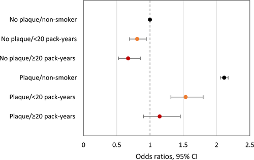
When considering the effect of lifetime tobacco exposure (pack-years), ORs for BOP were attenuated in particular in smokers with ≥ 20 pack-years (Figure 2, Table S1). As compared to plaque-free sites in non-smokers, the OR was 0.807 (0.689; 0.945) in smokers with < 20 pack-years and 0.671 (95% CI: 0.526-0.856) in smokers with ≥ 20 pack-years ( = 16.190, P = 3.05 × 10⁻⁴). In non-smokers, as compared to plaque-free sites, the OR of BOP was 2.115 (2.057; 2.175) for sites covered with plaque. In smokers, the association was likewise attenuated (Figure 2): The OR for BOP was 1.537 (1.314; 1.799) for smokers with < 20 pack-years, while it was 1.146 (0.901; 1.456) for smokers with ≥ 20 pack-years (
 =37.756, P = 6.33 × 10⁻⁹).

Table 3 presents ORs for BOP in different parts of the dentition in smokers as compared to non-smokers. Estimates of the model are listed in Table S2. As compared to non-smokers, the odds of BOP were reduced in all parts of the dentition, with ORs ranging between 0.685 (0.596; 0.787) for lower anterior teeth and 0.773 (0.675; 0.886) for lower posterior teeth ( = 32.043, P = 1.88 × 10⁻⁶). Interestingly, smokers had more plaque as compared to non-smokers in all parts of the dentition (
 = 15.234, P = .004, Table S3) with no difference between tooth types.
| Tooth type | OR | 95% CI | P-value |
|---|---|---|---|
| Upper anterior teeth | 0.710 | 0.616; 0.819 | 2.30 × 10⁻⁶ |
| Lower anterior teeth | 0.685 | 0.596; 0.787 | 9.44 × 10⁻⁸ |
| Upper posterior teeth | 0.725 | 0.631; 0.832 | 4.62 × 10⁻⁶ |
| Lower posterior teeth | 0.773 | 0.675; 0.886 | 2.19 × 10⁻⁴ |
4 DISCUSSION
The present analysis of data collected in a representative sample of adults in Northern Norway confirmed that smokers had less gingival bleeding upon probing to the bottom of the pocket/sulcus than non-smokers. The results are in line with site-specific analyses of data collected in a population-based epidemiological study conducted in the United States.16 In that study, authors had observed that the OR of bleeding upon probing was 0.53 in adults smoking even 10 cigarettes per day or less as compared to never smokers. It further decreased in heavy smokers. While the presence of plaque was not assessed in that study, authors reported a strong effect of sub- or supragingival calculus (in a way a proxy for plaque) on gingival bleeding in never smokers, which was gradually and largely attenuated in former, light, and heavy smokers. The effect of heavy smoking was, in fact, so strong that sites with calculus in heavy smokers showed less than or the same bleeding tendency as calculus-free sites in non-smokers.
It should be noted, however, that in the above study16 bleeding upon marginal probing was recorded rather than bleeding on probing to the bottom of the pocket. While in deep pockets bleeding on probing to the bottom of the pocket points to the presence of subgingival plaque, in the case of shallow pockets supragingival plaque may lead to bleeding. In the present material, only 4.5% of sites were 4 mm deep and 2.1% were 5 mm or deeper. Hence, the vast majority of sites (93.4%) were shallow (1-3 mm). All models were adjusted for PD anyway. A model confining the present material to shallow pockets only (see Table S6) indicated that estimates were notably not much different from those in Table 2, where the whole material was used. In the current classification of periodontal and peri-implant diseases and conditions, the extent of BOP (to the bottom of the sulcus/pocket) has generally been recommended for case definitions of periodontal health, (plaque-induced) gingivitis, and as an important sign for deciding whether the case of periodontitis is stable or unstable after periodontal treatment.27 BOP may be regarded as a rather simple, objective, and accurate means for the purpose of healthy and gingivitis case definitions. Recording of BOP is user-friendly and economic and requires minimal technology.28 Therefore, it has been recognized as a universally applicable means to describe local gingival inflammation in epidemiological studies as well.27
Our site-specific results strongly indicate that bleeding tendency after periodontal probing to the bottom of the pocket/sulcus is also influenced by smoking and smoking dose.
To what extent does smoking actually affect gingival bleeding response to supragingival plaque? As compared to sites without plaque, the odds of BOP were more than twice as large at plaque-covered sites in non-smokers. On the other hand, the OR for plaque-covered sites in smokers was only slightly increased to 1.45 (Figure 1), pointing to smoking as a strong effect modifier of the (causal) relationship between plaque and gingival inflammation. The extent of attenuation is remarkably similar to that observed in a randomized controlled trial 6 weeks after the introduction of triclosan-containing toothpaste in non-smoking young adults with mild gingivitis who had been asked not to change oral hygiene habits.29 In that study, as compared to plaque-free surfaces, the ORs for BOP were increased to 2.11-2.43 in volunteers using control toothpaste (depending on the Silness and Löe plaque index scores30). Respective ORs were between 1.07 and 1.86 in volunteers brushing with triclosan-containing toothpaste.
Interestingly, in the present study the bleeding response was not so much affected by smoking in younger adults (20- to 34-year-olds), a result that is in line with observations made in a 6-month longitudinal experiment in 19- to 30-year-old soldiers of the German Armed Forces who again had been asked not to change oral hygiene habits.18 A possible explanation for these observations could be that young smokers have not been exposed to tobacco long enough for it to affect the bleeding response. Moreover, when considering the lifetime exposure of tobacco in terms of pack-years in the present study, the bleeding response was attenuated with a dose-dependent effect for smokers with < 20 pack-years and those with ≥ 20 pack-years, also indicating that the effect of smoking may depend on the duration or amount of exposure.
The overall bleeding tendency of the gingiva, regardless of smoking status, was higher at lower anterior teeth as compared to other teeth when adjusted for plaque, PD, and subject-level covariates (Table S2). Tooth-type differences in gingival bleeding tendency were also reported in a retrospective study of dental patients in Italy.17 These authors found that the gingiva around posterior teeth was more likely to bleed upon probing than the gingiva at anterior teeth. Notably, differences were rather small and presence of plaque was not adjusted for.
In smokers, bleeding tendency was also lower in all parts of the dentition with no noticeable difference between tooth types as compared to non-smokers (Table 3). This is in agreement with results of the above-mentioned population-based study in the United States, where authors reported no difference in the effect of smoking on gingival bleeding tendency between different tooth groups or jaws.16
Our results also considered other factors associated with gingival bleeding. As mentioned before, the association of PD (a proxy for subgingival plaque) with BOP was very strong. With each millimeter increase, the odds of BOP increased almost threefold. This is consistent with results from previous studies where the OR of BOP was increased twofold per mm increase in PD,17 or when comparing sites with increased PD to healthy sites (PD 0-3 mm).16 Higher age (≥35 year) reduced the odds of bleeding by around 30%, apparently with a threshold effect, as gingival bleeding did not vary among persons 45 years old and older. A study of experimental gingivitis found that older persons developed more gingivitis than younger persons,31 while no difference in bleeding probability according to age was reported among Italian dental patients.17 In the present study, there was no difference in bleeding tendency between males and females. In previous site-specific analyses, differences in gingival bleeding between genders have been reported, however, in both directions.16, 17 In the present study, persons with higher education were less likely to bleed on probing, while income was not related to bleeding tendency. Previous studies have reported that people with lower income were more likely to show gingival bleeding16 and that lower education was related to more BOP or gingival inflammation.32, 33 In our study, overweight and obesity increased the bleeding tendency of the gingiva; however, higher body mass index was also associated with higher plaque levels. Obesity has been associated with periodontitis with several possible mechanisms proposed, that is, increased inflammatory response, change in dental plaque amount and composition, or both.34 Our results indicate that overweight/obesity is associated with more gingival bleeding and partly through increased levels of plaque. In particular, there was no noticeable interaction between plaque and overweight/obesity (Table S5), meaning BMI, in contrast to smoking, is not an effect modifier as regards the association between plaque and bleeding on probing.
The underlying mechanisms of smoking and its effect on gingival bleeding are somewhat unclear. There is limited evidence that tobacco smoke promotes gingival vasoconstriction in humans.35-39 There is some evidence of tobacco-induced suppressed angiogenesis, where a reduced number of gingival vessels or vessels of smaller caliber have been found in smokers relative to non-smokers.6, 40-42 Thermally induced nerve damage in the oral cavity of smokers43, 44 could potentially affect the microvascular response of the gingiva.2 Additionally, tobacco smoking alters the dental plaque composition.45 Findings from a large study of the human oral microbiome in US adults indicate that smoking promotes an anaerobic oral environment and a bacterial community with a reduced capability of degrading toxic components of cigarette smoke.45 Furthermore, it has been proposed that smoking can suppress oral pathogens’ production of short-chain fatty acid, which can influence components of immune and healing responses, thereby presenting an additional mechanism for reducing vascular response to dental plaque.46 Most importantly, cigarette smoking has been reported to affect the immune responses.47 For example, decreased levels of pro-inflammatory biomarkers in smokers with periodontitis suggest a reduced capacity to recruit inflammatory and immune cells, which may explain the enhanced susceptibility to periodontitis48 and the reduced bleeding response to plaque.
There are many factors, other than smoking, that can modify the gingival inflammatory response to plaque, which have not been controlled for in the current study. Such factors include pregnancy, diabetes, Down's syndrome, vitamin C deficiency, anti-microbial and anti-inflammatory agents, and conditions affecting the immune system (reviewed by Tatakis et al49). Moreover, as mentioned before, toothpaste containing the anti-bacterial compound triclosan was shown to attenuate the association between plaque and BOP in a randomized controlled trial.29 Additionally, studies have shown that diet, and especially vitamin D, can affect gingivitis.50, 51 Both smoking and obesity have been associated with lower levels of vitamin D in a population-based study in Northern Norway.52 Finally, the host-dependent variation in gingivitis susceptibility should be considered. In several studies, a subject-specific gingival inflammatory response has been reported, and “high and low responders”53 or “fast and slow responders” were identified.54
With increased focus on the inflammatory nature of periodontitis, host modulation therapy is an emerging treatment strategy for managing periodontitis, aiming to control the inflammation in order to control the infection.55 In this respect, smoking's effect on periodontal disease should be considered, where gingival inflammation is reduced, but periodontal destruction is enhanced. Smoking has on one hand toxic and on the other hand immunosuppressive effects.47 The latter might be the reason why incidence and/or severity of some inflammatory diseases have been reported to be reduced in smokers.56-58 Nicotine, the main immunosuppressive constituent of cigarette smoke, has even been suggested as a potential therapeutic agent in chronic inflammatory diseases such as dermatitis and ulcerative colitis.59, 60
As an epidemiological survey, the study has several limitations that need to be critically addressed. The study design was cross-sectional, so no causal relationship can be concluded. BOP and plaque were only measured at one time point, assuming a steady-state plaque environment.61 Due to the restrictions of the recording software, allowing only 4-site recordings of plaque, we had to collapse bleeding scores and PD originally measured at 6 sites per tooth to 4. This could have introduced bias with unknown effects on the results. Moreover, no pressure-controlled probe was used and examiners were not calibrated for measurements of the main outcome, BOP, as in a study of agreement and association of gingival bleeding after repeat probing it was found that the reliability of the rather invasive diagnostic, BOP, was generally low.62 In order to precisely assess the dose-dependent effect of smoking on the gingival bleeding response to plaque, information about amount and duration of smoking would be highly desirable. There was no objective measure of smoking, for example, measuring serum cotinine levels. Smoking history was self-reported in a questionnaire, presenting a potential source of imprecise smoking estimates. Nevertheless, reported smoking frequency was close to national estimates.63 Furthermore, pack-years was estimated from current number of cigarettes smoked per day. Number of daily cigarettes could have varied over participants’ years of smoking, meaning the estimated pack-years might not accurately depict their true cumulative dose of cigarettes. Some of the persons that reported as non-smokers could have been former smokers. Previous studies have reported a suppressive effect on gingival bleeding among former smokers, albeit small, as compared to smokers.16 In the present study, for models including all covariates, 166 participants had been excluded because of missing values in questions about education, income, smoking, and BMI. However, there were only small differences in BOP and plaque levels between the excluded and included participants.
Despite these limitations, this is, to the best of our knowledge, the first study to assess the influence of smoking on the gingival inflammatory response to supragingival plaque in a general adult population. Moreover, multilevel analysis confirms previous evidence of the attenuating effect of smoking on the inflammatory response to dental plaque at the site level.
In conclusion, analyses of data from a population-based epidemiological study in Northern Norway show that smoking reduces the general bleeding tendency of the gingiva but also attenuates the site-specific association between plaque and gingival bleeding. The extent of the attenuation is dependent on tobacco exposure, where smoking ≥ 20 pack-years further attenuates the association between gingival bleeding and plaque. The effect of smoking did not differ between different regions of the dentition. A reduced inflammatory response to dental plaque indicates that there might be a need for different strategies for periodontal inflammation control among smokers and non-smokers. BOP might not be a reliable measure of inflammation in smokers.


