Regional Cerebral Gray Matter Volume in HIV-Positive Patients with Executive Function Deficits
Corresponding Author
Diogo Goulart Corrêa
Department of Radiology, Hospital Universitário Clementino Fraga Filho, Federal University of Rio de Janeiro, Cidade Universitária, Ilha do Fundão, Rio de Janeiro, RJ, Brazil
Clínica de Diagnóstico por Imagem (CDPI), Avenida das Américas, 4666, 302A, 303, 307, 325, 326, Barra da Tijuca, Rio de Janeiro, RJ, Brazil
Correspondence: Address correspondence to Diogo Goulart Corrêa, Hospital Universitário Clementino Fraga Filho, Rua Rodolpho Paulo Rocco 255, Cidade Universitária, Ilha do Fundão, Rio de Janeiro, RJ 21941913, Brazil. E-mail: [email protected].Search for more papers by this authorNicolle Zimmermann
Department of Radiology, Hospital Universitário Clementino Fraga Filho, Federal University of Rio de Janeiro, Cidade Universitária, Ilha do Fundão, Rio de Janeiro, RJ, Brazil
Department of Psychology, Pontifical Catholic University of Rio Grande do Sul, Avenida Ipiranga, 6681, Partenon, Porto Alegre, RS, Brazil
Search for more papers by this authorTania Maria Netto
Department of Radiology, Hospital Universitário Clementino Fraga Filho, Federal University of Rio de Janeiro, Cidade Universitária, Ilha do Fundão, Rio de Janeiro, RJ, Brazil
Search for more papers by this authorGustavo Tukamoto
Clínica de Diagnóstico por Imagem (CDPI), Avenida das Américas, 4666, 302A, 303, 307, 325, 326, Barra da Tijuca, Rio de Janeiro, RJ, Brazil
Search for more papers by this authorNina Ventura
Department of Radiology, Hospital Universitário Clementino Fraga Filho, Federal University of Rio de Janeiro, Cidade Universitária, Ilha do Fundão, Rio de Janeiro, RJ, Brazil
Clínica de Diagnóstico por Imagem (CDPI), Avenida das Américas, 4666, 302A, 303, 307, 325, 326, Barra da Tijuca, Rio de Janeiro, RJ, Brazil
Search for more papers by this authorSarah de Castro Bellini Leite
Department of Radiology, Hospital Universitário Clementino Fraga Filho, Federal University of Rio de Janeiro, Cidade Universitária, Ilha do Fundão, Rio de Janeiro, RJ, Brazil
Search for more papers by this authorRafael Ferracini Cabral
Department of Radiology, Hospital Universitário Clementino Fraga Filho, Federal University of Rio de Janeiro, Cidade Universitária, Ilha do Fundão, Rio de Janeiro, RJ, Brazil
Clínica de Diagnóstico por Imagem (CDPI), Avenida das Américas, 4666, 302A, 303, 307, 325, 326, Barra da Tijuca, Rio de Janeiro, RJ, Brazil
Search for more papers by this authorRochele Paz Fonseca
Department of Radiology, Hospital Universitário Clementino Fraga Filho, Federal University of Rio de Janeiro, Cidade Universitária, Ilha do Fundão, Rio de Janeiro, RJ, Brazil
Department of Psychology, Pontifical Catholic University of Rio Grande do Sul, Avenida Ipiranga, 6681, Partenon, Porto Alegre, RS, Brazil
Search for more papers by this authorPaulo Roberto Valle Bahia
Department of Radiology, Hospital Universitário Clementino Fraga Filho, Federal University of Rio de Janeiro, Cidade Universitária, Ilha do Fundão, Rio de Janeiro, RJ, Brazil
Search for more papers by this authorEmerson Leandro Gasparetto
Department of Radiology, Hospital Universitário Clementino Fraga Filho, Federal University of Rio de Janeiro, Cidade Universitária, Ilha do Fundão, Rio de Janeiro, RJ, Brazil
Clínica de Diagnóstico por Imagem (CDPI), Avenida das Américas, 4666, 302A, 303, 307, 325, 326, Barra da Tijuca, Rio de Janeiro, RJ, Brazil
Search for more papers by this authorCorresponding Author
Diogo Goulart Corrêa
Department of Radiology, Hospital Universitário Clementino Fraga Filho, Federal University of Rio de Janeiro, Cidade Universitária, Ilha do Fundão, Rio de Janeiro, RJ, Brazil
Clínica de Diagnóstico por Imagem (CDPI), Avenida das Américas, 4666, 302A, 303, 307, 325, 326, Barra da Tijuca, Rio de Janeiro, RJ, Brazil
Correspondence: Address correspondence to Diogo Goulart Corrêa, Hospital Universitário Clementino Fraga Filho, Rua Rodolpho Paulo Rocco 255, Cidade Universitária, Ilha do Fundão, Rio de Janeiro, RJ 21941913, Brazil. E-mail: [email protected].Search for more papers by this authorNicolle Zimmermann
Department of Radiology, Hospital Universitário Clementino Fraga Filho, Federal University of Rio de Janeiro, Cidade Universitária, Ilha do Fundão, Rio de Janeiro, RJ, Brazil
Department of Psychology, Pontifical Catholic University of Rio Grande do Sul, Avenida Ipiranga, 6681, Partenon, Porto Alegre, RS, Brazil
Search for more papers by this authorTania Maria Netto
Department of Radiology, Hospital Universitário Clementino Fraga Filho, Federal University of Rio de Janeiro, Cidade Universitária, Ilha do Fundão, Rio de Janeiro, RJ, Brazil
Search for more papers by this authorGustavo Tukamoto
Clínica de Diagnóstico por Imagem (CDPI), Avenida das Américas, 4666, 302A, 303, 307, 325, 326, Barra da Tijuca, Rio de Janeiro, RJ, Brazil
Search for more papers by this authorNina Ventura
Department of Radiology, Hospital Universitário Clementino Fraga Filho, Federal University of Rio de Janeiro, Cidade Universitária, Ilha do Fundão, Rio de Janeiro, RJ, Brazil
Clínica de Diagnóstico por Imagem (CDPI), Avenida das Américas, 4666, 302A, 303, 307, 325, 326, Barra da Tijuca, Rio de Janeiro, RJ, Brazil
Search for more papers by this authorSarah de Castro Bellini Leite
Department of Radiology, Hospital Universitário Clementino Fraga Filho, Federal University of Rio de Janeiro, Cidade Universitária, Ilha do Fundão, Rio de Janeiro, RJ, Brazil
Search for more papers by this authorRafael Ferracini Cabral
Department of Radiology, Hospital Universitário Clementino Fraga Filho, Federal University of Rio de Janeiro, Cidade Universitária, Ilha do Fundão, Rio de Janeiro, RJ, Brazil
Clínica de Diagnóstico por Imagem (CDPI), Avenida das Américas, 4666, 302A, 303, 307, 325, 326, Barra da Tijuca, Rio de Janeiro, RJ, Brazil
Search for more papers by this authorRochele Paz Fonseca
Department of Radiology, Hospital Universitário Clementino Fraga Filho, Federal University of Rio de Janeiro, Cidade Universitária, Ilha do Fundão, Rio de Janeiro, RJ, Brazil
Department of Psychology, Pontifical Catholic University of Rio Grande do Sul, Avenida Ipiranga, 6681, Partenon, Porto Alegre, RS, Brazil
Search for more papers by this authorPaulo Roberto Valle Bahia
Department of Radiology, Hospital Universitário Clementino Fraga Filho, Federal University of Rio de Janeiro, Cidade Universitária, Ilha do Fundão, Rio de Janeiro, RJ, Brazil
Search for more papers by this authorEmerson Leandro Gasparetto
Department of Radiology, Hospital Universitário Clementino Fraga Filho, Federal University of Rio de Janeiro, Cidade Universitária, Ilha do Fundão, Rio de Janeiro, RJ, Brazil
Clínica de Diagnóstico por Imagem (CDPI), Avenida das Américas, 4666, 302A, 303, 307, 325, 326, Barra da Tijuca, Rio de Janeiro, RJ, Brazil
Search for more papers by this authorConflicts of Interest: There is no conflict of interest to declare.
ABSTRACT
PURPOSE
To evaluate whether human immunodeficiency virus (HIV)-positive patients with and without executive functions deficits and healthy control subjects differ on cortical thickness and subcortical brain structures volume in vivo.
METHODS
In total, 34 HIV-positive patients with executive functions deficits were compared with 13 HIV-positive patients without executive functions deficits and 19 gender-, age-, and education-matched control subjects. Executive functions impairments were classified by performance on the Wisconsin card sorting test. T1 3-dimensional magnetization prepared rapid gradient echo-weighted imaging was performed using a 1.5 Tesla (magnetic resonance) MR scanner. FreeSurfer software was used to perform cortical reconstruction and volumetric segmentation of subcortical gray matter structures.
RESULTS
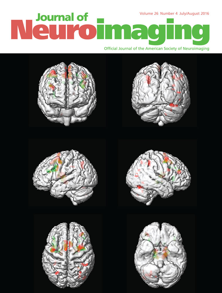
HIV-positive patients with executive functions deficits had smaller volumes in the right and left caudate compared with the HIV-positive patients without executive functions deficits and control groups. In addition, HIV-positive patients with executive functions deficits had smaller volumes in their left accumbens, right putamen, and globus pallidum compared with the control group. No significant differences in cortical thickness were observed between the groups.
CONCLUSION
HIV-positive patients with executive functions deficits have reduced volumes of several subcortical structures, primarily in the caudate nucleus.
References
- 1Ellis RJ, Calero P, Stockin MD. HIV infection and the central nervous system: a primer. Neuropsychol Rev 2009; 19: 144-51.
- 2Ernst T, Chang L, Jovicich J, et al. Abnormal brain activation on functional MRI in cognitively asymptomatic HIV patients. Neurology 2002; 59: 1343-9.
- 3Antinori A, Arendt G, Becker JT, et al. Updated research nosology for HIV-associated neurocognitive disorders. Neurology 2007; 69: 1789-99.
- 4Jurado MB, Rosselli M. The elusive nature of executive functions: a review of our current understanding. Neuropsychol Rev 2007; 17: 213-33.
- 5Cattie JE, Doyle K, Weber E, et al. Planning deficits in HIV-associated neurocognitive disorders: component processes, cognitive correlates, and implications for everyday functioning. J Clin Exp Neuropsychol 2012; 34: 906-18.
- 6Reger M, Welsh R, Razani J, et al. A meta-analysis of the neuropsychological sequelae of HIV infection. J Int Neuropsychol Soc 2002; 8: 410-24.
- 7Dawes S, Suarez P, Casey CY, et al. Variable patterns of neuropsychological performance in HIV-1 infection. J Clin Exp Neuropsychol 2008; 30: 613-26.
- 8Woods SP, Moore DJ, Weber E, et al. Cognitive neuropsychology of HIV-associated neurocognitive disorders. Neuropsychol Rev 2009; 19: 152-68.
- 9Aylward EH, Henderer JD, McArthur JC, et al. Reduced basal ganglia volume in HIV-1-associated dementia: results from quantitative neuroimaging. Neurology 1993; 43: 2099-104.
- 10Patel SH, Kolson DL, Glosser G, et al. Correlation between percentage of brain parenchymal volume and neurocognitive performance in HIV-infected patients. Am J Neuroradiol 2002; 23: 543-9.
- 11Di Sclafani V, Mackay RD, Meyerhoff DJ, et al. Brain atrophy in HIV infection is more strongly associated with CDC clinical stage than with cognitive impairment. J Int Neuropsychol Soc 1997; 3: 276-87.
- 12Ragin AB, Du H, Ochs R, et al. Structural brain alterations can be detected early in HIV infection. Neurology 2012; 11:79: 2328-34.
- 13Stout JC, Ellis RJ, Jernigan TL, et al. Progressive cerebral volume loss in human immunodeficiency virus infection: a longitudinal volumetric magnetic resonance imaging study. Arch Neurol 1998; 55: 161–8.
- 14Becker JT, Sanders J, Madsen SK, et al. Subcortical brain atrophy persists even in HAART-regulated HIV disease. Brain Imaging Behav 2011; 5: 77-85.
- 15Küper M, Rabe K, Esser S, et al. Structural gray and white matter changes in patients with HIV. J Neurol 2011; 258: 1066-75.
- 16Thompson PM, Dutton RA, Hayashi KM, et al. Thinning of the cerebral cortex visualized in HIV/AIDS reflects CD4+ T lymphocyte decline. Proc Natl Acad Sci USA 2005; 102: 15647-52.
- 17Kallianpur KJ, Kirk GR, Sailasuta N, et al. Regional cortical thinning associated with detectable levels of HIV DNA. Cereb Cortex 2012; 22: 2065-75.
- 18Becker JT, Maruca V, Kingsley LA, et al. Factors affecting brain structure in men with HIV disease in the post-HAART era. Neuroradiology 2012; 54: 113-21.
- 19Heaton RK, Chelune GJ, Talley JL, et al. Wisconsin Card Sorting Test Manual (revised and expanded). Odessa, FL: Psychological Assessment Resources; 1993.
- 20Cunha JA, Trentini CM, Argimon I, et al. Adaptação e padronização brasileira do Manual do Teste Wisconsin de Classificação de Cartas. São Paulo, SP: Casa do Psicólogo; 2005.
- 21Kavé G, Heled E, Vakil E, et al. Which verbal fluency measure is most useful in demonstrating executive deficits after traumatic brain injury? J Clin Exp Neuropsychol 2011; 33: 358-65.
- 22Gauthier L, Dehaut F, Joanette Y. The Bells Test: a quantitative and qualitative test for visual neglect. J Int Neuropsychol Soc 1989; 11: 49-54.
- 23Fonseca RP, Oliveira C, Gindri G, et al. Teste Hayling: um instrumento de avaliação de componentes das funções executivas. In: C Hutz, ed. Avaliação psicológica e neuropsicológica de crianças e adolescente. São Paulo, SP: Casa do Psicólogo; 2010: 337–64.
- 24 War Department Adjutant General's Office. Army individual test battery: manual of directions and scoring. Washington DC: War Department Adjutant General's Office; 1944.
- 25Wechsler D. Wechsler Adult Intelligence Scale – III. San Antonio, TX: The Psychological Corporation; 1997.
- 26Salgado JV, Malloy-Diniz LF, Abrantes SS, et al. Applicability of the Rey Auditory-Verbal Learning Test to an adult sample in Brazil. Revista Brasileira de Psiquiatria 2011; 33: 234–7.
- 27Fonseca RP, Salles JF, Parente MAMP. Instrumento de avaliação neuropsicológica breve NEUPSILIN. São Paulo, SP: Vetor; 2009.
- 28Fonseca RP, Parente MAMP, Côté H, et al. Bateria Montreal de Avaliação da Comunicação – Bateria MAC. São Paulo, SP: Pró-Fono; 2008.
- 29Fischl B, Dale AM. Measuring the thickness of the human cerebral cortex from magnetic resonance images. Proc Natl Acad Sci USA 2000; 97: 11050–5.
- 30Fischl B, Salat DH, Busa E, et al. Whole brain segmentation: automated labeling of neuroanatomical structures in the human brain. Neuron 2002; 33: 341-55.
- 31Desikan RS, Ségonne F, Fischl B, et al. An automated labeling system for subdividing the human cerebral cortex on MRI scans into gyral based regions of interest. Neuroimage 2006; 31: 968-80.
- 32Berger JR, Arendt G. HIV dementia: The role of the basal ganglia and dopaminergic systems. J Psychopharmacol 2000; 14: 214-21.
- 33Woods SP, Moore DJ, Weber E, et al. Cognitive neuropsychology of HIV-associated neurocognitive disorders. Neuropsychol Rev 2009; 19: 152-68.
- 34Churchill M, Nath A. Where does HIV hide? A focus on the central nervous system. Curr Opin HIV AIDS 2013; 8: 165-9.
- 35Gray F, Adle-Biassette H, Chretien F, et al. Neuropathology and neurodegeneration in human immunodeficiency virus infection: pathogenesis of HIV-induced lesions of the brain, correlations with HIV-associated disorders and modifications according to treatments. Clin Neuropathol 2001; 20: 146-55.
- 36Gouveia PAR, Brucki SMD, Malheiros SMF, et al. Disorders in planning and strategy application in frontal lobe lesion patients. Brain Cogn 2007; 63: 240-6.
- 37Royall DR, Lauterbach EC, Cummings JL, et al. Executive control function: A review of its promise and challenges for clinical research. J Neuropsychiatry Clin Neurosci 2002; 14: 377-405.
- 38Weidauer S, Nichtweiss M, Hattingen E. Differential diagnosis of white matter lesions: Nonvascular causes-Part II. Clin Neuroradiol 2014; 24: 93-110.
- 39Ances BM, Ortega M, Vaida F, et al. Independent effects of HIV, aging, and HAART on brain volumetric measures. J Acquir Immune Defic Syndr 2012; 59: 469-77.
- 40Ances BM, Roc AC, Wang J, et al. Caudate blood flow and volume are reduced in HIV+ neurocognitively impaired patients. Neurology 2006; 66: 862-6.
Citing Literature