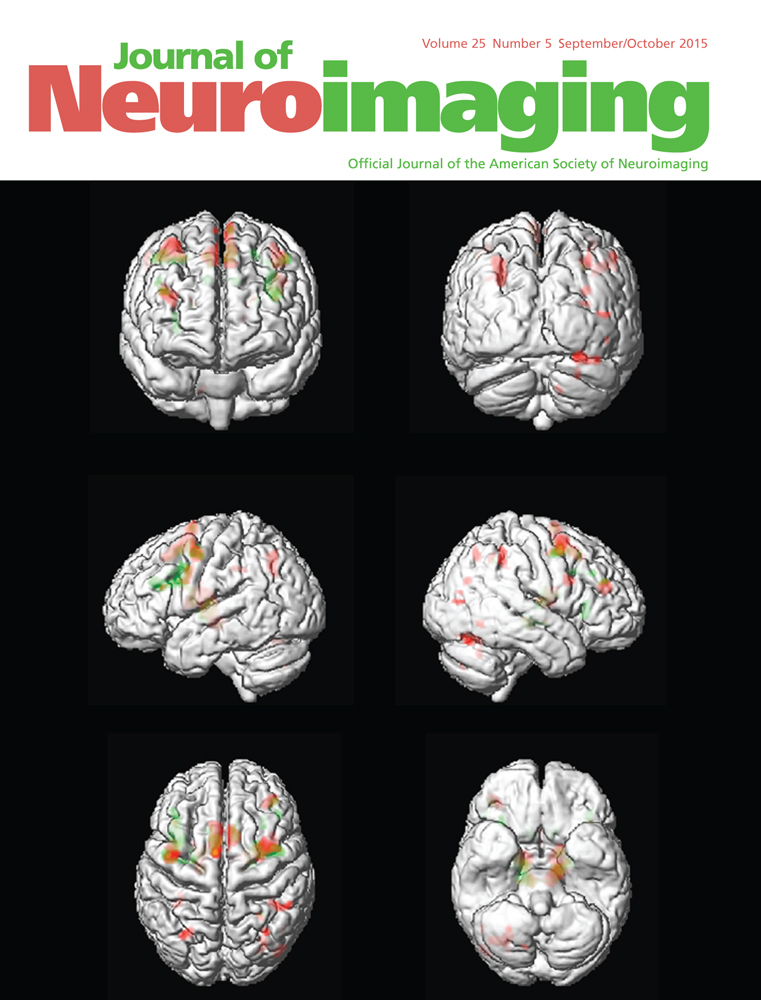
Predicting Prodromal Alzheimer's Disease in Subjects with Mild Cognitive Impairment Using Machine Learning Classification of Multimodal Multicenter Diffusion-Tensor and Magnetic Resonance Imaging Data
Corresponding Author
Martin Dyrba
German Center for Neurodegenerative Diseases, Rostock, Germany
Correspondence: Address correspondence to Martin Dyrba, German Center for Neurodegenerative Diseases (DZNE), c/o Zentrum für Nervenheilkunde, Gehlsheimer Str. 20, D-18147 Rostock, Germany. E-mail: [email protected]Search for more papers by this authorFrederik Barkhof
Department of Radiology and Nuclear Medicine, VU University Medical Center, Amsterdam, The Netherlands
Search for more papers by this authorAndreas Fellgiebel
Department of Psychiatry, University Medical Center Mainz, Mainz, Germany
Search for more papers by this authorMassimo Filippi
Neuroimaging Research Unit, Institute of Experimental Neurology, Division of Neuroscience, Scientific Institute and University Vita-Salute San Raffaele, Milan, Italy
Search for more papers by this authorLucrezia Hausner
Department of Geriatric Psychiatry, Central Institute of Mental Health, Medical Faculty Mannheim, University of Heidelberg, Mannheim, Germany
Search for more papers by this authorKarlheinz Hauenstein
Department of Radiology, University Medicine Rostock, Rostock, Germany
Search for more papers by this authorThomas Kirste
Mobile Multimedia Information Systems Group, University of Rostock, Rostock, Germany
Search for more papers by this authorStefan J. Teipel
German Center for Neurodegenerative Diseases, Rostock, Germany
Clinic for Psychosomatic and Psychotherapeutic Medicine, University Medicine Rostock, Rostock, Germany
Search for more papers by this authorthe EDSD study group
Search for more papers by this authorCorresponding Author
Martin Dyrba
German Center for Neurodegenerative Diseases, Rostock, Germany
Correspondence: Address correspondence to Martin Dyrba, German Center for Neurodegenerative Diseases (DZNE), c/o Zentrum für Nervenheilkunde, Gehlsheimer Str. 20, D-18147 Rostock, Germany. E-mail: [email protected]Search for more papers by this authorFrederik Barkhof
Department of Radiology and Nuclear Medicine, VU University Medical Center, Amsterdam, The Netherlands
Search for more papers by this authorAndreas Fellgiebel
Department of Psychiatry, University Medical Center Mainz, Mainz, Germany
Search for more papers by this authorMassimo Filippi
Neuroimaging Research Unit, Institute of Experimental Neurology, Division of Neuroscience, Scientific Institute and University Vita-Salute San Raffaele, Milan, Italy
Search for more papers by this authorLucrezia Hausner
Department of Geriatric Psychiatry, Central Institute of Mental Health, Medical Faculty Mannheim, University of Heidelberg, Mannheim, Germany
Search for more papers by this authorKarlheinz Hauenstein
Department of Radiology, University Medicine Rostock, Rostock, Germany
Search for more papers by this authorThomas Kirste
Mobile Multimedia Information Systems Group, University of Rostock, Rostock, Germany
Search for more papers by this authorStefan J. Teipel
German Center for Neurodegenerative Diseases, Rostock, Germany
Clinic for Psychosomatic and Psychotherapeutic Medicine, University Medicine Rostock, Rostock, Germany
Search for more papers by this authorthe EDSD study group
Search for more papers by this authorABSTRACT
BACKGROUND
Alzheimer's disease (AD) patients show early changes in white matter (WM) structural integrity. We studied the use of diffusion tensor imaging (DTI) in assessing WM alterations in the predementia stage of mild cognitive impairment (MCI).
METHODS
We applied a Support Vector Machine (SVM) classifier to DTI and volumetric magnetic resonance imaging data from 35 amyloid-β42 negative MCI subjects (MCI-Aβ42−), 35 positive MCI subjects (MCI-Aβ42+), and 25 healthy controls (HC) retrieved from the European DTI Study on Dementia. The SVM was applied to DTI-derived fractional anisotropy, mean diffusivity (MD), and mode of anisotropy (MO) maps. For comparison, we studied classification based on gray matter (GM) and WM volume.
RESULTS
We obtained accuracies of up to 68% for MO and 63% for GM volume when it came to distinguishing between MCI-Aβ42− and MCI-Aβ42+. When it came to separating MCI-Aβ42+ from HC we achieved an accuracy of up to 77% for MD and a significantly lower accuracy of 68% for GM volume. The accuracy of multimodal classification was not higher than the accuracy of the best single modality.
CONCLUSIONS
Our results suggest that DTI data provide better prediction accuracy than GM volume in predementia AD.
Supporting Information
Disclaimer: Supplementary materials have been peer-reviewed but not copyedited.
| Filename | Description |
|---|---|
| jon12214-sup-0001-Supmat.docx186 KB |
Table S1. Group characteristics and subject demographics for each center for MCI-Aβ42− versus MCI-Aβ42+. Table S2. Group characteristics and subject demographics for each center for MCI-Aβ42+ versus HC. Table S3. Scanner model and scan parameters for each center. Table S4. MK-SVM classification results for the multimodal analyses for MCI-Aβ42− versus MCI-Aβ42+. Table S5. MK-SVM classification results for the multimodal analyses for MCI-Aβ42+ versus HC. Table S6. Correlation of voxel intensity and diagnosis for MD for MCI-Aβ42+ versus HC. Table S7. Correlation of voxel intensity and diagnosis for GM volume for MCI-Aβ42+ versus HC. Figure S1. Mask for the DTI scans to restrict the analyses to WM areas (green) only. Frontal areas (red) were removed because of the strong susceptibility artifacts observed in those areas for some of the centers. |
Please note: The publisher is not responsible for the content or functionality of any supporting information supplied by the authors. Any queries (other than missing content) should be directed to the corresponding author for the article.
References
- 1Jack CR. Alliance for aging research AD biomarkers work group: structural MRI. Neurobiol Aging 2011; 32: S48-S57.
- 2Dubois B, Feldman HH, Jacova C, et al. Research criteria for the diagnosis of Alzheimer's disease: revising the NINCDS–ADRDA criteria. Lancet Neurol 2007; 6: 734-46.
- 3Brun A, Englund E. A white matter disorder in dementia of the Alzheimer type: a pathoanatomical study. Ann Neurol 1986; 19: 253-62.
- 4Roher AE, Weiss N, Kokjohn TA, et al. Increased A beta peptides and reduced cholesterol and myelin proteins characterize white matter degeneration in alzheimer's disease. Biochemistry 2002; 41: 11080-90.
- 5Neil JJ. Diffusion imaging concepts for clinicians. J Magn Reson Imaging 2008; 27: 1-7.
- 6Le Bihan D, Turner R, Douek P, et al. Diffusion MR imaging: clinical applications. AJR Am J Roentgenol 1992; 159: 591-9.
- 7Jack CR, Knopman DS, Jagust WJ, et al. Tracking pathophysiological processes in Alzheimer's disease: an updated hypothetical model of dynamic biomarkers. Lancet Neurol 2013; 12: 207-16.
- 8Takagi T, Nakamura M, Yamada M, et al. Visualization of peripheral nerve degeneration and regeneration: monitoring with diffusion tensor tractography. NeuroImage 2009; 44: 884-92.
- 9Concha L, Gross DW, Wheatley BM, et al. Diffusion tensor imaging of time-dependent axonal and myelin degradation after corpus callosotomy in epilepsy patients. NeuroImage 2006; 32: 1090-9.
- 10Teipel SJ, Stahl R, Dietrich O, et al. Multivariate network analysis of fiber tract integrity in Alzheimer's disease. NeuroImage 2007; 34: 985-95.
- 11Teipel SJ, Meindl T, Wagner M, et al. Longitudinal changes in fiber tract integrity in healthy aging and mild cognitive impairment: a DTI follow-up study. J Alzheimers Dis 2010; 22: 507-22.
- 12Cui Y, Wen W, Lipnicki DM, et al. Automated detection of amnestic mild cognitive impairment in community-dwelling elderly adults: a combined spatial atrophy and white matter alteration approach. NeuroImage 2012; 59: 1209-17.
- 13Dyrba M, Ewers M, Wegrzyn M, et al. Robust automated detection of microstructural white matter degeneration in Alzheimer's disease using machine learning classification of multicenter DTI data. PLoS One 2013; 8: e64925.
- 14Douaud G, Jbabdi S, Behrens TEJ, et al. DTI measures in crossing-fibre areas: increased diffusion anisotropy reveals early white matter alteration in MCI and mild Alzheimer's disease. NeuroImage 2011; 55: 880-90.
- 15Pfefferbaum A, Adalsteinsson E, Sullivan EV. Replicability of diffusion tensor imaging measurements of fractional anisotropy and trace in brain. J Magn Reson Imaging 2003; 18: 427-33.
- 16Vollmar C, O'Muircheartaigh J, Barker GJ, et al. Identical, but not the same: intra-site and inter-site reproducibility of fractional anisotropy measures on two 3.0T scanners. NeuroImage 2010; 51: 1384-94.
- 17Teipel SJ, Reuter S, Stieltjes B, et al. Multicenter stability of diffusion tensor imaging measures: a European clinical and physical phantom study. Psychiatry Res: Neuroimaging 2011; 194: 363-71.
- 18Zhu T, Hu R, Qiu X, et al. Quantification of accuracy and precision of multi-center DTI measurements: a diffusion phantom and human brain study. NeuroImage 2011; 56: 1398-1411.
- 19Cortes C, Vapnik V. Support-vector networks. Mach Learn 1995; 20: 273-97.
- 20Klöppel S, Stonnington CM, Chu C, et al. Automatic classification of MR scans in Alzheimer's disease. Brain 2008; 131: 681-9.
- 21Plant C, Teipel SJ, Oswald A, et al. Automated detection of brain atrophy patterns based on MRI for the prediction of Alzheimer's disease. NeuroImage 2010; 50: 162-74.
- 22Abdulkadir A, Mortamet B, Vemuri P, et al. Effects of hardware heterogeneity on the performance of SVM Alzheimer's disease classifier. NeuroImage 2011; 58: 785-92.
- 23Cuingnet R, Gerardin E, Tessieras J, et al. Automatic classification of patients with Alzheimer's disease from structural MRI: a comparison of ten methods using the ADNI database. NeuroImage 2011; 56: 766-81.
- 24Haller S, Nguyen D, Rodriguez C, et al. Individual prediction of cognitive decline in mild cognitive impairment using support vector machine-based analysis of diffusion tensor imaging data. J Alzheimer's Dis 2010; 22: 315-27.
- 25Graña M, Termenon M, Savio A, et al. Computer aided diagnosis system for Alzheimer disease using brain diffusion tensor imaging features selected by Pearson's correlation. Neurosci Lett 2011; 502: 225-9.
- 26Wee C-Y, Yap P-T, Zhang D, et al. Identification of MCI individuals using structural and functional connectivity networks. NeuroImage 2012; 59: 2045-56.
- 27O'Dwyer L, Lamberton F, Bokde ALW, et al. Using support vector machines with multiple indices of diffusion for automated classification of mild cognitive impairment. PLoS One 2012; 7: e32441.
- 28Sonnenburg S, Rätsch G, Schäfer C, et al. Large scale multiple kernel learning. J Mach Learn Res 2006; 7: 1531-65.
- 29Dyrba M, Ewers M, Wegrzyn M, et al. Combining DTI and MRI for the automated detection of Alzheimer's disease using a large European multicenter dataset. In: P-T Yap, T Liu, D Shen, C-F Westin, L Shen, eds. Multimodal Brain Image Analysis. Berlin/ Heidelberg: Springer; 2012: 18-28.
10.1007/978-3-642-33530-3_2 Google Scholar
- 30Hinrichs C, Singh V, Xu G, et al. Predictive markers for AD in a multi-modality framework: an analysis of MCI progression in the ADNI population. NeuroImage 2011; 55: 574-89.
- 31Zhang D, Wang Y, Zhou L, et al. Multimodal classification of Alzheimer's disease and mild cognitive impairment. NeuroImage 2011; 55: 856-67.
- 32Teipel SJ, Wegrzyn M, Meindl T, et al. Anatomical MRI and DTI in the diagnosis of Alzheimer's disease: a European multicenter study. J Alzheimers Dis 2012; 31: S33-S47.
- 33Fischer FU, Scheurich A, Wegrzyn M, et al. Automated tractography of the cingulate bundle in Alzheimer's disease: a multicenter DTI study. J Magn Reson Imaging 2012; 36: 84-91.
- 34Kilimann I, Grothe M, Heinsen H, et al. Subregional basal forebrain atrophy in Alzheimer's disease: a multicenter study. J Alzheimers Dis. 2014; 40: 687-700.
- 35Hampel H, Blennow K. CSF tau and β-amyloid as biomarkers for mild cognitive impairment. Dialogues Clin Neurosci 2004; 6: 379-90.
- 36Hampel H, Teipel SJ, Fuchsberger T, et al. Value of CSF beta-amyloid1–42 and tau as predictors of Alzheimer's disease in patients with mild cognitive impairment. Mol Psychiatry 2004; 9: 705-10.
- 37Mulder C, Verwey NA, van der Flier WM, et al. Amyloid- (1–42), total tau, and phosphorylated tau as cerebrospinal fluid biomarkers for the diagnosis of Alzheimer disease. Clin Chem 2010; 56: 248-53.
- 38Smith SM, Jenkinson M, Woolrich MW, et al. Advances in functional and structural MR image analysis and implementation as FSL. NeuroImage 2004; 23(Suppl 1): S208-S219.
- 39Gaser C, Volz H-P, Kiebel S, et al. Detecting structural changes in whole brain based on nonlinear deformations—application to schizophrenia research. NeuroImage 1999; 10: 107-13.
- 40Witten IH, Frank E. Data Mining: practical machine learning tools and techniques. The Morgan Kaufmann Series in data management systems. 2nd ed. San Francisco, CA: Morgan Kaufmann Publishers, 2005.
- 41Jaccard P. Étude comparative de la distribution florale dans une portion des Alpes et des Jura. Bulletin de la Société Vaudoise des Sciences Naturelles 1901; 37: 547-79.
- 42Good PI. Permutation tests: a practical guide to resampling methods for testing hypotheses. 2nd ed. New York: Springer, 2000.
10.1007/978-1-4757-3235-1 Google Scholar
- 43Ojala M, Garriga GC. Permutation tests for studying classifier performance. J Mach Learn Res 2010; 11: 1833-63.
- 44Rasmussen PM, Madsen KH, Lund TE, et al. Visualization of nonlinear kernel models in neuroimaging by sensitivity maps. NeuroImage 2011; 55: 1120-31.
- 45Smith ED, Szidarovszky F, Karnavas WJ, et al. Sensitivity analysis, a powerful system validation technique. Open Cybernetics System J 2007; 2: 39-56.
10.2174/1874110X00802010039 Google Scholar
- 46Talairach J, Tournoux P. Co-planar stereotaxic atlas of the human brain. 1st ed. Stuttgart: Thieme, 1988: 122.
- 47Zhang Y, Schuff N, Camacho M, et al. MRI markers for mild cognitive impairment: comparisons between white matter integrity and gray matter volume measurements. PLoS One 2013; 8: e66367.
- 48Wee C-Y, Yap P-T, Shen D, for the Alzheimer's Disease Neuroimaging I. Prediction of Alzheimer's disease and mild cognitive impairment using cortical morphological patterns. Hum Brain Mapp 2013; 34: 3411-25.
- 49Müller MJ, Greverus D, Dellani PR, et al. Functional implications of hippocampal volume and diffusivity in mild cognitive impairment. NeuroImage 2005; 28: 1033-42.
- 50Fellgiebel A, Dellani PR, Greverus D, et al. Predicting conversion to dementia in mild cognitive impairment by volumetric and diffusivity measurements of the hippocampus. Psychiatry Res.: Neuroimaging 2006; 146: 283-7.
- 51Müller MJ, Greverus D, Weibrich C, et al. Diagnostic utility of hippocampal size and mean diffusivity in amnestic MCI. Neurobiol Aging 2007; 28: 398-403.
- 52Agosta F, Pievani M, Sala S, et al. White matter damage in alzheimer disease and its relationship to gray matter atrophy. Radiology 2011; 258: 853-63.
- 53Canu E, McLaren DG, Fitzgerald ME, et al. Mapping the structural brain changes in Alzheimer's disease: the independent contribution of two imaging modalities. J Alzheimers Dis 2011; 26(Suppl 3): 263-74.
- 54Oishi K, Mielke MM, Albert M, et al. DTI analyses and clinical applications in Alzheimer's disease. J Alzheimers Dis 2011; 26(Suppl 3): 287-96.
- 55Zhuang L, Sachdev PS, Trollor JN, et al. Microstructural white matter changes, not hippocampal atrophy, detect early amnestic mild cognitive impairment. PLoS One 2013; 8: e58887.
- 56Cui Y, Liu B, Luo S, et al. Identification of conversion from mild cognitive impairment to Alzheimer's disease using multivariate predictors. PLoS One 2011; 6: e21896.
- 57Wolz R, Julkunen V, Koikkalainen J, et al. Multi-method analysis of MRI images in early diagnostics of Alzheimer's disease. PLoS One 2011; 6: e25446.
- 58Young J, Modat M, Cardoso MJ, et al. Accurate multimodal probabilistic prediction of conversion to Alzheimer's disease in patients with mild cognitive impairment. NeuroImage: Clin 2013; 2: 735-45.
- 59Stonnington CM, Tan G, Klöppel S, et al. Interpreting scan data acquired from multiple scanners: a study with Alzheimer's disease. NeuroImage 2008; 39: 1180-5.


