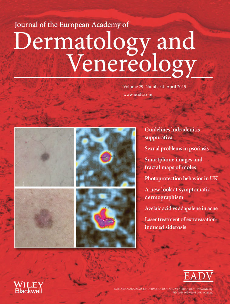
Accuracy of a smartphone application using fractal image analysis of pigmented moles compared to clinical diagnosis and histological result
Conflicts of interest:
T.M. and A.U. have served as consultant to SkinVision B.V. concerning the clinical study protocol. A.U. has served as consultant to SkinVision B.V. concerning the image analysis and classification algorithm. D.K., A.R., K.S., C.B. and T.R. have no conflict of interests to report.
Funding sources:
This study was supported in part by SkinVision B.V. for the design and conduct of the study and by a post-doctoral scholarship (POSDRU/159/1.5/S/138963) to A.U.
Abstract
Background
Lately, various smartphone applications have been introduced as diagnostic self-monitoring tools in the evaluation of pigmented moles, but most of these techniques have not been evaluated systematically.
Objectives
The purpose of this study was to evaluate prospectively the sensitivity and specificity of a recently developed smartphone application using fractal image analysis for the risk evaluation algorithm in the diagnosis of malignant melanoma compared to clinical diagnosis and histopathological result.
Methods
Consecutive patients with melanocytic lesions were recruited and clinical and dermoscopical diagnosis was documented by two dermatologists independently. Imaging and analysis with the smartphone application was performed prior to excision of lesions. The findings were compared to the histological results as gold standard.
Results
Of 195 included lesions histopathological analysis revealed 40 melanomas, 42 dysplastic nevi and 113 benign nevi. The sensitivity of the diagnosis melanoma by fractal image analysis using smartphone images was 73%, the specificity was 83% compared to a sensitivity of 88% and specificity of 97% regarding the clinical diagnosis by the dermatologists.
Conclusion
The smartphone application using fractal analysis might be a promising tool in the pre-evaluation of pigmented moles by laypersons, while it is to date inferior to the diagnostic evaluation by a dermatologist.