Optical coherence tomography is helpful in the diagnosis of foveal hypoplasia
Abstract.
Purpose: To investigate whether optical coherence tomography (OCT) is helpful in the diagnosis of foveal hypoplasia in children.
Methods: Children with albinism and aniridia were examined with Stratus OCT 3 software 4.0.1 (Carl Zeiss Meditec, Dublin, California, USA). A qualitative examination of the macular area was performed with a 128 A-scans/second-single-scan. Macular thickness was measured quantitatively with an automatic fast macular map protocol. The average thickness/volume of the macula was presented as numerical values and as a false colour code in nine modified early treatment of diabetic retinopathy study (ETDRS) areas (A1–A9). A previously collected control group of children was used for comparison.
Results: Macular thickness in 13 children with albinism and three children with aniridia was measured with OCT. Comparison with healthy children in the same population was performed. Patients with albinism and aniridia had significantly thicker central macula (A1) and foveola than children in the control group.
Conclusion: OCT was found to be useful in the diagnosis of foveal hypoplasia in children.
Introduction
Foveal hypoplasia is well known in aniridia and albinism and is also described as an isolated finding, without additional malformations (Nettleship 1906; Seefelder 1909; Usher 1920; Oliver et al. 1987). However, a clinical diagnosis of foveal hypoplasia in children is not always easy because fundus changes may be subtle and difficult to detect, especially if nystagmus is present.
Foveal hypoplasia has been diagnosed previously by funduscopy and fluorescein angiography (Oliver et al. 1987). During the last decade, optical coherence tomography (OCT) has been used increasingly as a method to visualize the various layers of the retina. OCT images correlate with the morphology of the foveal and parafoveal regions (Usher 1920). A standard protocol for measurements of macular thickness was developed by Hee et al. (1998). The macula is divided into nine areas, of which the central 1000-μm region comprises the fovea. Since its introduction for clinical use in 1996, OCT has become widely used to diagnose retinal conditions and to follow the progression of a disease and its response to therapy in adults. However, this method has not been used to a great extent in the diagnosis of retinal disease in children, possibly because of the lack of normal reference values in children.
The aim of this study was to investigate whether OCT could be of use in the diagnosis of foveal hypoplasia in children. We compared values with those of healthy children (Eriksson et al. 2008).
Materials and Methods
This study included children with albinism and children with hereditary aniridia. The children were recruited from the Low Vision Centre in Uppsala County and from the Department of Ophthalmology at Uppsala University Hospital. Iris transillumination and reduced visual acuity (VA) were required for the diagnosis of albinism.
Best-corrected optotype VA was assessed monocularly. Cycloplaegic retinoscopy was performed after dilating the pupil with a mixture of phenylephrine 1.5% and cyclopentholate 0.85%. Biomicroscopy of the anterior segment and ophthalmoscopy of the posterior pole of the eye were performed.
All OCT examinations were performed by one of the authors (U.E.). Stratus OCT 3 (Carl Zeiss Meditec, Dublin, California, USA) with software 4.0.1 was used. The OCT technique has been described extensively elsewhere (Huang et al. 1991; Hee et al. 1995; Puliafito et al. 1995). A qualitative examination of the macular area was performed in all patients with a 128 A-scans/second-single-scan. This scan gives a low resolution but a fast acquisition time, in order to reduce motion artefacts and scan displacements in eyes with poor fixation. Macular thickness was then measured quantitatively with a fast macular map protocol, which is a topographic two-dimensional thickness map constructed from six repetitive transversal OCT scans. This protocol is quick and examines the macular area automatically in 1.92 seconds. In eyes with low VA but good fixation, this is a useful method. In seven eyes with poor fixation and nystagmus, the macular thickness map protocol was used. In this program the six scans are obtained manually one by one, which gives the examiner better control of the scan placement. The settings in this program were 128 A-scans/second, for the reasons described earlier. The posterior pole was manually scanned horizontally with a 6 mm single line to look for signs of a foveal depression.
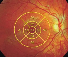
The retinal thickness is defined as the distance between two boundaries – the vitreoretinal interface and the anterior surface of the pigment epithelium – and is calculated automatically by the instrument algorithm. The average thickness/volume of the macula is presented as numerical values and as a false colour code in nine modified early treatment of diabetic retinopathy study (ETDRS) areas (Fig. 1). The central macula (A1) has a diameter of 1 mm, the inner circle (A2–5) a diameter of 3 mm and the outer circle (A6–9) a diameter of 6 mm. The foveal minimum is defined as the central point where the six radial scans intersect. This study was approved by the Ethics Committee of Uppsala University, Sweden.

The average thickness/volume of the macula presented as numerical values and as a false colour code in nine modified ETDRS areas.
Statistical Methods
Right (RE) and left (LE) eyes were analysed separately in the patients with albinism and aniridia; the values were compared with normal values in healthy children (Eriksson et al. 2008). Continuous data were analysed using the Kruskal–Wallis analysis of variance by ranks, followed by Dunn’s multiple comparison test. Spearman’s rho test was used for bivariate correlations.
Results
This study included 13 of 14 known children with albinism in Uppsala County, together with three children with a diagnosis of aniridia. The age, gender, VA and refraction of the study participants are given in Table 1.
| Patient number | Diagnosis | Gender | Age (years) | VA, RE | VA, LE | Refraction, RE | Refraction, LE | Nystagmus |
|---|---|---|---|---|---|---|---|---|
| 1 | Aniridia | Female | 5 | 0.63 | 0.63 | +3.5 = –0.5 cyl | +3.5 = −0.5 cyl | No |
| 2 | Aniridia | Male | 12 | 0.7 | 0.7 | +0.75 = −0.75 cyl | +1.5 = −1.5 cyl | No |
| 3 | Aniridia | Female | 17 | 0.5 | 0.1 | +1.25 = −2.5cyl | −1.75 = −1.25cyl | No |
| 4 | Albinism | Male | 10 | 0.2 | 0.2 | +4.5 = −2.0 cyl | +4.0 = −3.0 cyl | Yes |
| 5 | Albinism | Male | 12 | 0.2 | 0.2 | +4.5 = −1.5 cyl | +3.25 = −2.75cyl | Yes |
| 6 | Albinism | Male | 6 | 0.2 | 0.3 | +0.5 = −0.5 cyl | +0.5 = −0.5 cyl | Yes |
| 7 | Albinism | Male | 6 | 0.4 | 0.4 | ±0 | ±0 | No |
| 8 | Albinism | Male | 12 | 0.2 | 0.2 | +1.0 = −1.25cyl | +1.75 = −3.5cyl | Yes |
| 9 | Albinism | Male | 7 | 0.4 | 0.5 | +5.0 = −1.0 cyl | +5.0 = −1.0 cyl | No |
| 10 | Albinism | Female | 9 | 0.5 | 0.5 | +2.5 = −0.5 cyl | +2.5 = −0.5 cyl | No |
| 11 | Albinism | Female | 15 | 0.2 | 0.1 | +8.0 = −4.5 cyl | +9.5 = −4.0 cyl | Yes |
| 12 | Albinism | Female | 13 | 0.2 | 0.2 | +1.5 = −1.75cyl | +2.0 = −2.25cyl | Yes |
| 13 | Albinism | Male | 6 | 0.3 | 0.3 | +4.0 = −2.0 cyl | +5.5 = −2.5 cyl | Yes |
| 14 | Albinism | Female | 17 | 0.2 | 0.1 | +6.0 = −4.0 cyl | +6.0 = −4.5 cyl | Yes |
| 15 | Albinism | Female | 8 | 0.1 | 0.1 | +3.5 = −0.75 cyl | +3.5 = −1.0 cyl | Yes |
| 16 | Albinism | Male | 5 | 0.1 | 0.1 | +6.25 = −2.5 cyl | +6.0 = −3.0 cyl | Yes |
- VA, visual acuity; RE, right eye; LE, left eye.
All eyes had clear media. Iris transillumination was found in all 13 patients with albinism. Nystagmus was found in 10 of the patients with albinism, but in none of those with aniridia (Table 1). There were four pairs of siblings among the patients with albinism. In one of the pairs there was an x-linked heredity, while the other three pairs had no relatives with known albinism. The remaining five children had no heredity of albinism or nystagmus.
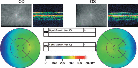
The values of macular thickness in albinism and aniridia are presented in Table 2 and were compared with values in healthy children (Eriksson et al. 2008). Patients with albinism and aniridia had significantly thicker central macula (A1) (RE p < 0.001, LE p < 0.001) and foveal minimum (RE p < 0.001, LE p < 0.001) compared with children in the control group (Fig. 2). There was a significant difference between the groups regarding the inner macular circle (A2–A5) (RE p < 0.001, LE p < 0.001) and the outer macular circle (A6–A9) (RE p < 0.001, LE p = 0.001). The difference was explained by a lower retinal thickness in the children with albinism (see Table 2). Furthermore, there was a reduced total macular volume in the children with albinism compared with the controls (RE p < 0.001, LE p = 0.001). No difference was found between the few children with aniridia and the controls regarding the retinal thickness of the inner and outer circle or the total macular volume. No correlation between VA and central macular thickness (A1) was found.
| Aniridia (n = 3) | Albinism (n = 13) | Controls (n = 55)* | ||||||||
|---|---|---|---|---|---|---|---|---|---|---|
| Right eye | Left eye | Right eye | Left eye | |||||||
| Mean | Median (IQR) | Mean | Median (IQR) | Mean | Median (IQR) | Mean | Median (IQR) | Mean | Median (IQR) | |
| Foveal minimum (μm) | 252 | 259 (45) | 244 | 259 (67) | 246 | 243 (17) | 248 | 245 (34) | 166 | 167 (22) |
| Central macula (A1) (μm) | 259 | 262 (38) | 255 | 262 (47) | 247 | 245 (22) | 250 | 248 (34) | 204 | 205 (23) |
| Average inner circle (A2–A5) (μm) | 272 | 274 (19) | 271 | 272 (34) | 244 | 242 (18) | 247 | 246 (32) | 279 | 278 (20) |
| Average outer circle (A6–A9) (μm) | 242 | 238 (17) | 240 | 238 (34) | 227 | 229 (16) | 228 | 224 (21) | 245 | 243 (18) |
| Macular volume (mm3) | 7.06 | 7.01 (0.45) | 7.03 | 7.00 (0.92) | 6.56 | 6.62 (0.40) | 6.59 | 6.52 (0.59) | 7.11 | 7.10 (0.53) |
- IQR, interquartile range.
- * 55 randomized eyes from 5–16-year-old healthy children (Eriksson et al. 2008).

Optical coherence tomography image of a child with foveal hypoplasia (albinism).
Discussion
OCT confirmed a foveal hypoplasia in all our patients, including three with aniridia and 13 with albinism. Unlike the healthy population of children, a foveal pit was lacking or reduced in size and the appearance of the macular area resembled that of the rest of the retina. In children with albinism, the inner and outer macular areas were generally thinner than in those with aniridia and in the healthy group. All children cooperated well.
Foveal hypoplasia characterizes both albinism and aniridia. In albinism, foveal hypoplasia is well known and has been confirmed histologically (Nettleship 1906; Usher 1920; O’Donnell et al. 1976; Fulton et al. 1978; Mietz et al. 1992). The foveal area is described as a continuous ganglion cell layer without a rod-free area (Fulton et al. 1978). A detailed funduscopy is often difficult to perform in patients with albinism because of nystagmus and light sensitivity. Hence, a clinical diagnosis of foveal hypoplasia is not always easy to perform.
Aniridia is another condition comprising hypoplasia of the fovea and replacement with a continuous ganglion cell layer that has also been confirmed histologically (Seefelder 1909). Classical aniridia is a dominantly inherited condition with PAX6 mutations as the underlying mechanism (Prosser & van Heyningen 1998). Furthermore, aniridia is a heterogenous condition and a wide variety of phenotypes are described, from total aniridia to only very subtle iris hypoplasia (Nelson et al. 1984; Edén et al. 2008; Lee et al. 2008). Therefore, objective confirmation of foveal hypoplasia can be helpful in the diagnosis of aniridia in some patients.
Fluorescein angiography has been used previously in the investigation of suspected foveal hypoplasia (Oliver et al. 1987). However, it is an invasive method and it is usually not possible to perform it in children under the age of 10 years. OCT, on the other hand, is a non-invasive and quick method that can be used in many children. The method has been found to be useful in adults with foveal hypoplasia (Recchia et al. 2002; McGuire et al. 2003) and also in a 10-year-old girl with oculocutaneous albinism (Meyer et al. 2002).
In our cohort of children with albinism, no association between VA and foveal thickness was found. Recently, Seo et al. (2007) found that OCT was helpful in correlating VA with grading of foveal hypoplasia in 13 young patients with albinism. However, in a reply to comments by Harvey et al. (2008), Seo et al. (2008) conclude that their case numbers were not enough to analyse the role of foveal depression as a visual prognostic factor. Moreover, they state that the visual outcome is probably affected more by multifactorial causes than by foveal depression alone.
In the present study, OCT examinations revealed a continuity of retinal layers and a lack of foveal depression in patients with albinism, which is well in line with previous OCT studies (Meyer et al. 2002; Recchia et al. 2002; McGuire et al. 2003). A general thinning of the inner and outer macular area was also detected in the children with albinism, but not in the three children with aniridia. An association between reduced macular thickness and myopia has been described (Mrugacz et al. 2004; Pierre-Kahn et al. 2005) but this was not the case in our children with albinism, who were all hypermetropic.
Albinism and aniridia are two genetically determined conditions. The genetics of albinism are complex, but the condition is most often caused by mutations in genes affecting the melanogenesis, while aniridia is caused by mutations in the PAX6 gene (Prosser & van Heyningen 1998; Oetting et al. 2003). It can be speculated that the thinner inner and outer macular areas in albinism may reflect different aetiological mechanisms of foveal hypoplasia in albinism and aniridia. However, the small number of children in our study means that its results have to be interpreted with caution. Further studies are needed to elucidate this finding.
It is hereby shown that OCT makes a helpful contribution in the investigation of retinal disease in children. We have previously reported on its use in x-linked retinoschizis in children (Eriksson et al. 2004). This article shows that OCT was useful in confirming a foveal hypoplasia. Furthermore, it may be helpful in the investigation of children with impaired or subnormal vision of unknown aetiology.


