Rarebit perimetry and fovea test before and after cataract surgery
ABSTRACT.
Purpose: To evaluate the effect of cataract on rarebit perimetry and the fovea test.
Methods: Twenty-five consecutive patients scheduled for cataract surgery (mean age 63.0 ± 7.9 years) were examined prior to and after cataract surgery with a complete ophthalmological examination. In addition, the rarebit perimetry (RBP) and the rarebit fovea test (RFT) were performed.
Results: Best-corrected visual acuity [BCVA, expressed in minimum angle of resolution (MAR)], RBP and RFT mean hit rate (MHR) improved significantly after cataract surgery. The relative pre–postsurgery difference was larger in the RFT [2.1 standard deviations (SDs)] compared to in BCVA (0.78 SDs). Seven patients had good BCVA (≤ 1.25) and RBP (83–99%) but low RFT (0–66%) before surgery. One patient with low preoperative BCVA (2.5) had a normal RFT (94%).
Conclusion: Cataract influenced both the RFT and RBP test, albeit the former more than the latter. The influence of cataract on RFT results, even when visual acuity is decreased only moderately, has to be taken into account when evaluating foveal function in patients with cataract. The larger relative change in RFT compared to BCVA values is thought to indicate that RFT is more sensitive for the effect of cataract. Therefore, RFT appears to be a sensitive test for visual disturbance and can presumably provide additional information at the preoperative evaluation of the patient.
Introduction
Senile cataract is a vision-impairing disease characterized by gradual thickening and progressive loss of transparency of the lens, strongly associated with ageing and often coexisting with other systemic or ophthalmic diseases like diabetes, glaucoma and age-related macular degeneration (AMD) (Ninn-Pedersen 1986–1990). Glaucoma treatment may also contribute to cataract progression (Heijl et al. 2002). Follow-up examinations using perimetry and foveal function tests are important in the management of patients with glaucoma and AMD. However, the interpretation of results from these examinations can be problematic in the presence of cataract: perimetry results from conventional (Hayashi et al. 2001) and frequency-doubling technology (FDT) (Siddiqui et al. 2005), as well as from high-pass resolution perimetry (Martin 1997), are influenced by lens opacities.
The rarebit perimetry (RBP) (Frisén 2002), relying on the detection of very small and bright dots, has been shown to be sensitive to early damage in glaucoma (Martin & Wanger 2004; Brusini et al. 2005). The test is simple and fast to perform, and no learning effect has been reported (Salvetat et al. 2007). The rarebit system also includes a test of foveal function, the rarebit fovea test (RFT). The RFT gives information about the most central visual field (VF) not obtained by other tests, for instance visual acuity at high and low contrast (Malmer & Martin 2005; Nilsson et al. 2006).
Salvetat et al. (2007) recently studied RBP in normal patients regarding the influence of cataract. They found that the RBP VFs improved significantly after cataract extraction and that the decrease in retinal sensitivity caused by the cataract tended to be more pronounced in the central zone compared to the midperipheral and peripheral zones. To the best of our knowledge, there are no previous studies on the influence of cataract on the RFT results, testing within the 4 × 3º central VF. The aim of the current study was to evaluate the effect of cataract on RBP and RFT results.
Materials and Methods
Twenty-five consecutive patients (16 women and nine men) with a mean age of 63.0 ± 7.9 years (range 44–74) scheduled for cataract surgery were recruited from the Department of Anterior Segment Surgery at St Eriks Eye Hospital, Stockholm, Sweden. Inclusion criteria were no previous known ocular disease other than senile cataract and age ≤ 75 years. Exclusion criterion was intraocular pressure (IOP) > 21 mmHg. Moreover, no patient in the study used any medication known to elicit VF defects of any kind. Eleven patients were treated for systemic hypertension without signs of retinopathy, and one patient had diabetes mellitus but no sign of diabetic retinopathy.
All patients were examined 1–2 weeks before and 4 weeks after cataract extraction. They underwent a slit-lamp ophthalmic examination in dilation by one of the authors (O.A.), including fundus evaluation. Cataract was defined as nuclear, cortical, posterior subcapsular or mixed (i.e. a combination of two or three types in the same eye). Best-corrected visual acuity [BCVA, expressed in minimum angle of resolution (MAR)] according to the Early Treatment Diabetes Retinopathy Study (ETDRS), IOP measurement and the RBP and RFT examinations were performed before pupil dilation.
The cataract surgery was performed by one of the authors (C.G.L.) using a corneal incision of less than 3 mm, capsulorhexis, phacoemulsification of the lens nucleus and implantation of an acrylic foldable intraocular lens (IOL) in the capsular bag. There were no peroperative complications. All patients used dexametasone eyedrops following surgery three times daily for 1 week, two times daily for 1 week and one time daily in the third and final week.
RBT and RFT
The RBP evaluates a 30 × 20º VF and has been described extensively elsewhere (Frisén 2002; Salvetat et al. 2007). No stimuli are presented within the central 5º VF. This central area is tested by a special program, the RFT (Malmer & Martin 2005; Nilsson et al. 2006), which covers the central 4 × 3º VF.
During the test, 10 stimuli were presented in each of 24 rectangular test areas in the RBP program and 10 test areas in the RFT program. The test stimuli were presented on a 17′′ LCD screen with a test distance of 0.5 m for the outer RBP field, 1 m for the inner RBP field and 2 m for the RFT.
In both tests, the stimuli were presented one or two at a time, randomly mixed with blank presentations, which were used for control purposes. Patients were instructed to indicate the number of dots seen (one, two or none) by clicking on a computer mouse once, twice or not at all after each presentation. The results are presented as mean hit rate (MHR), i.e. the number of dots perceived in relation to dots presented. In RBP, the normal MHR is reported to be between 78% and 100%, median 97% (Martin & Wanger 2004) and between 78% and 99%, mean 91%, standard deviation (SD) 5.7 (Salvetat et al. 2007). In RFT, the normal median MHR is 100% (range 97–100%) in patients aged less than 65 years and 88% (range 34–98%) in patients aged 65 years or more (Malmer & Martin 2005; Nilsson et al. 2006, 2007). Reliability is assessed by subjective evaluation by the examiner as reliable, borderline or not reliable, and also by automatic counting of errors, i.e. false-positive responses during the examination. Three errors or less was considered acceptable (Frisén 2002).
Stimulus presentation time is short (less than 200 ms) in order to make detection by refixation difficult (Leigh & Zee 2006). Version 4.0 of the tests was used. The learning effect in RBP was reported to be low, and the mean MHR test–retestvariability was 2.9% in the study by Salvetat et al. (2007). The intraindividual variability in RFT is low (Nilsson et al. 2006). In a pilot study of the repeatability in RFT by examining eight healthy individuals eight times within 2 hr and a further two times 1 week later, the coefficient of variation was 1.1% (Nilsson 2008).
During both tests, trial lenses were used to correct for the total refractive error and test distance. All patients underwent a short training session, lasting until the examiner was convinced that they fully understood the task. The study was approved by the local ethical committee and was performed according to the Declaration of Helsinki. Written informed consent was obtained from all participants.
Statistics
For correlation and comparison between BCVA and RBP and RFT mean hit rates, the Spearman rank correlation and the Mann–Whitney tests were used. A p-value of 0.05 was regarded as significant. In order to compensate for the differences in scale, the relative improvement after surgery in BCVA and RFT was calculated by dividing the pre–postsurgery difference by the SDs from the presurgery values. Statistical analyses were performed using graphpad instat, version 3.0 (GraphPad Software, San Diego, CA, USA).
Results
All 25 patients were included in the analyses of the RBP result but one patient did not perform the follow-up RFT test, leaving 24 patients for the analysis of RFT data. All patients showed reliable results (i.e. three errors or less during all examinations). Two patients had some problems with double-clicking the computer mouse, and their performances were judged as borderline by the examiner. The individual pre–post-surgery differences in examination results are summarized in Table 1. The spherical equivalent refraction prior to surgery was −0.5 D ± 2.9 (range −7.5 to 5.5); 1 month after cataract surgery, it was −0.5 D ± 1.2 (range −3.5 to 2). Eighteen patients had mixed cataract, three had nuclear cataract and four had posterior subcapsular cataract.
| Preoperative | Postoperative | p-value | |
|---|---|---|---|
| BCVA MAR (n = 25) | |||
| Median | 2 | 0.83 | < 0.0001 |
| Range | 1–8.33 | 0.62–1.61 | |
| IQD | 0.62–1.61 | 0.83–1 | |
| RBP MHR (n = 25) | |||
| Median | 77 | 93 | 0.0006 |
| Range | 0–99 | 58–90.5 | |
| IQD | 60–98 | 89.5–96 | |
| RFT MHR (n = 24) | |||
| Median | 8 | 93.5 | 0.0001 |
| Range | 0–94 | 47–100 | |
| IQD | 0–52 | 85–96.75 | |
- BCVA, best-corrected visual acuity; MAR, minimum angle of resolution; RBP, rarebit perimetry; RFT, rarebit fovea test; MHR, mean hit rate, expressed as percentage.
Postoperatively, one patient showed mild transient anterior uveitis. Six patients had minor peripheral after-cataract and one had a fibrosis of the posterior capsule. Five patients had minor macular abnormalities, four had age-related retinal pigment epithelial defects, one had drusen and two had suspect excavated optic discs. These abnormalities did not change during follow-up time.
BCVA, RBP MHR and RFT MHR improved significantly (Mann–Whitney test) after cataract surgery (Table 1). The relative pre–postsurgery difference was 2.1 SDs in the RFT compared to 0.78 SDs in BCVA, i.e. 2.7 times larger.
There was a significant correlation between BCVA and RBP result both before (p = 0.0011, r = 0.62; Spearman rank correlation) and after (p = 0.004, r = −0.55; Spearman rank correlation) cataract surgery. Before surgery the correlation between BCVA and RFT results was weak (r = −0.48, p = 0.0161); however, after surgery the correlation was significant (r = −0.65, p = 0.0005).
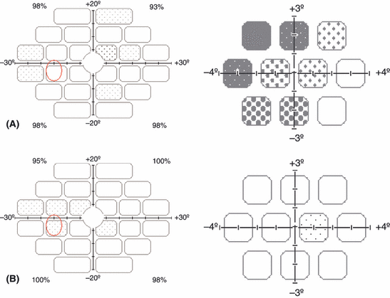
A weak (r = 0.48, p = 0.02) correlation was also observed between the preoperative BCVA and the increase in RBP MHR after cataract surgery. Seven patients had good decimal BCVA (≤ 1.25) and RBP (83–99%) but low RFT (0–66%) before surgery; for an example, see Fig. 1A,B. One patient with low preoperative BCVA (2.5) had normal age-corrected RFT (94%).

(A) Rarebit perimetry (RBP) [mean hit rate (MHR) 97%] to the left and rarebit fovea test (RFT) (MHR 52%) to the right from a 64-year-old woman with mixed cataract. Best-corrected visual acuity (BCVA) was 1.25 prior to cataract surgery. (B) RBP (MHR 98%) to the left and RFT (MHR 99%) to the right from the same patient. BCVA was 0.8 after cataract surgery.
There were no significant differences in RBP or RFT postoperative results in the patients with and without after-cataract. Six patients did not reach normal RBP and RFT values after surgery. Three of these had minor macular changes, one had borderline performance, one was referred for further examination, and in one case no reason was found for subnormal results. These three patients were the only ones not reaching BCVA 1.0 or better after surgery. Figure 1A,B shows RBP and RFT printouts, respectively, from one of the examined patients. In the RBP both the examination time and the reaction time decreased significantly after cataract surgery (p= 0.004 and 0.002, respectively).
Discussion
Nineteen of 25 patients reached normal values in RBP and RFT after cataract extraction and IOL implantation. BCVA was MAR 1.0 or better in all but three patients. None of these patients reached normal results on the rarebit tests after surgery. Cataract influenced both the RFT and RBP tests, albeit the former more than the latter. The larger relative change in RFT compared to BCVA values is thought to indicate that RFT is a more sensitive test for the effect of cataract. In fact, seven patients had good decimal BCVA and RBP but low RFT before surgery. One can assume that those patients brought forward symptoms other than low visual acuity (e.g. decreased contrast sensitivity, problems with glare) that influenced the decision to perform surgery. The clinical relevance of this observation could be assessed by comparing BCVA and RFT, respectively, to an analysis of subjective visual complaints in cataract patients – for example using the NEI-VFQ-25 (Mangione et al. 2001) or the NIKE (Lundström et al. 2006) instruments.
In one patient with low BCVA, both the RBP and the RFT were normal. Thus, a simple heuristic for establishing indication for cataract surgery could be low BCVA and/or low RFT. It was not possible to evaluate the effect of different types of cataract on the RBP and RFT in this study because most patients had the mixed type.
The results in the current study are in agreement with those presented by Salvetat et al. (2007), who noted larger effect of cataract on the more central part of the RBP (i.e. between 5 and 10º). Because the test targets used in the rarebit tests are adjusted in relation to the size of the receptive fields, they get progressively smaller when presented in the more central parts of the VF. It is likely that smaller stimuli will be influenced by lens opacities to a greater extent and therefore the results will show a greater decrease in more central parts of the VF. In analogy, lens opacities could be expected to have a still larger influence on the RFT testing within the 4º VF, as observed in the current study.
In conclusion, cataract seems to have a large influence on RFT, even when visual acuity is decreased only moderately. This finding must be taken into account when evaluating foveal function in patients with cataract. Patients with IOL and without other ocular abnormalities had normal RFT results, and an IOL does not seem to be a hindrance for the use of rarebit tests when evaluating the VF. RFT appears to be a sensitive test for visual disturbance and might be useful in the preoperative evaluation of patients with subjective complaints and minor lens opacities.


