Oculometry findings in high myopia at adult age: considerations based on oculometric follow-up data over 28 years in a cohort-based Danish high-myopia series
Abstract.
Purpose: To present and discuss oculometry data in a series of adults with high myopia followed between the ages of 26 and 54 years. Emphasis is on axial length (AL) findings and corneal curvature radius (Crad).
Methods: Thirty-four out of the 39 individuals recruited as teenagers from a Copenhagen 1948 birth cohort with myopia of at least 6 D have had current follow-up exams, to include AL measurements (by ultrasound, 1974–2002; the latter year also with the Zeiss IOLMaster) and keratometry. The cross-sectional and longitudinal analyses are based primarily on the eyes with high myopia; however, the fellow eye is also assessed in unilateral cases.
Results: At age 54 years, the maximum myopia in the series was −26 D; the highest AL value was 35.4 mm. The myopia had increased in most, with an increase from the 26-year oculometry baseline averaging 1.0 D [standard deviation (SD) 1.84]. Ultrasound measurements over the 28 years gave a significant correlation between axial eye elongation and myopia progression of adult age (r = 0.65). The regression line was y = 0.43 + 0.36x, with myopia increase on the x-axis. Throughout sessions, the association between AL and refraction was given by correlation coefficients numerically above 0.8, whereas AL and Crad had r-values of 0.3–0.5. However, a mean Crad in the sample of 7.66 (SD 0.28) mm meant that the more general expectancy of rather flat corneas in high myopia was not fulfilled. Our data further suggested a reduction in lens power over the study period.
Conclusion: In relation to refraction, AL and Crad remain the two main oculometry parameters. Apparently the correlation patterns regarding the cornea that are broadly valid for axial ametropia in the population cannot be extended to the marginal high myopia tail of the distribution. A significant proportion of eyes with high myopia thus had steeper corneas than expected, as a so-called index contribution (albeit a small one) to the marginal refractive error.
Introduction
This article discusses adult oculometric observations in high myopia observed over 28 years in a Danish cohort-based sample recruited as teenagers (Goldschmidt & Fledelius 2005). The study has two main objectives: (i) to investigate the axial eye length and its apparently established role as main refractive parameter (Donders 1864; Fledelius 1971, 1982; Delmarcelle et al. 1976; Curtin 1985; Mondon & Metge 1994); and (ii) to discuss the occurrence of steep corneas in high myopia, a less acknowledged issue in the literature (Francois & Goes 1973; Delmarcelle et al. 1976).
Materials and Methods
In 1962, 39 14 year olds were identified with myopia of at least 6 D. The sample was recruited from a Copenhagen 1948 birth cohort of 9243 individuals (Goldschmidt 1968). The high myopia was unilateral in ten and bilateral in 29 participants. At baseline the 39 adolescents (16 male and 23 female), were generally in good health and had no recorded eye diseases.
In 1964, at age 16 years, the 39 participants had a comprehensive ophthalmological examination and were invited to attend for follow-ups at age 26, 36, 47 and 54 years, then also to include ultrasound oculometry. Since baseline one man had died, two women had emigrated and two men were excluded because of eye pathology (retinitis pigmentosa/trauma of head and eye), thus reducing the original sample to 34 participants.
Comparing the findings from 1974 and 2002, the present analysis will deal with the oculometry profiles at first and last status and focus on the changes over time. In 1974, 29 participants attended (11 male and 18 female). In 2002, 33 participants attended (13 male and 20 female).
At all sessions refraction (recorded in D as spherical equivalent value, i.e. sphere + half cylinder value) was determined from best spectacle correction for Snellen acuity testing. From 1984, automated refractometry was added as guidance (Topcon, Tokyo, Japan, Nidek ARK 2000S, Aichi, Japan) and autokeratometry replaced the classical optical Javal–Schiøtz principle for assessing corneal power/curvature radius.
Ultrasound A-scan oculometry (Kretztechnik 7000, immersion technique, Kretztechnik, Zipf, Austria) was used in 1974. At later attendances, it was replaced by a Sonometrics 400 equipment (Sonometrics Inc, New York, NY, USA) with a solid tip transducer (contact method), which implied a drop of local anaesthesia, also serving as moistening agent and reducing corneal applanation by contact to a minimum (Fledelius & Goldschmidt 1979; Goldschmidt et al. 1981; Shammas 1984; Fledelius et al. 1988; Olsen & Nielsen 1989; Fledelius & Goldschmidt 2000). All A-mode evaluations were performed manually by the same researcher (H.C.F.).
As a supplement, in 2002 axial ocular measurements were performed by the non-contact IOLMaster (Carl Zeiss Meditec AG, Jena, Germany). Paired measurements in the series by the two methods showed differences ranging from −0.4 to 0.7 mm; the mean difference was 0.09 mm [standard deviation (SD) 0.22], with the acknowledged trend of the IOLMaster axes being slightly longer than by ultrasound.
For the cross-sectional status in 2002, we used the axial length (AL) measurements generated by the IOLMaster; otherwise, the ultrasonically measured ALs were included. In particular, this held for the longitudinal 1974/2002 oculometry evaluations, with 57 data sets available. Twenty-nine participants had measurements in 1974 and 2002; one blind eye was excluded from analyses because of retinal detachment.
As an indirect measure of the lens power, we calculated the power of the hypothetical intraocular lens (IOL) refractively required to replace the natural lens using the Haigis IOL formula (A constant 118.4) (Haigis et al. 2000). The research followed the tenets of the Declaration of Helsinki.
Statistics
Parametric statistics were used for appropriate data (mean values, SD, correlation and regression lines); otherwise non-parametric calculations (χ2, Mann–Whitney, Wilcoxon, binomial test) were used [spss 12.0 (SPSS, Chicago, IL, USA)]. A P-value < 0.05 was considered significant.
Results
The results are presented as
- 1
Cross-sectional data over the period, subdivided by gender.
- 2
Longitudinal changes between the ages of 26 and 54 years.
- 3
Analysis of anisometropia in the series at age 54 years.
- 4
Correlations and regression statistics.
Gender, cross-sectional data
In Table 1 all highly myopic eyes are presented, subdivided by sex. Ranges, mean values and SDs are given for refraction at the ages of 16, 26 and 54 years, and for the main oculometry parameters [corneal curvature radius (Crad), AL), anterior chamber depth (ACD) and lens thickness (LT)] at the ages of 26 and 54. The differences according to gender were significant at both ages regarding AL (p = 0.001) and Crad (p = 0.002), and also at age 54 for LT (p < 0.001). In general, the values thus indicated a longer eye in men, with a flatter cornea and a thinner lens.
| Mean (SD)range | p-value | ||
|---|---|---|---|
| Male eyes | Female eyes | ||
| Age 16 years, 58 eyes (21 male, 37 female) | |||
| Refraction (D)† | −8.58 (2.61)−5 to −14 | −8.01 (1.91)−5 to −13 | 0.34 |
| Age 26 years, 51 eyes (18 male, 33 female) | |||
| Refraction (D)† | −10.38 (4.04)−5.75 to −21.25 | −9.62 (2.50)−6 to −16 | 0.41 |
| AL (mm) | 27.6 (1.3)25.6–30.8 | 26.3 (1.1)24.7–29.6 | 0.000* |
| Crad (mm) | 7.85 (0.22)7.50–8.15 | 7.60 (0.27)7.12–8.30 | 0.002* |
| ACD (mm) | 3.92 (0.22)3.4–4.3 | 3.86 (0.27)3.3–4.3 | 0.49 |
| LT (mm) | 3.75 (0.28)3.4–4.4 | 3.84 (0.20)3.5–4.6 | 0.18 |
| Age 54 years, 55 eyes (20 male, 35 female) | |||
| Refraction (D) | −12.36 (6.12)−5 to −26 | −10.21 (3.06)−5 to −16.7 | 0.09 |
| AL (mm) | 28.8 (2.7)25.9–35.4 | 26.8 (1.5)24.5–30.7 | 0.001* |
| Crad (mm) | 7.79 (0.23)7.42–8.25 | 7.55 (0.30)7.10–8.29 | 0.002* |
| ACD (mm) | 3.65 (0.32)3.2–4.3 | 3.53 (0.27)2.9–4.0 | 0.18 |
| LT (mm) | 4.29 (0.31)3.7–4.8 | 4.58 (0.30)4.1–5.1 | 0.001* |
- Baseline refraction is presented for all 58 eyes with high myopia at inclusion in the study.
- *Statistically significant differences.
- †The actual refractive range bottom value of −5 D reflects that at teenage inclusion a few fellow eyes with only marginal high myopia were grouped as ‘bilateral’ because of expectation of progression, which did not occur in all cases.
- SD, standard deviation; AL, axial length; Crad, corneal curvature radius; ACD, anterior chamber depth; LT, lens thickness.
Longitudinal changes
The available 50 eyes with high myopia had shown an average increase in refraction of 2.0 D between the ages of 16 and 26, and a further 1.0 D increase between the ages of 26 and 54 years. For the latter period, the maximum refractive change was 7 D towards more myopia, and the maximum AL increase was 4.6 mm.
The oculometry data are apparent from Table 2. Changes over the 28 years in the 50 highly myopic eyes were statistically significant for all refractive components. Using the ultrasonic AL data from both occasions, the 1 D mean increase of myopia was accompanied by a mean axial elongation of 0.79 mm, and a slightly steeper cornea (mean Crad 0.05 mm lower). LT increased 0.66 mm by age, and ACD decreased correspondingly (by 0.29 mm).
| Mean (SD) | p-value | ||
|---|---|---|---|
| Age 26 years | Age 54 years | ||
| High myopia, 50 eyes | |||
| Refraction (D) | −9.84 (3.13) | −10.85 (4.20) | 0.000* |
| AL (mm) | 26.7 (1.3) | 27.5 (2.1) | 0.000* |
| Crad (mm) | 7.68 (0.27) | 7.63 (0.31) | 0.000* |
| ACD (mm) | 3.88 (0.26) | 3.59 (0.31) | 0.000* |
| Lens thickness (mm) | 3.82 (0.24) | 4.48 (0.34) | 0.000* |
| Lens power (D) | 23.4 (2.2) | 21.7 (1.9) | 0.000* |
| Fellow eyes (7) | |||
| Refraction (D) | −2.55 (1.97) | −2.25 (2.53) | 0.442 |
| AL (mm) | 24.2 (1.4) | 24.5 (2.5) | 0.041* |
| Crad (mm) | 7.71 (0.29) | 7.68 (0.28) | 0.115 |
| ACD (mm) | 3.83 (0.23) | 3.71 (0.25) | 0.059 |
| LT (mm) | 3.79 (0.17) | 4.33 (0.21) | 0.000* |
| Lens power (D) | 23.0 (2.4) | 20.8 (1.8) | 0.001* |
- *Statistically significant differences (Wilcoxon, non-parametric comparison of paired data).
- SD, standard deviation; AL, axial length; Crad, corneal curvature radius; ACD, anterior chamber depth; LT, lens thickness.
The average LT had increased 0.54 mm in men and 0.74 mm in women (p = 0.14). This observation is in accordance with the established continued growth of the lens throughout life. On the assumptions chosen, the lens power approximations for the 50 eyes with high myopia had an endpoint mean value of 21.7 D (1.93), with an apparent reduction in lens power parallel to the adult-age increase in degree of myopia. Parametric regression suggested a trend of lower lens power with increasing length of the eye, although this was not significant (p = 0.06).
For comparison, the seven fellow eyes with paired data are included in Table 2. There was a significant mean AL increase of 0.3 mm, whereas refractive level had not changed on average. Over the 28 years, the LT mean values had increased from 3.79 to 4.33 mm, and calculated lens power decreased by 2.2 D.
Anisometropia
In Table 3 oculometry results at age 54 in unilateral high myopia (n = 10) are compared to those of their fellow eyes. Intra-pair ALs differed significantly (p < 0.001) whereas Crad, ACD and LT appeared to be equal (with P-values ranging from 0.77 to 0.97). Significant anisometropia, here defined as a side difference in refraction above 1 D, further appeared in 12 of those with bilateral high myopia. Table 4 lists the 22 participants with anisometropia in the full series.
| Mean (SD) | p-value | ||
|---|---|---|---|
| High myopia (n = 10 eyes) | Fellow eye (n = 10 eyes) | ||
| Refraction (D) | −15.98 (6.96) | −2.05 (2.60) | 0.000* |
| AL (mm) | 30.1 (3.6) | 24.5 (1.5) | 0.000* |
| Crad (mm) | 7.70 (0.26) | 7.70 (0.25) | 0.97 |
| ACD (mm) | 3.71 (0.32) | 3.68 (0.23) | 0.80 |
| LT (mm) | 4.31 (0.11) | 4.29 (0.20) | 0.77 |
- *Intra-pair difference was statistically highly significant.
- SD, standard deviation; AL, axial length; Crad, corneal curvature radius; ACD, anterior chamber depth; LT, lens thickness.
| Bilateral myopia (n = 12) | Unilateral myopia (n = 10) | |
|---|---|---|
| Anisometropia (D) | ||
| 1.0–2.5 | 8 | |
| 2.5–5 | 3 | |
| 5.0–10 | 1 | 3 |
| Above 10 | 7 | |
Correlation statistics
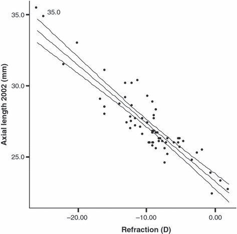
The scattergram in Fig. 1 illustrates the strong association between refraction and AL in the series (r = −0.90). With a view to the significant intra-pair variation, as given by the above anisometropia data, the regression statistics were based on all 65 eyes with data available at age 54. The refractive ranges were from −0.3 to −25 D for 32 right eyes and from +2.0 to −26 D for 33 left eyes, with corresponding AL ranges of 22.3–34.8 and 22.6–35.4 mm, respectively. The correlation between the two parameters had coefficients of −0.90 in both sets of eyes, and the regression lines calculated were remarkably close (right eye: y = 23.22–0.40x; left eye: y = 23.31–0.41x). In the subgroup of 22 participants with anisometropia exceeding a side difference of 1 D (Table 4), there was a high correlation between individual side differences in refraction and AL (r = −0.97). One mm of relative axial elongation resulted in an average of 2.24 D more minus refraction.

Scattergram showing the association between refractive value (D, x-axis) and axial length (mm, y-axis) based on cross-sectional data from 65 eyes at the age of 54 years. Regression line given by y = 23.28 + 0.41x (r = −0.90).
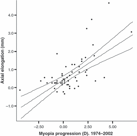
A Pearson regression calculation was performed regarding the changes in the same two parameters over the 28 years, as based on all 57 eyes with longitudinal measurements. With myopia increase on the x-axis (thus given with a positive sign), a full sample regression line of y = 0.43 + 0.36x was calculated (r = 0.65; n = 57, Fig. 2). This corresponds to an AL change of 2.78 D/mm.

The association between adult myopia progression (x-axis, given with a positive sign) and axial ocular elongation (y-axis; longitudinal data sets from 57 eyes). Regression line given by y = 0.43 + 0.36x (r = 0.65).
Finally, Table 5 presents the correlation coefficients in the series that indicated a significant relationship between parameters. Because of its close and constant association to AL, vitreous length measurement was omitted from all tables (correlation coefficient r = 0.97; Goldschmidt et al. 1981).
| Correlation coefficient (r) | ||
|---|---|---|
| Cross-sectional, age 54† | ||
| AL | Refraction | −0.90 |
| AL | Crad | 0.32 |
| ACD | LT | −0.36 |
| ACD | Lens power | −0.59 |
| ACD | Crad | −0.27* |
| Longitudinal, changes in parameters‡ | ||
| AL | Myopia increase | 0.64 |
| LT | ACD | −0.48 |
| AL | Lens power | −0.55 |
- *p < 0.05. The remaining Pearson correlation r-values all expressed P-values < 0.01.
- †Cross-sectional comparison of refraction and oculometry data in all 65 eyes.
- ‡Correlations between changes over time, as based on the 57 longitudinal data sets.
- AL, axial length; Crad, corneal curvature radius; ACD, anterior chamber depth; LT, lens thickness.
Among the minor associations, we emphasize the positive correlation between AL and Crad (r = 0.32). In general, a longer eye will be more myopic and have a flatter cornea. A thicker lens by age was associated with a lower ACD value (r = −0.36), and the negative correlation between ACD and Crad (r = −0.27, p < 0.05) should indicate a slightly deeper chamber in case of a more peaked cornea.
Discussion
The main results of this study were that the descriptive statistics and correlations indicate that myopic eyes are long eyes with flatter corneae, and that adult-age myopia increase is associated with further axial elongation. Regarding size differences according to gender and LT increase by age, both events appeared independent of the above main refractive parameter associations. Parallel to the age-related LT increase, there is an axial anterior chamber shallowing, all supporting the hypothesis that mid lens position remains by and large the same. There is some indication that lens power decreases by age, as nature’s means of possibly reducing the myopia increase that axial elongation should have inferred. In the following sections, the two main refractive parameters are discussed separately.
AL
Myopia has proved to be axial throughout our series. Despite variation in attendance over time, numerically the correlation coefficients between refractive value and AL uniformly never came under 0.80. The association is further demonstrated by the scattergram in Fig. 1 and by the anisometropia data given in Table 3. The apparent indication of AL as the factor underlying the side difference in refraction in anisometropic participants is in accordance with previous studies (Fledelius 1981, 1982; Höh 1992; Rendu 1992).
From the age of 26 years, the evaluation of eventual AL change in eyes with high myopia (n = 50) over the 28-year follow-up period was compared to the refractive change recorded. The correlation coefficient of r = 0.65 indicated a rather strong association between adult-age increase of myopia and axial elongation, with the calculated regression line as given earlier. The intercept value of 0.43 mm mathematically suggested that with increasing years the adult eye without myopic change would elongate by this measure, and the slope that each dioptre of myopia increase was on average associated with 0.36 mm axial elongation. Over the age span (26–54 years) this corresponds to 1 mm elongation per 2.78 D myopia increase, whereas the anisometropia regression analysis suggested a value of 2.24 D.
Although the intercept value indicating axial adult eye elongation of 0.43 mm may be questioned, we have ample evidence that AL increase per se is not confined merely to childhood and adolescence. Parallel to the continued increase in myopia degree in most participants, we found an axial elongation of adult age, timely in succession of what is regarded as the basic or refraction-neutral growth of the teenage eye.
The scattergram in Fig. 2 illustrates that about one third of the eyes with high myopia had pretty static refraction and size during the adult-age observation period, whereas other eyes elongated markedly parallel to the increase of myopia. This constitutes a biological contrast to most other outgrown body measures in young adults (e.g. head circumference and body height; Fledelius and Stubgaard 1986a, 1986b, 1986c). Scleral distension with renewed axial elongation of adult age has also been confirmed in emmetropic adolescents who after the late teenage years acquired adult-onset myopia (Fledelius & Stubgaard 1986a; McBrien & Millodot 1987). It may be hypothesized that the current tissue cata-/anabolism in such eyes is not an exact remodelling, but directional: with sclera disappearing from the inside and being built up on the outer scleral sphere.
Crad
In our analyses, the Crad value showed no mathematical influence on refractive value. However, with r-values fluctuating about 0.4, there was a weak positive correlation to AL – a correlation generally in accordance with literature results also taken from refractively unselected samples (Stenström 1946; Delmarcelle et al. 1976; Fledelius 1988). Thus, in a longer eye the trend in the population will be towards a slightly flatter cornea.
Given the normal range of corneal curvature radius (7.1–8.6 mm), the prototype eye with high myopia should have a relatively flat cornea. In general, the corneal curvature has come to its endpoint measure in early pre-school years, and in previous investigations of refractively unselected Danish children aged 10 years male and female mean values were 7.94 mm and 7.81 mm, respectively (Fledelius 1976, 1982, 1995). Mean values from a Swedish group of young adults were on a similar level (7.90 mm and 7.77 mm, respectively), with a correlation coefficient between AL and Crad of 0.31 (Stenström 1946). All considered, in a high myopia sample on average we should expect a Crad of about 8.0 mm.
However, the possibility exists that some eyes with high myopia do not follow the normative course for growth and evolution, as outlined here. Regarding the cornea, this was suggested from data by Francois & Goes (1973). They investigated Belgian adolescents and presented mean Crads of 7.70, 7.80 and 7.61 mm for hypermetropia, emmetropia and myopia, respectively. In a previous evaluation, the same researchers reported a mean value of 7.57 mm in participants with myopia between 6 and 15 D (n = 63). Likewise, our present Crad mean figures (men 7.81 mm, women 7.56 mm, cf. Table 1; for the pooled high myopia sample 7.66 mm) were lower than what was expected from the above keratometry data considered normative for Danish adolescents, thus apparently supporting the Belgian findings. The deviation from expected relative flatness with increased AL is best explained by assuming that a fraction of the eyes with high myopia simply do not adhere to the rules of morphology and correlation that seem valid for ordinary juvenile myopia.
To further classify our keratometry results into normal, relatively steep and very steep corneas, we chose split values of 7.70/7.45 mm in men and 7.57/7.32 mm in women. The limits were suggested from our previous normative teenage Crad values according to gender, with 1 or 2 SDs (∼ 0.25 mm) deducted, respectively. Sixteen had peaked corneas, 13 in both eyes and three unilaterally. Nineteen of the 29 peaked corneas were within 1–2 SD below average, and 10 eyes came under the –2 SD limit value. Compared to the hypothetical distribution into normal, relatively peaked and very peaked according to an SD-based Crad subdivision, the trend in the myopia sample towards a peaked cornea was highly significant (χ2 = 13.86, df = 2, p < 0.001).
Searching the literature, a slight trend towards a stronger refracting cornea in the more myopic eye was indicated from findings in Höh’s anisometropia series of 1992, although this was not statistically significant. Mean Crad values of 7.73 and 7.75 mm were reported in the more myopic eye [mean refractive value −11.3 D (SD 5.4)] and in the less myopic eye [−3.1 D (SD 4.3)], respectively. The trend could not be confirmed in our 10 unilateral high myopia cases [mean side difference in refraction 13.9 D (5.70)], nor in the full anisometropia subsample (n = 22). Comparing the more myopic to the less myopic eye, mean values were 7.70 in both eyes in the unilateral high myopia sample, and 7.71 mm in the extended anisometropia series.
On a casuistic level, a hereditary component was suggested from one of our most ‘peaked’ corneal cases, a woman with myopia degree −0.25 D in her right eye and −7.25 D in her left. She had relatively short eyes (AL: 22.2 mm and 24.6 mm, respectively) and quite low Crad values (about 7.13 mm). Her sister (1 year younger, not in the trial) had right- and left-eye myopia values of −5 D and −7.75 D, AL values of 23.8 mm and 24.3 mm and even lower Crad values of 7.12 mm and 6.99 mm, respectively. Corneal surfaces were regular in both sisters, and with no features of keratoconus. – With reference to environment, and here with a specific view to the more curved corneas of eyes eventually presenting with myopia of prematurity (Fledelius 1976, 1982, 1995), we found no anamnestic or clinical evidence of such a background in our series. For birth year 1948, marked prematurity was usually incompatible with survival, and Denmark had not had its first case of retrolental fibroplasia yet.
Lens power
The 1 D relative myopization by 0.79 mm mean axial elongation denoted less longitudinal dioptric change than the about 1.9 D as cross-sectionally to be expected from such an AL increase. The minor changes according to Crad measurements, and according to possible shifts in cardinal refractive planes caused by lens thickening by age, could hardly be held responsible for the apparent discrepancy regarding dioptric equivalent to axial elongation. We therefore suggest a modifying influence of the lens power, to indicate a decrease in refractive power over time. Here, reference is made to the so-called lens paradox (Koretz & Handelman 1986), as quoted in further investigations by Dubbelman & Van der Heide (2001). Despite lenses getting thicker and more curved with age, the established refractive trend in the population aged 35 years or older is towards hypermetropia, and not a relative myopization (Fledelius 1988). Correcting for Scheimpflug image distortion (caused by oblique incidence) and axial IOLMaster measurement data, Dubbelman et al. (2001) reported on a fall in refractive index on behalf of the lens in the order of 1.43 to 1.42, as relevant for the age span of our high-myopia series.
Conclusions
High myopia is predominantly axial by nature, and adult ocular elongation even parallels the late increase of myopia that was seen in two out of three participants in our unselected long-term high myopia series followed between the ages of 26 and 54 years.
In the population refractive distribution, there is correlation evidence that longer eyes have flatter corneas. This correlation appears reduced or partly eliminated in high myopia, giving some place for an index component (corneal power) as an alternative to axial elongation as sole or main event. In the present sample, the proportion of cases with steep corneal curvatures was thus found to be significantly increased. Furthermore, there was an indication of decreased lens power over time in the series.
Acknowledgements
This study was supported by grants from the Bagenkop Myopia Foundation and the Danish Eye Health Society.


