COMPUTED TOMOGRAPHY OF THE PHARYNX IN A CLOSED VS. OPEN MOUTH POSITION
Abstract
The pharynx is anatomically complex and evaluation can be difficult even with cross-sectional imaging. Eight animals had computed tomography (CT) studies of the head performed with the mouth open and closed. The studies were anonymized and evaluated by four radiologists for visibility of six anatomic regions (dorsal wall of nasopharynx, lumen of nasopharynx, dorsal margin of the soft palate, ventral margin of the soft palate, oropharynx, and laryngopharynx) and for certainty of a normal or abnormal diagnosis of four different anatomic regions (nasopharynx, soft palate, oropharynx, and laryngopharynx). Mean visual scores differed significantly between mouth positions and were improved when the mouth was open. The ability of radiologists to classify anatomic regions as normal or abnormal vs. unsure also varied between mouth positions, and there was greater uncertainty when the mouth was closed. In addition, estimated volume of the air-filled nasopharynx differed significantly as a function of mouth position and was greater when the mouth was open (mean=1.187 cm3, SE=0.177) vs. closed (mean=0.584 cm3, SE=0.116). Computed tomographic evaluation of the pharynx can be improved with the mouth open.
Introduction
Computed tomography (CT) is being used more widely for investigation of pharyngeal disease in animals. Chronic oropharyngeal foreign bodies, nasopharyngeal stenosis, and cysts can be diagnosed and managed with the assistance of CT.1–3 The complex anatomy of the pharynx, however, is difficult to evaluate due to the silhouetting of multiple structures of similar attenuation.
The pharynx is a common passage for both the respiratory and digestive systems. It can be divided into three major regions: the nasopharynx, oropharynx, and laryngopharynx. The nasopharynx is dorsal to the soft palate, extending from the choanae of the nasal cavity to the intrapharyngeal ostium. The oropharynx extends from the fauces to the intrapharyngeal ostium, and is bounded by the soft palate dorsally, the root of the tongue ventrally and the tonsillar fossa laterally. The laryngopharynx is the portion of the pharynx dorsal the larynx.4,5
Our objective was to determine whether there was improved CT visualization of pharyngeal structures with the mouth in an open compared with a closed position. In addition, the level of confidence of the radiologist regarding identification of abnormalities was compared between open and closed mouth CT studies. We hypothesized that CT performed with the mouth open would provide better visualization of pharyngeal structures and that the radiologist would be more confident determining that structures were normal or abnormal.
Materials and Methods
Eight animals scheduled for CT evaluation of the head were studied. The owner consented to two CT examinations and the application of two separate CT studies was judged not to be detrimental. There were six cats (two domestic longhair, two domestic shorthair, one Persian, and one Maine Coon) and two dogs (one Italian Greyhound and one mixed breed). The age ranged from 1 to 10 years, with a mean (± SD) of 3.4 ± 3.2 years. There were four neutered males, two intact males, one neutered female, and one intact female. The animals' weight ranged from 3.5 to 9.8 kg, with a mean of 5.3 ± 2.2 kg. One cat's weight was unknown.
Each animal had a CT examination of the head acquired with the mouth closed using a third generation single slice CT scanner* with a kVp of 120 and mA of 100–150. Examinations consisted of contiguous 1–3 mm collimated transverse images of the head with the patient in sternal recumbency and under general anesthesia. All patients had an endotracheal tube placed and received inhalant anesthesia. Images were acquired from the nares to the caudal aspect of the larynx with the mouth in a closed position. A closed position was defined as the mouth in a neutral position with the endotracheal tube in place, and was at an approximate angle of 5°–15°. Next, the mouth was propped open at approximately 30–45° angle with gauze, tape roll, or portions of a plastic syringe or syringe case. Images were again acquired from the nares to the caudal aspect of the larynx. All images were acquired using a bone algorithm, except for two cats, whose studies were acquired with a standard soft tissue algorithm. The smallest field of view that allowed inclusion of the entire head was used. One cat received intravenous contrast medium.†
Images from each study were anonymized, and the open and closed mouth images were separated from each other. Each study was limited to images of the pharyngeal structures, beginning just rostral to the choanae and extending caudal to the larynx. The anonymized studies were evaluated by three Diplomates of the American College of Veterinary Radiology (A.Z., M.S., W.L.) and one senior radiology resident (D.C.). DICOM images were viewed using commercially available software,‡ and the evaluators were allowed to alter window or level and to use available tools such as multiplanar reconstruction or zoom. Six anatomic regions, including the dorsal wall of nasopharynx, lumen of nasopharynx, dorsal margin of the soft palate, ventral margin of the soft palate, oropharynx, and laryngopharynx, were assessed for visibility with the following scale: poor (score=0), satisfactory (score=1), or good (score=2). In addition, four anatomic regions, including the nasopharynx, soft palate, oropharynx, and laryngopharynx, were assessed as being normal, abnormal, or unsure. For statistical analysis, a score of 0 was attributed to the regions assessed as normal or abnormal and a score of 1 was attributed to the region classified as unsure.
The CT images were downloaded from the PACS server to a remote workstation. The gas-filled volume of the nasopharynx was estimated for each animal in an open and closed mouth position utilizing a software package designed for volumetric analysis of CT data.§ Hand-drawn regions of interest were made approximately from the choanae to the intrapharyngeal ostium of the pharynx.
Differences in visual scores between mouth positions were evaluated with a mixed effect model containing mouth position, radiologist, anatomic region, and the interaction between anatomic region and mouth position as fixed effects. A random effect for each animal was included to account for the correlation of observations from the same animal. Within each mouth position, we tested for differences among anatomic regions with a mixed effect model containing anatomic region and radiologist as fixed effects and animal as a random effect. Where anatomic region was a significant factor, we evaluated all pairwise comparisons with t-tests based on least squares means and a Tukey's adjustment to maintain the family-wise error rate at 0.05. To determine if the certainty of classification varied as a function of mouth position and region, a mixed effect logistic model was used to model the probability of an unsure designation as a function of mouth position, radiologist, and anatomic region. This model was fit using a marginal generalized estimating equations approach6 through the alternating logistic regressions algorithm.7 The correlation among observations from the same animal was modeled as an exchangeable log odds ratio structure. Within each mouth position, we also tested for differences among anatomic regions with a mixed effect model containing anatomic region and radiologist as fixed effects and animal as a random effect. For significant main effects, Wald χ2-tests were used to evaluate all pairwise comparisons with a Bonferroni's adjustment to maintain the type I error rate at 0.05. To determine if estimated nasopharynx volume differed between mouth positions, estimated volumes were compared with a paired sample t-test. A significance level of 0.05 was used for all tests and statistical analyses were conducted using SAS version 9.2.
Results
Visual scores differed significantly between mouth position (F1, 376=140.50, P<0.0001), radiologist (F3, 376=7.36, P<0.0001), and across anatomic regions (F5, 376=7.13, P<0.0001). Mean visual scores were higher for the open mouth position (open: 1.70 ± 0.036, closed: 1.08 ± 0.053, n=216). The interaction between mouth position and anatomic region also was significant (F5, 376=7.27, P<0.0001), indicating that visualization of regions was affected differentially by mouth position. We investigated differences in visualization scores further by anatomic region and mouth position. Visual scores did not differ among anatomic regions with the mouth in the open position (F5, 184=0.16, P=0.98) but did with a closed mouth position (F5, 184=13.48, P<0.0001). For the oropharynx and ventral margin of the soft palate, visual scores were significantly lower than for the other four regions when the mouth was closed (Table 1). The significance of anatomic region as a main effect resulted from the low visual scores for the oropharynx and ventral margin of the soft palate with a closed mouth position.
| Anatomic region | Mouth position | |
|---|---|---|
| Open | Closed | |
| Dorsal wall of nasopharynx | 1.84 ± 0.07 | 1.50 ± 0.10b |
| Lumen of nasopharynx | 1.78 ± 0.07 | 1.41 ± 0.10b |
| Dorsal margin of the soft palate | 1.84 ± 0.07 | 1.41 ± 0.10b |
| Ventral margin of the soft palate | 1.84 ± 0.07 | 0.81 ± 0.14a |
| Oropharynx | 1.81 ± 0.07 | 0.72 ± 0.14a |
| Laryngopharynx | 1.81 ± 0.07 | 1.37 ± 0.12b |
| Overall mean | 1.82 ± 0.03 | 1.20 ± 0.05 |
- Overall mean visual scores were higher when the mouth was open.
- Visual scores did not differ significantly between anatomic regions with the mouth open, but did for the mouth closed. For the oropharynx and the ventral margin of the soft palate, visual scores were significantly lower than for the other four regions when the mouth was closed. Values with different letters differed significantly at a Tukey's adjusted P-value of 0.05. Visibility scores were as follows: poor (score=0), satisfactory (score=1), or good (score=2).
We next investigated factors influencing classification certainty. The odds of an unsure classification differed significantly between mouth positions (χ2=5.30, P=0.021, df=1) and was nearly significantly different among anatomic regions (χ2=7.51, P=0.057, df=3). Similar to the visual score findings, classification certainty was greater with the animal's mouth in the open than closed position. With the closed mouth position, 28.9% of the determinations were unsure, compared with only 2.3% for an open mouth position. The odds of an unsure classification was significantly greater when the animal's mouth was closed than open (odds ratio=27.53 [95% CI 3.12, 243.05]). Although, overall the closed mouth position resulted in greater classification uncertainty than with an open mouth position, classification certainty varied among anatomic regions for the closed mouth position. The odds of an unsure classification was significantly higher for the soft palate and oropharynx than for nasopharynx and laryngopharynx with a closed mouth position (Table 2). Few classifications were unsure for the open mouth position, which precluded assessment of the interaction between open mouth position and anatomic region.
| Anatomic region | Odds ratio |
|---|---|
| Nasopharynx | 0.057 [0.012, 0.272] |
| Soft palate | 1.162 [0.432, 3.126] |
| Oropharynx | 1.013 [0.287, 3.575] |
| Laryngopharynx | 0.056 [0.004, 0.768] |
- The odds of an unsure classification was significantly higher for the soft palate and oropharynx than for nasopharynx and laryngopharynx with a closed mouth position. A Bonferroni's adjustment was used to construct confidence intervals to maintain a family-wise error rate of 0.05.
Finally, estimated nasopharynx volume differed significantly between mouth positions (t=−2.45, df=7, P=0.044). The estimated volume was significantly greater when the mouth was open (mean=1.187 cm3, SE=0.177) vs. closed (mean=0.584 cm3, SE=0.116).
Discussion
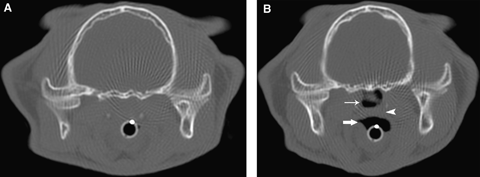
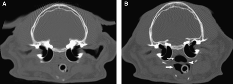
Visualization of pharyngeal anatomy was improved significantly with the mouth open compared with closed (1, 2). In addition, radiologists were more likely to classify a region of the pharynx as definitively normal or abnormal when the mouth was open compared with an unsure classification when the mouth was closed. When the mouth is propped open, there is increased luminal gas within the pharyngeal structures, particularly the nasopharynx. This results in opening of pharyngeal spaces that are compressed when the mouth is closed and allows for visualization of the soft tissue structures that otherwise silhouette with each other.

(A) Closed mouth position at the level of the temporomandibular joints. (B) Same patient in Fig. 1A with the mouth open. Note the increased gas within the pharynx when the mouth is open. The soft tissue structure in the nasopharynx is an inflammatory polyp, which was not visualized on the closed mouth study. Oropharynx (large arrow), nasopharynx (small arrow), and soft palate (arrowhead).

(A) Closed mouth position at the level of the tympanic bullae. (B) Same patient in Fig. 2A with the mouth open. Note the increased gas within the pharynx and improved visualization of the soft palate when the mouth is open. Oropharynx (large arrow), nasopharynx (small arrow), and soft palate (arrowhead).
Improved opening of the pharynx is supported further by volumetric estimates of the gas-filled portion of the nasopharynx that were measured in each animal, which increased significantly with the mouth open. As the soft tissue structures pull away from one another, the air filled volume of the pharynx increases. Although not the purpose of this study, this implies that measurement of pharyngeal area is possible using CT with the mouth open. This could be useful in the assessment of nasopharyngeal stenosis and in measuring for nasopharyngeal stents. Additional studies with specific guidelines for degree of mouth opening are needed to establish normal pharyngeal volume.
Some differences in anatomic region were also identified. For the oropharynx and ventral margin of the soft palate, visual scores were significantly lower when the mouth was closed. Also, the estimated odds of an unsure classification were higher for the soft palate and oropharynx, especially for a closed mouth position. When the mouth is closed, the oropharynx is one of the first regions to collapse with loss of the luminal gas providing contrast to the pharynx. Visibility of the soft palate, especially the ventral aspect, is likely affected similarly since it represents one margin of the oropharynx. In some animals, the nasopharynx and laryngopharynx can maintain luminal gas even with the mouth closed.
It was not possible to assess the effect of mouth position on pharyngeal imaging in conjunction with administration of contrast medium because only one animal received intravenous contrast medium. Many of the normal oral and pharyngeal tissues are highly contrast enhancing, and contrast medium administration may have improved visualization of this region, even with the mouth closed. Tumors, abscesses, or inflammatory polyps are also likely to have improved conspicuity with contrast medium administration.
The technique used to prop the mouth open was not standardized, and tape rolls, syringe cases, and gauze were all used to open the mouth at the level of the canine teeth to approximately a 30–45° angle. Patients with objects, such as gauze, in the caudal aspect of the oropharynx were excluded since this can cause compression of the soft palate and collapse of the nasopharynx. In addition, metallic or other highly attenuating objects cause beam hardening and edge gradient artifacts that would reduce the quality of the study. Further studies with a mouth gag standardized to body size could be performed to determine the ideal method and degree to prop the mouth open.
All animals were under general anesthesia, and had an endotracheal tube present for both the open and closed mouth examinations. The presence of the endotracheal tube can alter visibility of pharyngeal structures, since it extends from the oropharynx through the laryngopharynx and may compress the nasopharynx. Temporary removal of the endotracheal tube may improve CT examination of the pharynx, but may not be advised due to the clinical status of the patient. Further studies evaluating CT of the pharynx with and without an endotracheal tube should be considered to determine the benefit of this procedure.
Although the radiologists were unaware of the patient and mouth position, some of these variables may have been identified. For this reason, images rostral to the choanae were eliminated so that the mouth gag was not identified in those animals with the mouth propped open. In addition, species differences and disease processes may have allowed patient identification and reduced anonymity. The animals in this study were clinically abnormal, and some of the final diagnoses included rhinitis, otitis media, nasopharyngeal stenosis, and inflammatory polyps. Comparisons of nasopharyngeal volume were only compared within the same animal to reduce effects of the patient and disease variability.
In conclusion, CT examination with the mouth open may improve diagnostic evaluation in comparison to having the mouth closed. There is significant increase in the size of the air-filled portion of the nasopharynx with the mouth open, providing better delineation of the surrounding structures.
Footnotes
ACKNOWLEDGMENTS
The authors would like to thank Winnie Y. Lo and Jason Peters for their assistance and contributions to this manuscript. Statistical support was made possible by Grant Number UL1 RR024246 from the National Center for Research Resources (NCRR), a component of the National Institutes of Health (NIH) and NIH Roadmap for Medical Research. Its contents are solely the responsibility of the authors and do not necessarily represent the official view of NCRR or NIH. In formation on Re-engineering the Clinical Research Enterprise can be obtained from http://nihroadmap.nih.gov/clinicalresearch/overview-translational.asp.


