RELATION BETWEEN AGE, BODY WEIGHT, AND MEDIAL RETROPHARYNGEAL LYMPH NODE SIZE IN APPARENTLY HEALTHY DOGS
Abstract
The aims of this study were to determine the size of the medial retropharyngeal lymph nodes in apparently healthy dogs using ultrasonography and to investigate relationships between body weight (1.8–59 kg), age (1.0–15 years), and medial retropharyngeal lymph node sizes (width, height, and length). The sample population consisted of 100 apparently healthy, volunteered, adult dogs. The data were normally distributed, thus mean, SD, and Pearson's correlation were used. Repeatability of ultrasound measures was assessed as the percentage of differences between duplicate measures that were within 2 SDs of the differences: all measures were at least 93% repeatable (differences typically were ≤0.25 cm and always <1 cm). No difference between sexes was observed. The medial retropharyngeal lymph node increased in size with increased body weight (r=0.46 to 0.59) and decreased in size with increased age (r=−0.30 to −0.50). Although statistically significant, the actual variation is not likely clinically important due to the small range of sizes, measurement error, and various combinations of age and body weight. Therefore, regardless of body weight or age, the average width is 1.0 cm, height is 0.5 cm, and length is 2.5 cm and maximum width is 2 cm, height is 1 cm, and length is 5 cm. Based on the maximal difference between duplicate measures (with some exception), any change ≥0.4 cm with width or height, or ≥1.0 cm in length, in a follow up measurement probably represents a true biological change rather than measurement error.
Introduction
We occasionally observe large medial retropharyngeal lymph nodes in dogs undergoing computed tomography (CT) of the head for radiation-therapy planning. These lymph nodes are rarely detected during palpation even when palpation was repeated after CT and the node was considered enlarged. In humans, the sensitivity and specificity of diagnosing regional lymph node metastasis to the neck by palpation is poor (approximately 60–70%).1 Therefore, imaging is often performed to reduce diagnostic uncertainty about regional lymph node metastasis.1
The retropharyngeal lymph center consists of the medial retropharyngeal lymph node and occasionally the lateral retropharyngeal lymph node. The normal appearance and size of the medial retropharyngeal lymph node of dogs has been reported for magnetic resonance (MR) imaging but not for ultrasonography.2 In veterinary medicine, ultrasonography is frequently used to assess lymph node size and structure. Therefore, one aim of this study was to use ultrasonography to determine the size of the medial retropharyngeal lymph node in apparently healthy dogs. We questioned whether alterations in the size of the medial retropharyngeal lymph node were associated with body weight or with age. Therefore, the other aim of this study is to assess the relationship between body weight and age with medial retropharyngeal lymph node size (width, height, and length).
Methods
The population consisted of 100 adult (>1 year old) dogs that were not excessively overweight, having a body condition score not exceeding 6 on a scale of 1–9 determined by physical examination. Dogs were apparently healthy and owners volunteered the dog's enrollment in the study between March 30, 2007 and June 10, 2007. The Institutional Animal Care and Use Committee of Cornell University approved the study. Aggressive or uncooperative dogs were not studied.
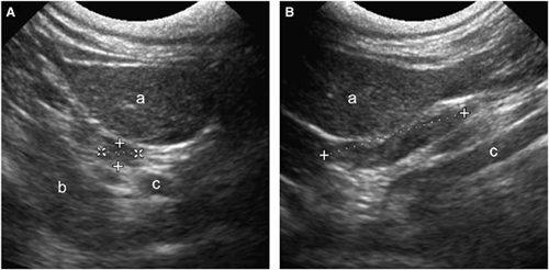
For each dog, the age, gender, breed, and body weight (kg) were recorded. All lymph node measurements were performed by the same examiner (G.O.B.), who is a third-year resident, using the same ultrasound machine* and a curved 8.5-MHZ transducer. Dogs were in dorsal or lateral recumbency, and alcohol or coupling gel was applied to the skin without clipping the hair; one dog was examined while standing. Dogs were awake and manually restrained. Whether the left or right lymph nodes were examined first was determined by coin toss. Useful landmarks for identifying the medial retropharyngeal lymph node were the longus colli muscle, mandibular salivary gland, and common carotid artery. The entire lymph node was scanned in two planes from the ventrolateral aspect of the neck: a transverse or transverse–oblique plane was used to produce a short-axis sonogram of the medial retropharyngeal lymph node, and a sagittal or sagittal–oblique plane was used to produce a long-axis sonogram. Width, height, and length were measured using electronic calipers that were part of the ultrasound machine. Width and height were measured on the short-axis sonogram and length on the long-axis sonogram (Fig. 1). Width was defined as the left–right measurement; height, the dorsoventral measurement; length, the craniocaudal measurement. Duplicate measures of the same lymph node were made by repeating the examination on another frozen image. The examiner was not aware of the ultrasound measures as a card was placed over the display. Images with the measurements were recorded digitally and printed.

Short-axis (A) and long-axis (B) sonograms of the right medial retropharyngeal lymph node in an apparently healthy, 15 kg, 8-year old, neutered female, mixed-breed dog: width 0.59 cm, height 0.40 cm, length 2.23 cm. The medial retropharyngeal lymph node (calipers) is located craniodorsal to the mandibular salivary gland (a), ventral to the longus colli muscle (b) and lateral to the common carotid artery (c). 67 × 34 mm (300 × 300 DPI).
Statistical analyses were performed using commercially available computer software.†,‡ Because measurements of the left and right medial retropharyngeal lymph nodes were not independent, differences and relationships in measurements of the left and right medial retropharyngeal lymph nodes were considered separately. Duplicate measures were averaged before analysis to improve measurement precision. Kolmogorov–Smirnov tests confirmed that the distributions of the data were normal for age, body weight, and lymph node measurements. Therefore, the data were described using mean, SD, minimum value, and maximum value. Additionally, Pearson's correlation coefficient (r) was used to investigate the null hypothesis that there was no linear association between variables. Statistical significance was set at 5% (two sided). We also calculated 95% confidence interval (CI) and the coefficient of determination (r2), which describes the proportion of variation in one variable that is explained by the variation in the other variable.
Repeatability of ultrasound measures was assessed by obtaining the SD of the differences between duplicate measures per dog and comparing them to the difference between duplicate measures. The method was considered repeatable if 95% of (the absolute values of) differences between duplicate measures were <2 SDs of the differences.
Results
The sample of 100 dogs contained mixed- and pure breed dogs weighing between 1.8 and 59 kg. There were six intact females, 47 neutered females, two intact males, and 45 neutered males (Table 1). Descriptive statistics for body weight, age and lymph node sizes are shown (Table 1). There was no difference between genders when we compared the 95% CIs on the means for all measurements.
| Statistic | BodyWeight (kg) | Age(years) | Left MRLN (cm) | Right MRLN (cm) | ||||
|---|---|---|---|---|---|---|---|---|
| Width | Height | Length | Width | Height | Length | |||
| Minimum | 1.8 | 1.0 | 0.34 | 0.23 | 0.85 | 0.32 | 0.26 | 0.79 |
| Mean | 21.2 | 5.6 | 0.88 | 0.52 | 2.44 | 0.88 | 0.52 | 2.52 |
| Maximum | 59.0 | 15.0 | 1.82 | 0.99 | 4.60 | 1.65 | 1.00 | 5.09 |
| 95% CI for mean | 18.8, 23.6 | 5.0, 6.3 | 0.82, 0.94 | 0.49, 0.55 | 2.27, 2.61 | 0.83, 0.94 | 0.49, 0.55 | 2.35, 2.69 |
| SD | 12.1 | 3.3 | 0.31 | 0.16 | 0.84 | 0.29 | 0.15 | 0.86 |
- MRLN, medial retropharyngeal lymph node; CI, confidence interval; SD, standard deviation.
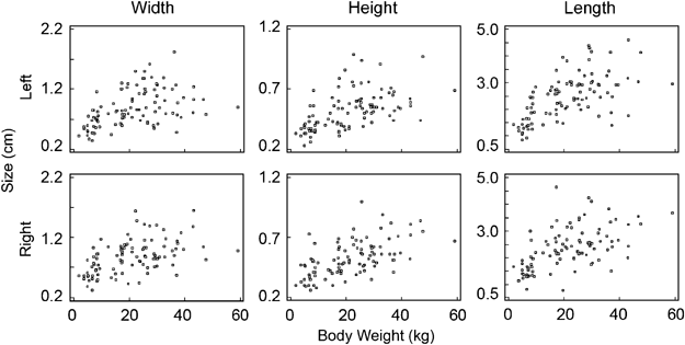
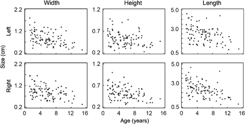
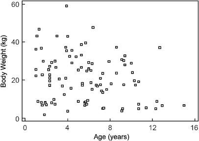
Scatter plots for body weight and medial retropharyngeal lymph node size, age and medial retropharyngeal lymph node size, and age and body weight are shown (2-4). A fair-to-good, positive, correlation between body weight and medial retropharyngeal lymph node size (width, height, and length) and a fair, negative, correlation between age and medial retropharyngeal lymph node size (width, height, and length) were observed (Table 2). In other words, the medial retropharyngeal lymph node increased in size slightly with increased body weight and, to a lesser extent, decreased in size with increased age. Additionally, there was no association between age and body weight (r=−0.19; CI=−0.38 to 0.00; P=0.053).


Scatter plot for body weight and age in dogs in Table 1. There was no association (r=−0.19) between age and body weight. 98 × 73 mm (300 × 300 DPI).
| Statistic | Left MRLN | Right MRLN | ||||
|---|---|---|---|---|---|---|
| Width | Height | Length | Width | Height | Length | |
| Body weight | ||||||
| r | 0.46 | 0.54 | 0.61 | 0.52 | 0.59 | 0.57 |
| P-value | <0.0001 | <0.0001 | <0.0001 | <0.0001 | <0.0001 | <0.0001 |
| 95% CI for r | 0.29, 0.60 | 0.38, 0.67 | 0.41, 0.72 | 0.36, 0.65 | 0.44, 0.70 | 0.42, 0.69 |
| r2 | 0.21 | 0.29 | 0.37 | 0.27 | 0.35 | 0.33 |
| Age | ||||||
| r | −0.48 | −0.30 | −0.40 | −0.40 | −0.39 | −0.50 |
| P-value | <0.0001 | 0.0027 | <0.0001 | <0.0001 | <0.0001 | <0.0001 |
| 95% CI for r | −0.62, −0.32 | −0.47, −0.11 | −0.55, −0.22 | −0.56, −0.22 | −0.55, −0.21 | −0.63, −0.34 |
| r2 | 0.23 | 0.09 | 0.16 | 0.16 | 0.15 | 0.25 |
- CI, confidence interval.
Ultrasound measures of medial retropharyngeal lymph node size were considered repeatable (Table 3) for width and length of the left medial retropharyngeal lymph node and height and length for the right medial retropharyngeal lymph node because at least 95% of differences between duplicate measures were within 2 SDs of the differences. Ultrasound measures for height of the left medial retropharyngeal lymph node (93%) and width of the right medial retropharyngeal lymph node (94%) did not meet our criterion for repeatability.
| Statistic | Left | Right | ||||
|---|---|---|---|---|---|---|
| Width | Height | Length | Width | Height | Length | |
| Mean of the differences of duplicate measures (cm) | 0.00 | 0.00 | 0.01 | −0.01 | 0.00 | 0.05 |
| SD of the differences of duplicate measures (cm) | 0.16 | 0.09 | 0.24 | 0.09 | 0.09 | 0.26 |
| % of dogs with difference between duplicate measures ≤2 SDs | 97 | 93 | 96 | 94 | 95 | 95 |
| Maximum difference (absolute value) between duplicate measures (cm) | 0.93 | 0.25 | 0.93 | 0.35 | 0.35 | 0.81 |
- SD, standard deviation.
Discussion
Body weight and age each contributed approximately 25% of the variability in lymph node sizes (width, height, and length) based on the coefficient of determination. Additionally, statistically significant relationships were shown between body weight, age, and all lymph node sizes based on the correlation coefficient. As such, it is reasonable to generalize that old, small-breed dogs tend to have smaller medial retropharyngeal lymph nodes than young, large-breed dogs. In between these extremes, however, the differences between medial retropharyngeal lymph node sizes relative to age and body weight are not likely to be clinically important due to the small range of sizes observed, measurement error, and various possible combinations of age and body weight Therefore, regardless of body weight or age, it is reasonable for clinical use to consider the average medial retropharyngeal lymph node width is 1.0 cm, height is 0.5 cm, and length is 2.5 cm. And, as a rule of thumb, the medial retropharyngeal lymph node may be considered enlarged when the width is ≥2.0 cm, height is ≥1.0 cm, and length ≥5.0 cm. These findings are consistent with previous descriptions of medial retropharyngeal lymph node size.2,3 Furthermore, based on our maximal difference between duplicate measures (with some exception), any change ≥0.4 cm with width or height, or ≥1.0 cm in length, in a follow up measurement probably represents a true biological change rather than measurement error.
On average, the sizes of the left and right medial retropharyngeal lymph node were symmetric. In some individuals, however, asymmetry was observed especially for length. The asymmetry might be due to biologic variation of the individual, measurement error due to movement of the head and neck between measurements, or error created by different orientation of the probe between measurements. Subjectively, we do not believe there is an association between the shape of the head and lymph node size. The negative association between the age and medial retropharyngeal lymph node size may be similar to senescent atrophy, which is seen with other lymphatic tissue like the thymus.
It is our belief that the sample population of 100 dogs in this study is a reasonable representation of the target population of all apparently healthy dogs and not subjected to undue bias. In addition, we believe that the measurements are repeatable because most of the times the measurements were within 2 SD (of the difference) of each other.


