Implant design and interface force transfer
Implantatdesign und Kraftübertragung auf den Knochen: eine photoelastische Analyse und Untersuchung mit Dehnmessstreifen
A photoelastic and strain-gauge analysis
Abstract
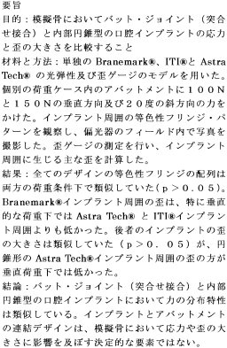
enObjectives: To compare stress and strain magnitudes of butt-joint and internal-cone oral implants in a bone simulant.
Material and methods: Photoelastic and strain-gauged models of solitary Brånemark®, ITI® and Astra Tech® implants were obtained. Vertical and 20° oblique forces of 100 and 150 N were applied on the abutments in separate load cases. Isochromatic fringe patterns around implants were observed and photographed in the field of a polariscope. Strain-gauge measurements were performed and principal strains induced around implants were calculated.
Results: Isochromatic fringe orders of all designs were similar under both loading conditions (P>0.05). Strains around Brånemark® implants were lower than around Astra Tech® and ITI® implants, particularly under vertical loads. The latter implants had similar strain magnitudes (P>0.05), although strains around the conical Astra Tech® implant were lower under vertical load.
Conclusions: Butt-joint and internal-cone oral implants have similar force distribution characteristics. The implant–abutment mating design is not a decisive factor affecting stress and strain magnitudes in a bone simulant.
Résumé
frLe but de cette étude a été de comparer les amplitudes de stress et de tension au niveau des implants buccaux joint par les bouts et ceux avec un cone interne dans un stimulateur osseux. Des modèles de photo-élastique et de jauge de tension d'implants solitaires ad modum Branemark® ITI® et Astra Tech® ont été obtenus. Des forces verticales et obliques à 20° de 100 et 150 N ont été appliquées sur les piliers dans des cas de charges séparées. Les modèles de frange isochromatique autour des implants ont été observés et photographiés dans le champs dapos;un polariscope. Des mesures-jauge quotidiennes ont été effectuées et les tensions principales induites autour des implants ont été calculées. Les ordres de bordure isochromatique de tous les modèles étaient semblables sous les deux conditions de mise en charge (p>0,05). Les forces autour des implants ad modum Brånemark®étaient inférieures à celles autour des implants Astra Tech® et ITI®, particulièrement sous les charges verticales. Ces derniers implants avaient des amplitudes de tensions semblables (p>0,05), bien que les tensions autour de l'implant Astra Tech® conique étaient inférieurs sous charge verticale. Les implants buccaux joints par les bouts et ceux avec un cone interne possèdent les caractéristiques de distribution des forces semblables. Le modèle d'accouplement implant-pilier n'est pas un facteur décisif affectant les amplitudes de stress et de tension dans un simulateur osseux.
Zusammenfassung
deZiel: Ein Vergleich von Kraft- und Dehnspitzen bei Implantaten in einer knochenähnlichen Substanz, deren Sekundärteile mit einem “Druckknopfsystem” oder einem Innenkonus befestigt sind.
Material und Methode: Man verwendete Modelle, die eine photoelastische Analyse zulassen und mit Dehnmessstreifen versehen sind. In diese Modelle implantierte man Einzelimplantaten vom Typ Brånemark®, ITI® und Astra Tech®. Auf die Sekundärteile trafen Schläge von 100 und 150 N in vertikaler Richtung und in einem Einfallswinkel von 20° auf. Man untersuchte die isochromatischen Spannungslinien um die Implantate und fotographierte sie mit Polarisationsfiltern. Die Werte der Dehnmessstreifen zeichnete man auf und errechnete daraus die Hauptstossrichtungen dieser Kräfte um die Implantate.
Resultate: Die Anordnung der isochromatischen Spannungslinien war bei allen Implantattypen und bei beiden Belastungstypen ähnlich (p>0.05). Die Dehnkräfte um Brånemark-Implantate waren kleiner als um Astra Tech®- und ITI®-Implantate, insbesondere bei vertikaler Belastung. Diese zwei Implantate zeigten bei der Aufzeichnung der Dehnung ähnliche Werte (p>0.05), sie waren aber um das konische Astra Tech®-Implantat bei vertikaler Belastung etwas kleiner.
Zusammenfassung: Orale Implantate mit einem “Druckknopf-” oder “Innenkonus-System” zeigen ähnliche Kräfteverteilungsmuster. In einer knochenähnliche Substanz ist somit das Design der Implantat-Sekundärteilverbindung bezüglich Kraft- und Dehngrössen kein entscheidender Faktor.
Resumen
esObjetivos: Comparar las magnitudes de estrés y de tensión de implantes con juntas de cobertura y de cono interno en un simulador óseo.
Material y Métodos: Se obtuvieron modelos foto-elásticos y con indicador de la tensión de implantes solitarios Brånemark, ITI® y Astra Tech®. Se aplicaron fuerzas verticales y oblicuas de 20° de 100 y 150 N sobre los pilares en casos separados de carga. Se observaron y fotografiaron los patrones isocromáticos marginales alrededor de los implantes en el campo de un polariscopio. Se llevaron a cabo mediciones del indicador de la tensión y se calcularon las tensiones principales alrededor de los implantes.
Resultados: Los ordenamientos isocromáticos marginales de todos los diseños fueron similares bajo ambas condiciones de carga (p<0.05). Las tensiones alrededor de los implantes Brånemark® fueron menores que alrededor de los implantes Astra Tech® e ITI®, particularmente bajo cargas verticales. Estos últimos implantes tuvieron magnitudes similares de tensión (p<0.05), aunque las tensiones alrededor del implante cónico Astra Tech® fue menor bajo carga vertical.
Conclusiones: Los implantes orales de junta de cobertura y de cono interno tienen unas características de distribución de fuerzas similares. El diseño de emparejamiento implante-pilar no es un factor decisivo que afecte a las magnitudes de estrés y de tensión en un simulador óseo.