Relationship Between Cocaine Use and Coronary Artery Disease in Patients With Symptoms Consistent With an Acute Coronary Syndrome
Disclosures: Drs. Hollander and Litt have received research funding from Siemens. The other authors have no disclosures to report.
Supervising Editor: Stephen Smith, MD.
Abstract
ACADEMIC EMERGENCY MEDICINE 2011; 18:1–9 © 2011 by the Society for Academic Emergency Medicine
Objectives: Observational studies of patients with cocaine-associated myocardial infarction have suggested more coronary disease than expected on the basis of patient age. The study objective was to determine whether cocaine use is associated with coronary disease in low- to intermediate-risk emergency department (ED) patients with potential acute coronary syndrome (ACS).
Methods: The authors conducted a cross-sectional study of low- to intermediate-risk patients < 60 years of age who received coronary computerized tomographic angiography (CTA) for evaluation of coronary artery disease (CAD) in the ED. Patients were classified into three groups with respect to CAD: maximal stenosis <25%, 25% to 49%, and ≥50%. Prespecified multivariate modeling (generalized estimating equations) was used to assess relationship between cocaine and CAD.
Results: Of 912 enrolled patients, 157 (17%) used cocaine. A total of 231 patients had CAD ≥ 25%; 111 had CAD ≥ 50%. In univariate analysis, cocaine use was not associated with a lesion 25% or greater (12% vs. 14%; relative risk [RR] = 0.89, 95% confidence interval [CI] = 0.5 to 1.4) or 50% or greater (12% vs. 11%; RR = 1.15, 95% CI = 0.6 to 2.3). In multivariate modeling adjusting for age, race, sex, cardiac risk factors, and Thrombosis in Myocardial Infarction (TIMI) score, cocaine use was not associated with the presence of any coronary lesion (adjusted RR = 0.95, 95% CI = 0.69 to 1.31) or coronary lesions 50% or greater (adjusted RR = 0.78, 95% CI = 0.45 to 1.38). There was also no relationship between repetitive cocaine use and coronary calcifications or between recent cocaine use and CAD.
Conclusions: In symptomatic ED patients at low to intermediate risk of an ACS, cocaine use was not associated with an increased likelihood of coronary disease after adjustment for age, race, sex, and other risk factors for coronary disease.
Approximately 2.4 million people habitually use cocaine in the United States, making it the second most widely used illicit drug.1 In 2005, cocaine was the most common illicit drug leading to emergency department (ED) visits and accounted for over 30% of all drug related encounters.2 Chest pain is a common complication of cocaine use, accounting for 40% of patients who present to the ED after cocaine use.3 Although reported rates of acute myocardial infarction (AMI) vary widely (1%–31%),4–10 those studies specifically designed to determine the rate of AMI in ED patients with cocaine-associated chest pain have found rates of approximately 6%;9,10 one out of every four AMIs in people aged 18 to 45 years can be linked to cocaine use.11
The pathophysiology of MI in cocaine users is multifactorial.12 Acutely, cocaine causes increased myocardial oxygen demand, coronary vasoconstriction, platelet aggregation, and in situ thrombus formation,12 which can lead to AMI even in patients with normal coronary arteries.13 Autopsy reports and small observational studies have suggested that chronic cocaine users may develop coronary artery disease (CAD) at younger than expected ages.14–17 Early development of atherosclerosis in this cohort is especially concerning, because cocaine-induced coronary vasoconstriction is enhanced at sites of coronary atherosclerosis.18 However, no large studies have directly compared the prevalence of clinically relevant coronary disease in cocaine users and nonusers.
The relationship between cocaine use and coronary disease is particularly important in symptomatic patients where management and disposition of patients should be based upon likelihood of underlying CAD.19 Therefore, we evaluated the relationship between cocaine use and CAD in patients presenting with symptoms consistent with a potential acute coronary syndrome (ACS).
Methods
Study Design
This study was a retrospective analysis of prospectively collected data for an institutional review board (IRB)-approved continuous quality improvement (CQI) project. To assess the relationship between CAD and cocaine use, we conducted a cross-sectional study on patients with chest pain who received coronary computerized tomographic angiography (CTA) from May 2005 to December 2008. All patients provided verbal consent to receive coronary CTA according to standard radiology department procedure. Written informed consent was not required since the test was ordered as part of routine care; CT imaging at our institution does not require written informed consent. Additional IRB approval was obtained for the record review portion of the study assessing cocaine use, as this was not part of the initial IRB submission for the CQI project.
Study Setting and Population
The study was conducted in the Hospital of the University of Pennsylvania, which is an urban tertiary referral center with an annual ED census of approximately 57,000 during the enrollment period. Patients who presented to the ED with a chief complaint of chest discomfort (or the equivalent) prompting an electrocardiogram (ECG) for evaluation of potential ACS who also received coronary CTA as part of the evaluation were prospectively identified and included in the analysis. Although our cardiac CQI projects use prospective data collection, the IRB does not require written informed consent since there is no specific intervention. Our department has prospectively collected data for cardiac clinical pathway CQI projects for over a decade. The decision to obtain a coronary CTA as part of standard patient care was made by the treating physician and is a widely used clinical pathway within our ED. Patients who receive coronary CTA are generally low to intermediate risk for an ACS, but high enough risk that they would otherwise be admitted to the observation unit or hospital to have ACS excluded. These are usually patients who have a presentation concerning for ACS with a Thrombosis in Myocardial Infarction (TIMI) score less than 3 or a TIMI score 3 or higher with a recent stress test that did not reveal reversible ischemia.
Patients were excluded if they had chest pain explained by local trauma or radiographic abnormalities or they were at such low risk that they would otherwise be discharged home without admission or a provocative test to exclude ACS. In our institution, patients at very low risk of ACS (approximately 30% to 40% of all patients who present with chest pain syndromes) do not receive any admission or objective cardiac testing.
High-risk patients with an initial ECG diagnostic of ischemia or AMI, transient ST-segment elevation or depression ≥ 1 mm that persisted for at least 1 minute, elevated cardiac markers, recurrent ischemic chest pain, or hemodynamic instability were also excluded as these patients were not candidates for coronary CTA. Patients were also excluded if they had a contraindication to coronary CTA (such as iodinated contrast allergy or glomerular filtration rate < 60 mL/min) or if coronary CTA was not available. Coronary CTA was available weekday daytime hours and approximately every third weekend from December 2005 through December 2007 and from 7 AM to 10 PM weeknights as well as weekend mornings after January 2008. Patients who presented outside of these hours were enrolled if they received an overnight stay in the observation unit and received coronary CTA in the morning.
Study Protocol
Initial Evaluation. Patients receiving a coronary CTA were identified prospectively by trained personnel who were present in the ED from 7 AM to midnight, 7 days a week. Structured data collection was performed using the Standardized Reporting Guidelines20 in accordance with the Key Definitions21 and were documented by the treating physician. The Standardized Reporting Guidelines20 were developed through a multidisciplinary panel of representatives of the American Heart Association (AHA), American College of Cardiology (ACC), Society for Academic Emergency Medicine (SAEM), and American College of Emergency Physicians (ACEP) to better standardize criteria that should be reported in risk stratification studies of patients with potential ACS. This information includes self-reported demographic characteristics; cardiac risk factors; chest pain characteristics; associated symptoms; medications; and initial vital signs, ECG, treatment, and disposition. The Key Definitions21 were similarly determined through representation of the AHA, ACC, and ACEP, as well as other societies and organizations. It provides specific definitions for many of the items in the Standardized Reporting Guidelines.20
Assessment of CAD. We used coronary CTA to assess the presence of coronary disease, since coronary CTA results correlate with coronary angiography, and coronary CTA provides a noninvasive measurement of coronary stenoses.22 The protocol for coronary CTA included patients with an initial TIMI risk score of 0–2 (approximately 50%–70% of patients being evaluated for ACS).23,24 Patients with tachycardia were treated with beta-blockers, benzodiazepines, or diltiazem at the discretion of the emergency physician caring for the patient. Beta-blockers, which are usually used for heart rate control in coronary CTA, were not used for patients with recent cocaine use because of the potential adverse effects of unopposed alpha-adrenergic receptors.12,25 As a result of the need to possibly modify medication administration in the setting of cocaine use to control the heart rate while the patient was in the CT scanner, the radiologists were not blinded to cocaine use within the last 72 hours. They were blinded to cocaine use history other than acute use (for example, they did not know whether patient had a chronic cocaine use history). Coronary CTA was performed using predominantly 64-slice or dual-source scanners (Siemens Medical Solutions, Malvern, PA), which have 98% sensitivity and 92% specificity for detection of coronary stenosis on a per patient basis.22 The study began with a low-dose non-contrast ECG-triggered acquisition through the entire chest for the purpose of calcium scoring and evaluation of lung abnormalities. This was followed by a weight-based intravenous injection of 80–120 mL of nonionic iodinated contrast (Omnipaque 350, GE Amersham, Milwaukee, WI) with bolus tracking in the descending aorta. After the appropriate scan delay, an ECG-gated acquisition from the pulmonary artery bifurcation through the inferior heart border was performed. Studies were reviewed on dedicated 3D workstations (Leonardo and MMWP, Siemens Medical Solutions, and AdvantageWindows4.2, GE) using axial, multiplanar reformatted, and thin-slab maximum-intensity projection images in an interactive display. Image data were reconstructed at multiple phases of the cardiac cycle and postprocessed and analyzed on independent workstations. The degree of any observed stenosis was measured with an electronic caliper by comparing the lumen diameter with the diameter of a proximal reference segment.26 Studies were read by attending radiologists with subspecialty cardiovascular imaging training; all met ACC/AHA Level 3 training guidelines for cardiac CT.27 As all readers were Level 3 credentialed and demonstrated the required interrater reliability for this certification, we did not assess inter- or intrarater reliability again for this study. Interpretations were provided for all studies. No technical limitations resulted in a patient being excluded from the analysis.
Cocaine Use Assessment. While patients were in the ED, they were directly questioned about cocaine use within the preceding week. To assess repetitive cocaine use, medical records for all visits within in our health system (before and after the index presentation) were reviewed by a trained research assistant for all patients in the study.
Patients were then classified into one of the following categories: no documentation of presence or absence of cocaine use during any visit, documented absence of history of cocaine use, documented repetitive cocaine use, or documented single episode of cocaine use. Based upon review of these visits, patients were classified into the major categories of no cocaine use ever, single use of cocaine, or repetitive cocaine use. Unless it was specifically noted that the patient had used cocaine less than five times, patients were considered repetitive users because it has been shown that ED patients who report cocaine use are seldom first-time users, and more than 99% of patients with cocaine-associated chest pain are chronic users.10 A sample of 100 medical records was independently reviewed by a second observer to assess interrater reliability of the assessment of cocaine use. Agreement was 100% with a kappa of 1.0. Patients were also classified as recent cocaine use or not, which was defined as cocaine use within the past week or a positive urine toxicology screen.
Data Analysis
Our main outcome was presence of CAD. Patients were classified into three groups with respect to CAD: <25%, 25% to 49%, and 50% or greater maximal stenosis. The relationship between CAD and cardiac risk factors and cocaine use was assessed using chi-square or Fisher’s exact test for categorical data and Student’s t-test for continuous variables. To assess the relationship between cocaine use and CAD, an adjusted relative risk (RR) of CAD was calculated using a generalized linear model with a log link, Gaussian error, and robust estimates of the standard errors of the model coefficients was used.28 We chose this method because the outcome of interest (CAD) was present more than 25% of the time and the odds ratio does not approximate the RR well when the outcome is this common.28 The model controlled for age, race, sex, tobacco use, family history of premature coronary disease (<55 years), diabetes mellitus, hypercholesteremia, hypertension, and TIMI risk score. This list of possible confounders was determined a priori, and there were no stepwise techniques used to select variables. Data for these analyses are presented as RRs with 95% confidence intervals (CIs). We performed a secondary analysis to evaluate the RR of cocaine use for coronary calcium. We also examined the relationship between recent cocaine use and CAD using a similar model. All analyses were performed using SAS statistical software (Version 9.1, SAS Institute, Cary NC).
Results
There were 976 visits by 959 patients during which the patient received a coronary CTA for evaluation of possible ACS. Only the coronary CTA from the first visit was included for the 17 patients with repeat studies. An additional 38 patients had no documentation of the presence or absence of cocaine use on this or prior visits and were excluded from subsequent analysis. Finally, the nine patients with a calcium score > 400, in whom no contrast enhanced imaging was performed, were also excluded. There were 5,423 visits to the health system by the 912 patients that were reviewed for cocaine use assessment. Overall, 157 patients had prior cocaine use and 755 had documentation of no prior cocaine use. Only two patients claimed a single episode of cocaine use. Seventy-three (8.1%) reported cocaine use in the preceding week. There were 110 patients who had urine toxicology testing performed: 34 patients tested positive, 33 (97%) of whom had self-reported cocaine use. Of the nine patients excluded due to calcium score > 400 without coronary CTA imaging, two had documented cocaine use.
Of 912 enrolled patients, 231 (25%) had coronary disease with a maximal stenosis ≥ 25% in at least one vessel, and 111 patients (12%) had at least one vessel with ≥ 50% stenosis. Compared to patients without coronary disease, patients with some coronary disease (Table 1) were older and had more traditional cardiac risk factors such as tobacco use (44% vs. 33%), hypercholesterolemia (28% vs. 16%), and hypertension (55% vs. 43%). For the 38 patients excluded due to lack of documentation of cocaine use, the rate of coronary disease was similar to that of the overall cohort (≥25%, 7/38 [18%]; and ≥50%, 5/38 [11%]). Compared to noncocaine users, cocaine users (Table 2) were similar with respect to age and most traditional cardiac risk factors; however, they had more tobacco use (66% vs. 29%).
| Characteristic | No CAD (n = 681) | CAD ≥ 25% (n = 231) | CAD ≥ 50% (n = 111) |
|---|---|---|---|
| Age, yr (mean ± SD) | 46.0 ± 8.1 | 51.6 ± 8.3 | 53.2 ± 9.3 |
| Male, n (%) | 272 (40) | 121 (52) | 60 (54) |
| Race | |||
| Black of African American | 447 (71) | 136 (63) | 63 (61) |
| White | 150 (24) | 75 (35) | 37 (36) |
| Other/unknown | 39 (6) | 6 (3) | 4 (4) |
| Cardiac risk factors* | |||
| Hypertension | 294 (43) | 127 (55) | 66 (59) |
| Hypercholesterolemia | 109 (16) | 65 (28) | 36 (33) |
| Family history of CAD | 123 (18) | 53 (23) | 29 (26) |
| Diabetes mellitus | 93 (14) | 36 (16) | 20 (18) |
| Current tobacco use | 222 (33) | 101 (44) | 54 (49) |
| Cocaine use in last week | 56 (8) | 17 (7) | 9 (8) |
| Cocaine use | 117 (17) | 40 (17) | 18 (16) |
| Mean (± SD) number of cardiac risk factors | 1.2 ± 1.0 | 1.7 ± 1.1 | 1.8 ± 1.2 |
| Past medical problems | |||
| CAD | 7 (1.0) | 4 (1.7) | 3 (2.7) |
| Myocardial infarction | 9 (1.3) | 3 (1.3) | 3 (2.7) |
| Congestive failure | 4 (0.6) | 3 (1.3) | 3 (2.7) |
| TIMI risk score | |||
| 0 | 433 (65) | 112 (49) | 47 (42) |
| 1 | 185 (27) | 82 (36) | 37 (33) |
| 2 | 56 (8) | 29 (13) | 21 (19) |
| ≥ 3 | 6 (1) | 8 (3) | 6 (5) |
- Data are reported as number (%).
- CAD = coronary artery disease; CTA = computerized tomographic angiography; TIMI = Thrombolysis in Myocardial Infarction.
- *Cardiac risk factors were determined based upon patient self-report and review of records available at the time of the ED evaluation.
| Characteristic | No Cocaine Use (n = 755) | Cocaine Use (n = 157) |
|---|---|---|
| Age, yr (mean ± sd) | 47.6 ± 8.8 | 46.2 ± 6.4 |
| Male | 302 (40) | 91 (58) |
| Race | ||
| Black or African American | 451 (64) | 132 (88) |
| White | 209 (30) | 16 (11) |
| Other/unknown | 43 (6) | 2 (1) |
| Cardiac risk factors * | ||
| Hypertension | 356 (47) | 65 (42) |
| Hypercholesterolemia | 159 (21) | 15 (10) |
| Family history of CAD | 148 (20) | 28 (18) |
| Diabetes mellitus | 103 (14) | 26 (17) |
| Current tobacco use | 219 (29) | 104 (66) |
| Mean (± SD) number of cardiac risk factors | 1.3 ± 1.1 | 1.5 ± 1.2 |
| Medical history | ||
| Known CAD | 8 (1) | 3 (2) |
| Myocardial infarction | 6 (1) | 6 (4) |
| Congestive failure | 5 (1) | 2 (1.2) |
| TIMI risk score | ||
| 0 | 457 (61) | 88 (56) |
| 1 | 219 (29) | 48 (31) |
| 2 | 66 (9) | 19 (12) |
| ≥ 3 | 12 (2) | 2 (1) |
- Data are reported as number (%).
- CAD = coronary artery disease; TIMI = Thrombolysis in Myocardial Infarction.
- *Cardiac risk factors were determined based upon patient self-report and review of records available at the time of the ED evaluation.
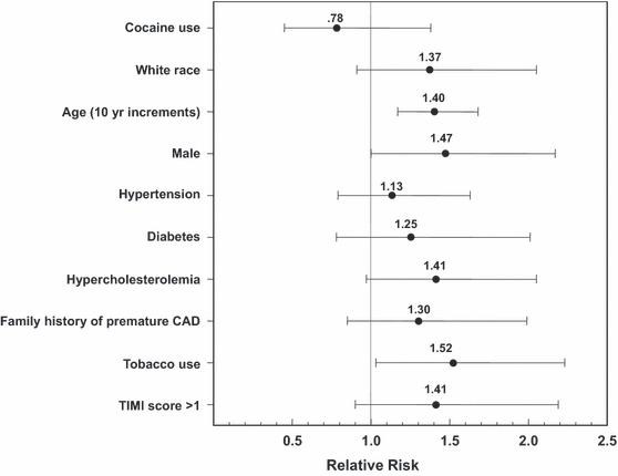
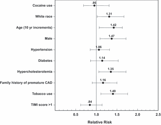
We did not find that cocaine users were at increased risk of having coronary disease, whether it was defined as a lesion 25% or greater (12% vs. 14%, RR = 0.89, 95% CI = 0.5 to 1.4) or 50% or greater (12% vs. 11%, RR 1.15, 95% CI = 0.6 to 2.3). In our multivariate modeling (1, 2) adjusting for age, race, sex, cardiac risk factors, and TIMI score, repetitive cocaine use was not associated with the presence of a coronary lesion ≥ 25% (adjusted RR = 0.95, 95% CI = 0.69 to 1.31) or coronary lesions ≥ 50% (adjusted RR 0.78, 95% CI = 0.45 to 1.38). Repetitive cocaine use was also not related to coronary calcium (adjusted RR = 1.04, 95% CI = 0.73 to 1.48). We did not detect a relationship between recent cocaine use and the presence of coronary lesions ≥ 25% (adjusted RR = 0.92, 95% CI = 0.58 to 1.45) or coronary lesions ≥ 50% (adjusted RR = 0.96, 95% CI = 0.46 to 2.01).

Adjusted RR for CAD ≥50%. CAD = coronary artery disease; RR = relative risk.

Adjusted RR for CAD ≥25%. CAD = coronary artery disease; RR = relative risk.
Discussion
Cocaine increases myocardial oxygen demand while decreasing blood flow through coronary vasoconstriction,18,25,29 platelet aggregation,12,30,31 and thrombosis.17,32–34 These effects of cocaine can cause myocardial ischemia even in patients with normal coronary arteries.13,35 The greatest risk for cocaine-induced myocardial infarction is within the first hour following use;36 however, delayed or recurrent coronary vasoconstriction occurs as the concentrations of cocaine metabolites rise later.37
Although initial reports of cocaine-associated MI were focused on patients without coronary disease,13 most patients with cocaine-associated MI actually have CAD.14,15 Hollander et al., 14,38 in a multicenter study of 136 patients (mean ± SD age, 38 ± 10 years) with cocaine-associated MI, found that 49 of the 70 patients (70%) evaluated for coronary disease had a clinically significant stenosis. Kontos et al.15 examined 90 patients (mean ± sd age, 42 ± 8 years) with cocaine use who received coronary angiography for symptoms compatible with ischemia and found that 50% had at least single-vessel disease (50% or greater stenosis). Of the patients with proven myocardial infarction, 77% had coronary disease.15 Autopsy reports in small cohorts of young cocaine users also found higher rates of atherosclerosis than would be expected on the basis of age.16,17,35 The observation of CAD in these young patients raised concern about a relationship between cocaine use and early development of atherosclerosis.
In vivo animal models and in vitro studies have suggested a mechanism for these observations. Accelerated atherosclerosis secondary to cocaine use has been demonstrated in an animal model where rabbits were fed a high-cholesterol diet.39 The group that received cocaine developed more plaque in the intima of the aorta.39 Plaque consisted of both foam cells and smooth muscle proliferation; cholesterol levels were similar between the cocaine and control groups.39 In vitro studies have shown that cocaine causes defects in the endothelial cell barrier and increases its permeability to low-density lipoprotein, the expression of endothelial adhesion molecules, and mast cell migration.40 These studies suggest biologic plausibility, but do not provide evidence that cocaine use actually causes clinically significant atherosclerosis in humans.
We did not find an association between cocaine use and clinically significant CAD in symptomatic low- to intermediate-risk patients evaluated with coronary CTA. The few comparative studies attempting to address this relationship included small numbers of patients, included isolated subsets of patients, focused on coronary calcium alone, or did not distinguish between repetitive (chronic) cocaine use and recent use. Patrizi et al. 41 reported a series of 51 patients with ST-segment elevation MI. Cocaine users and nonusers had similar numbers of critical stenoses (76% vs. 70%); however, the cocaine users had higher prevalence of multivessel disease (65% vs. 32%).41 These results contrast with those of Weber et al., 34 who found that patients with cocaine-associated MI had impairments in both epicardial and microvascular flow, but that epicardial flow was actually better in patients with cocaine-associated MI than in patients with ST-segment elevation MI unrelated to cocaine. In the current study of symptomatic low to intermediate patients without ST-segment deviation, we were unable to find a significant increase in epicardial coronary disease in cocaine users who presented with symptoms of a potential ACS.
The Coronary Artery Risk Development in Young Adults (CARDIA) group evaluated coronary calcifications in an inception cohort of over 3,000 healthy patients, of whom 35% reported cocaine exposure.42 After adjustment for age, sex, ethnicity, socioeconomic status, family history, tobacco use, and alcohol use, the investigators found no relationship between cocaine exposure and coronary calcium. We found no relationship between cocaine and coronary calcifications in a symptomatic cohort. Because patients with cocaine-associated MI are typically younger, it is plausible that atherosclerotic plaques may have not yet calcified and therefore would not have been identified by the CARDIA group. Therefore, we also examined the relationship between cocaine use and coronary disease (with or without calcifications) and did not find an association.
Bamberg et al.43 compared 44 symptomatic cocaine users with 132 nonusers in an ED-based nested cohort study. The patients were matched for age and sex. Although cocaine users had a sixfold increased risk for ACS, they had similar amounts of plaque (both calcified and noncalcified) detected on coronary CTA. We confirmed and extended these findings to patients with both repetitive cocaine use and recent cocaine use in a larger cohort of patients. While a single episode of cocaine use will not lead immediately to development of coronary disease, cocaine causes enhanced coronary vasoconstriction in areas of atherosclerosis.18 Imaging of the coronary arteries shortly after cocaine use might theoretically have found more significant stenosis in patients with recent use; however, we found no such relationship.
To our knowledge, our study is the largest direct comparison of cocaine users and nonusers with respect to coronary disease. An additional difference between our study and others is that our patients were symptomatic. This is the cohort of patients where knowledge of underlying coronary disease is very important to ensure appropriate disposition and treatment.19 After adjustment for sex, race, tobacco use, cardiac risk factors, and TIMI score, we were unable to find a difference in the prevalence of CAD in patients with and without cocaine use.
Although we did not find a relationship between cocaine use and coronary disease, we did find a relationship between coronary lesions ≥ 25% and white race, age, male sex, hypercholesterolemia, and tobacco use. Only age and tobacco use were found to be significant for coronary disease ≥ 50%. We believe that the reason for this observation is that the larger number of patients with a ≥ 25% stenosis provided adequate power to demonstrate the difference. Although not statistically significant, the point estimates of RR for coronary disease found for most cardiac risk factors were similar in the ≥ 25 and ≥ 50% groups.
Limitations
We enrolled only symptomatic patients from a single center. A larger sample size would have provided more power to detect an association between cocaine use and coronary disease; however, it is worth noting that the adjusted RR for cocaine (1, 2) was less than 1, whereas it was greater than 1 for all traditional risk factors. Although an epidemiologic study investigating the relationship between cocaine and underlying coronary disease might better address whether cocaine use leads to subclinical atherosclerosis, it might not be as relevant to the clinical setting. ACC/AHA guidelines for patients with ACS recommend risk stratification of patients based upon likelihood of underlying obstructive coronary disease.19 This is specifically the patient population that we studied: symptomatic patients being evaluated for potential ACS. It should also be noted that we only evaluated low- to intermediate-risk patients receiving coronary CTA. Although it is possible that we could have missed a disproportionate number of high-risk cocaine-using patients with coronary disease, this appears unlikely as our prevalence of cocaine use in this study is actually higher than in more broad-based studies from our institution.8 Similarly, we cannot eliminate the possibility of selection bias since not all low- to intermediate-risk patients received coronary CTA.
With respect to recent cocaine use, only 110 patients had urine toxicology testing performed. Only one patient who denied recent cocaine use tested positive. Of the 34 patients who tested positive, 33 (97%) self-reported cocaine use. It is our opinion that the selection bias that might occur from mandatory urine toxicology testing would be more harmful to study validity than any misclassification bias that might result from lack of mandatory testing. With respect to cocaine use history, we used extensive methodology in an attempt to limit this potential bias. We examined 5,423 medical records to classify patients into cocaine use categories rather than just rely on information obtained from a single visit, as most prior studies have done. Additionally, we were unable to capture frequency or duration of use and therefore could not assess any dose–response relationship between cocaine use and likelihood of coronary disease.
Our study must be interpreted in the appropriate context. We cannot eliminate the possibility that a smaller association still exists. Whether or not chronic cocaine use is associated with development of coronary disease, it must also be kept in mind that cocaine increases myocardial oxygen demand while decreasing blood flow through coronary vasoconstriction, platelet aggregation, and thrombosis, even in patients with normal coronary arteries. Thus, although ACC/AHA guidelines recommend risk stratification of patients with potential ACS based upon likelihood of underlying obstructive coronary disease, the likelihood of coronary vasoconstriction must also be considered in patients with recent cocaine use.
Conclusions
After adjustment for other factors known to affect the development of coronary artery disease, we were unable to find an association between cocaine use and prevalence of coronary disease in hemodynamically stable symptomatic patients who presented to the ED with a potential acute coronary syndrome, but without electrocardiographic signs of ischemia on the initial ECG. This suggests that a prior cocaine use history should not be used to risk stratify symptomatic patients for likelihood of clinically significant coronary disease. The risk of coronary disease should be determined from other factors, as it appears to be independent of cocaine use. Validated strategies, such as short-term observation, for management of patients with potential cocaine-associated myocardial ischemia are still recommended,44 since cocaine use is associated with an increased risk of an acute coronary syndrome,43 and it can precipitate coronary vasoconstriction, even the absence of coronary disease.


