The therapeutic potential of stem cells in heart disease
All authors declare no conflicts of interest.
Abstract
Abstract. Coronary heart disease and chronic heart failure are common and have an increasing frequency. Although interventional and conventional drug therapy may delay ventricular remodelling, there is no basic therapeutic regime available for preventing or even reversing this process. Chronic coronary artery disease and heart failure impairs quality of life and are associated with subsequent worsening of the cardiac pump function. Numerous studies within the past few years have been demonstrated, that the intracoronary stem cell therapy has to be considered as a safe therapeutic procedure in heart disease, when destroyed and/or compromised heart muscle must be regenerated. This kind of cell therapy with autologous bone marrow cells is completely justified ethically, except for the small numbers of patients with direct or indirect bone marrow disease (e.g. myeloma, leukaemic infiltration) in whom there would be lesions of mononuclear cells. Several preclinical as well as clinical trials have shown that transplantation of autologous bone marrow cells or precursor cells improved cardiac function after myocardial infarction and in chronic coronary heart disease. The age of infarction seems to be irrelevant to regenerative potency of stem cells, since stem cells therapy in old infarctions (many years old) is almost equally effective in comparison to previous infarcts. Further indications are non-ischemic cardiomyopathy (dilative cardiomyopathy) and heart failure due to hypertensive heart disease.
INTRODUCTION
Background and rationale for stem cell therapy
Stem cells have the important properties of self-regeneration and differentiational plasticity (Allgöwer 1956; Krause et al. 2001; Jiang et al. 2002; Quaini et al. 2002). Thus, they are ideal candidates for regeneration of damaged myocardial tissue (Goodell et al. 2001), for example, in myocardial infarction or in congestive heart failure. When acute myocardial infarction occurs, heart muscle tissue is regionally destroyed (Fig. 1) (Pfeffer & Braunwald 1990; Ertl et al. 1993; Pfeffer 1995; Ren et al. 2002). By percutaneous coronary intervention, coronary restitution can be achieved and coronary perfusion may be normalized; however, regular heart muscle function may not be restored, so that remodeling and heart failure are not necessarily prevented. Prevention of remodeling, however, may possibly be realized by cell transplantation, which leads to myocardial restitution with the beneficial aim of restoring or normalizing compromised heart function.
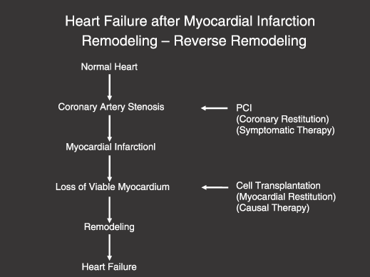
Development of heart failure (remodelling) after myocardial infarction. By percutaneous coronary interventions (PCI), coronary stenoses can be removed; however, loss of viable myocardium cannot be reversed. Myocardial restitution represents causal therapy and can be realized by stem cell transplantation to reverse remodelling.
One possible way of heart muscle repair is to transplant cells of primarily non-cardiac origin for cardiac regeneration, such as human bone marrow-derived mononuclear cells containing human stem cells (Fig. 2) (Ferrari et al. 1998; Blau et al. 2001). These cells may operate as a source of cardiac cells; that is, as precursor of heart muscle tissue and of coronary blood vessel cells. Human bone marrow contains CD34-positive haematopoietic and CD34-negative mesenchymal stem cells (Reyes et al. 2001) and both these types of stem cell may contribute to heart muscle repair. Haematopoietic stem cells are also progenitor cells; for example, for endothelial cells (Condorelli et al. 2001).
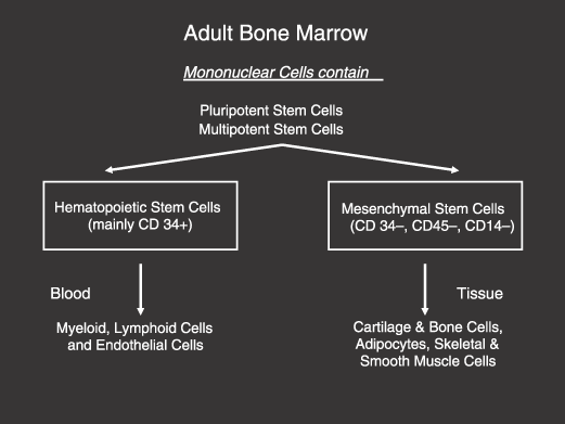
Adult bone marrow and its two major cell components for heart muscle repair.
The first steps and experimental cornerstones demonstrating mononuclear bone marrow stem cell differentiation to muscle cells in experimental heart disease, especially in experimental myocardial infarction, were realized by different investigators: demonstrating (i) regeneration of myocardium after infarction (Tomita et al. 1999, 2002; Orlic et al. 2001); (ii) reduction of infarct size (Kocher et al. 2001); and (iii) de novo expression of cardiac proteins by human bone marrow cells (Toma et al. 2000, 2002). Our Düsseldorf group performed clinical stem cell therapy for the first time in March 2001, when treating acute myocardial infarction by intracoronary cell transfer (Strauer et al. 2001). The aims of this procedure, which had not been achieved before, were:
- 1
To use the body's own stem cells from bone marrow for cardiac tissue repair; that is, to use all three fractions of mononuclear cells, haematopoietic, angioblastic and mesenchymal stem cells;
- 2
To facilitate and potentiate cell migration by artificial ischaemia, which is one of the most effective stimuli for stem cell differentiation, most probably via the CXCR4–SDF interrelationships. This has been achieved by repetitive intracoronary balloon dilatation within the occlusion of the former infarct-related artery; and
- 3
To enrich and to accumulate mononuclear cells within the infarct zone and the border zone by the intracoronary route of administration.
Clinical studies with direct intracoronary transplantation of adult stem cells until now focus on at least four clinically relevant situations:
- 1
therapy for acute myocardial infarction
- 2
long-term effects after myocardial infarction
- 3
therapy for old myocardial infarction (≥ 8 years) with heart failure
- 4
therapy for congestive heart failure (dilatative cardiomyopathy)
Methodological prerequisites for clinical stem cell treatment
Important conditions for clinical stem cell therapy are the precise and careful techniques of bone marrow cell preparation, availability of large cell concentrations within the area of interest (border zone of infarction), migration of stem cells into the apoptotic or necrotic myocardial area, and prevention of homing of transplanted cells to other extracardiac organs.
For stem cell transplantation in cardiological diseases, adult bone marrow (80–120 ml) is aspirated under local anaesthesia from the iliac crest. Mononuclear bone marrow cells then need to be isolated under good manufacturing practice (GMP) conditions by Ficoll density separation, before the erythrocytes are lysed with water. Either the mononuclear cells are used for cell therapy directly or they can be incubated overnight in Teflon bags before culture in X-Vivo 15 Medium supplemented with 2% heat-inactivated autologous plasma. The next day, bone marrow mononuclear cells are harvested, washed and finally resuspended in heparinized saline. Heparinization and filtration are carried out to prevent cell clotting and microembolization during intracoronary transplantation. During cell preparation, viability needs to be determined several times and finally must reach around 95%. All microbiological tests of the clinically used cell preparations must prove negative for extraneous contamination.
Intracoronary cell delivery
One of the most important and crucial methodological questions refers to the optimum mechanism of cell delivery to the heart. When given intravenously, only a very small fraction of infused cells can reach the infarct region after the following injection; assuming normal coronary blood flow of 80 ml/min per 100 g of left ventricular weight, a quantity of 160 ml per left ventricle (assuming a regular left ventricular mass of ≈ 200 g) will flow per minute. This corresponds to only around 3% of cardiac output (assuming a cardiac output of 5000 ml/min) (Gregg & Fisher 1963; Strauer 1979). Thus, intravenous application would require many circulation passages to enable infused cells to come in contact with the infarct-related artery. Throughout this long circulation and recirculation time, homing of cells to other organs could considerably reduce the numbers of cells dedicated to cell repair in the infarcted zone. Supplying the entire complement of cells by intracoronary administration obviously seems to be advantageous for tissue repair of infarcted heart muscle and may also be superior to intraventricular injection, as all cells are able to flow through the infarcted and peri-infarcted tissue during the immediate first passage (Fig. 3) (Fuchs et al. 2001; Wang et al. 2001; Galinanes et al. 2004). Accordingly, by this intracoronary procedure the infarct tissue and the peri-infarct zone can be enriched with maximum available numbers of cells at all times.
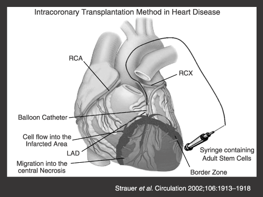
Procedure of cell transplantation into infarcted myocardium in humans. (a) The balloon catheter enters the infarct-related artery and is placed above the border zone of the infarction. It is then inflated and the cell suspension is infused at high pressure under stopflow conditions. (b) In this way, cells are transplanted into the infarcted zone via the infarct-related vasculature (red dots). Cells infiltrate the infarcted zone. Blue and white arrows suggest the possible route of migration. (c) A supply of blood flow exists within the infarcted zone. Cells are therefore able to reach both the border and the infarcted zone.
As stem cells differentiate into more mature types of progenitor cell, it is thought that a special microenvironment in so-called niches regulates cell activity by providing specific combinations of cytokines and by establishing direct cellular contact. For successful long-term engraftment, at least some stem cells must reach their niches, a process referred to as homing (Oh et al. 2003). Mouse experiments have shown that significant numbers of bone marrow cells (BMCs) appear in the liver, spleen, and bone marrow after intravenous injection (Hendrikx et al. 1996). To offer BMCs the best chance of finding their niche within the myocardium, a selective intracoronary delivery route has been developed (Strauer et al. 2001, 2002). Presumably, therefore, fewer cells would be lost by extraction toward organs of secondary interest by this first pass-like effect. To facilitate transendothelial passage and migration into the infarcted zone, cells are infused by high-pressure injection directly into the necrotic area, and the balloon is kept inflated for 2–4 min; cells are not washed away immediately under these conditions.
Cells are directly transplanted by the intracoronary administration route into the infarcted zone. This is accomplished by a balloon catheter, which is placed within the infarct-related artery (Fig. 3). After exact positioning at the site of the former infarct-vessel occlusion, percutaneous transluminal coronary angioplasty (PTCA) is performed four times for 2–4 min each. During this time, intracoronary cell transplantation via the balloon catheter is performed, using four fractional high-pressure infusions of 5 ml cell suspension, each of which contains 6–8 million mononuclear cells. PTCA thoroughly prevents backflow of cells and at the same time produces a stop-flow beyond the site of balloon inflation to facilitate high-pressure infusion of cells into the infarcted zone. Thus, prolonged contact time for cellular migration is allowed. This migration process is probably only present in injured and ischaemic tissue (Szilvassy et al. 1999). Myocardial ischaemia may be the best stimulus for a stem cell to find its optimum myocardial niche, probably due to SDF-1 and CXCR4 interrelations (Damås et al. 2002; Elmadbouh et al. 2007). Therefore, ischaemia-producing stimulus by balloon dilatation during bone marrow cell infusion seems to be absolutely necessary for the cells to home into the cardiac niche and for therapeutical effectiveness of cell migration (Sussmann 2001). Exact methodological standardization is mandatory for both effectiveness of stem cell therapy in clinical heart disease and comparability of the multiple varieties of multicentre stem cell studies. Up to now in Düsseldorf, we have treated more than 250 patients with acute and chronic myocardial infarction and with non-ischaemic heart failure; while worldwide, more than 2000 stem cell patients studies have been published.
CLINICAL RESULTS
Acute myocardial infarction
In acute myocardial infarction, a variety of studies has demonstrated longstanding (up to 3 years and more) improvement of ventricular performance after using stem cell therapy, resulting in increase in ejection fraction by 4–20 (mean 14%) and decreased infarct size by 3–30% (mean 37%) (Tables 1 and 2) (Strauer et al. 2001, 2002). These data show large variability that not only relates to the biological and specific haemodynamic situation of the infarcted heart, but may depend on different methodological procedures (e.g. the number of transplanted stem cells, mode of balloon-induced preconditioning, time interval between the acute infarct and cell transfer, kind of left ventricular volume determination). However, altogether, there is in all clinical studies unequivocal improvement of performance of the infarcted heart (ejection fraction and/or infarct size) after stem cell therapy of at least 6%, which is quantitatively more than the sum of the interventional measures (PTCA, stent) and which is achieved in addition to these therapeutic interventions (Fig. 4) (Assmus et al. 2002, 2006; Chen et al. 2004; Schachinger et al. 2006). Thus, autologous stem cell therapy represents an innovative and effective procedure for regeneration of destroyed heart muscle in the early phase after the infarct (Wollert et al. 2003; Aviles et al. 2004; Janssens et al. 2006; Lunde et al. 2006).
| Study | Patients | Cell application | Cell type after AMI | Number of cells | Results | Method |
|---|---|---|---|---|---|---|
| Controlled study for safety and feasibility | ||||||
| Strauer et al. 2001 | n = 1 | 5 days | BMCs | 12 × 106 | EF, +16% | GBP |
| Cardiac index, +30% | GBP | |||||
| Infarct size, –36% | SPECT | |||||
| Strauer et al. 2002 | n = 20 1 : 1 vs. controls | 7–9 days | BMCs | 28 × 106 | EF, +9% | LV angiogram |
| Infarct size, –60% | LV angiogram | |||||
| Assmus et al. 2002 | n = 59 | 4–6 days | Circ. Prog. BMCs | 16 × 106213 × 106 | EF, +16% | LV angiogram |
| ESV, –25% | LV angiogram | |||||
| Fernandez-Aviles et al. 2004 | n = 20 | 12–14 days | BMCs | 78 × 106 | EF, +10% | MRI |
| ESV, –16% | MRI | |||||
| Chen et al. 2004 | n = 69 1 : 1 vs. controls | 18 days | MSC from BM | EF, +36% | LV angiogram | |
| ESV, –53% | LV angiogram | |||||
| Infarct size, –60% | ||||||
| Randomized study at a single center | ||||||
| Wollert et al. 2004 | n = 60 1 : 1 random. | 4–6 days | BMCs vs. rand. controls | 2460 × 106 | EF, +13% | MRI |
| ESV, –2% | MRI | |||||
| Infarct size, –43% | MRI | |||||
| Janssens et al. 2006 | n = 67 1 : 1 random. Placebo double blind | < 24 h | BMCs vs. i.c. placebo | 304 × 106 | EF, +7% | MRI |
| ESV, –3% | MRI | |||||
| Infarct size, –50% | MRI | |||||
- AMI = acute myocardial infarction; BMCs = bone marrow cells; circ. Prog. = circulating progenitor cells; MSC = mesenchymal stem cells; BM = bone marrow.
| Study | Patients | Cell application after AMI | Cell type | Number of cells | Results | Method |
|---|---|---|---|---|---|---|
| Double blind, placebo-controlled study | ||||||
| Lunde et al. 2006 | n = 100 1 : 1 random. | 6 days | BMCs vs. rand. controls | 87 × 106 | EF, +19% | SPECT |
| Infarct size, –25% | SPECT | |||||
| Schachinger et al. 2006 | n = 204 1 : 1 random.Placebo double blind | 4 days | BMCs vs. i.c. placebo | > 230 × 106 | EF, +11% | LV-angiogram |
| ESV, –1% | LV angiogram | |||||
| Controlled Study for safety and feasibility | ||||||
| Strauer et al. 2005 | n = 36 1 : 1 vs. controls | 3 months till 9 years | BMCs | 28 × 106 | EF, +15% | LV angiogram |
| Infarct size, –30% | LV angiogram | |||||
| Schächinger et al. 2006 | n = 75 BMC-group Circ. Prog. Group Controls | > 3 months | BMCs Circ. Prog. No infusion | 205 × 106 | EF, +7% (BMC) | LV angiogram |
| 22 × 106 | ESV, –4% | LV angiogram | ||||
| Infarct size, –7% | LV angiogram | |||||
| Seth et al. 2006 | n = 24 | no infarction DCM | BMCs | 28 × 106 | EF, +5.4% | LV angiogram |
| ESV, –20% | ||||||
| EDV, ± constant | ||||||
- AMI = acute myocardial infarction; BMCs = bone marrow cells; circ. Prog. = circulating progenitor cells; DCM = dilative cardiomyopathy; EF = ejection fraction; ESV = endsystolic volume; EDV = enddiastolic volume; LV = left ventricular; SPECT = single photon emission computed tomography.
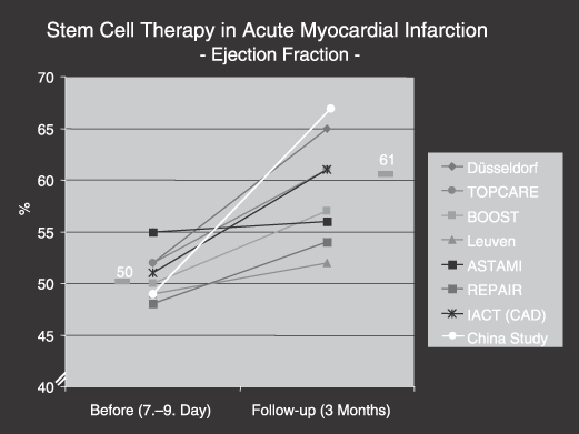
Ejection fraction (stroke volume/enddiastolic volume, %) in patients with stem cell therapy in acute myocardial infarction. Note the marked increase in ejection fraction three months after intracoronary cell transplantation, Düsseldorf. Transplantation of progenitor cells and regeneration enhancement in acute myocardial infarction, Intracoronary autologous bone-marrow cell transfer after myocardial infarction, clinical trial, performed in Leuven (article published by Janssens et al. 2006), autologous stem cell transplantation in acute myocardial infarction, Reinfusion of enriched progenitor cells and infarct remodeling in acute myocardial infarction, Intracoronary autologous bone marrow cell transplantation in chronic coronary artery disease.
Time point for delivery of stem cells after infarction
The time point for cell delivery as 7–8 days after infarction onset was chosen in most studies for the following reasons:
- 1
In dogs, infarcted territory becomes rich in capillaries and contains enlarged, pericyte-poor ‘mother vessels’ and endothelial bridges 7 days after myocardial ischaemia and reperfusion. By 28 days later, a significant muscular vessel forms. Thus, with such timing, cells may be able to reach the worst damaged parts and at the same time salvage tissue. Transendothelial cell migration may also be enhanced due to an adequate muscular coat not yet having been formed.
- 2
Until now, only one animal study has attempted to determine the optimum time for cardiomyocyte transplantation to maximize myocardial function after left ventricular injury. Adult rat hearts were cryo-injured and foetal rat cardiomyocytes were transplanted immediately, 2 weeks later, and 4 weeks later. The authors discussed the inflammatory process (strongest in the first days after infarction) as being responsible for negative results after immediate cell transplantation (Li et al. 2001), and they assumed that the best results seen after 2 weeks may have been due to transplantation before scar expansion. Until now, however, no systematic experiments have been performed with BMCs to correlate the results of transplantation with the length of such a time delay.
- 3
Another important variable is the inflammatory response in myocardial infarction, which seems to be a superbly orchestrated interaction of cells, cytokines, growth factors and extracellular matrix proteins mediating myocardial repair (Frangogiannis et al. 2002). In the first 48 h, debridement and formation of a fibrin-based provisional matrix predominates before a healing phase ensues. Vascular endothelial growth factor is at its peak concentration 7 days after myocardial infarction, and the decline of adhesion molecules (intercellular adhesion molecules, vascular cell adhesion molecules) does not take place before days 3 and 4 after myocardial infarction. We have assumed that transplantation of mononuclear BMCs within the ‘hot’ phase post-myocardial infarction inflammation might lead them to take part in the inflammation cascade rather than in the formation of functional myocardium and vessels (Soeki et al. 2000).
Taking all this into account, it may be concluded that cell transplantation within the first 5 days after acute infarction is not possible for logistical reasons and is not advisable because of the inflammatory process. Although the ideal time point for transplantation remains to be defined, it is most likely between days 7 and 14 after the onset of myocardial infarction.
Long-term action of stem cells
One important issue of stem cell efficacy is the long-term outcome of cell-transplanted patients. Cell therapy has a long-persistent action on infarcted heart muscle. After 3, 15 and 36 months, respectively, the regional infarct area decreases significantly from 34 to 14% (Fig. 5). This is well congruent with improved fluorodeoxyglucose (FDG) metabolism in positron emission tomography (PET) studies and with scintigraphic examination, and demonstrates contraction restoration of initially severely damaged myocardium (Hofmann et al. 2005; Caveliers et al. 2007). The left ventricular ejection fraction also increased over the time of 3 years, showing significant increases from 55 to 66%, respectively. This was mostly due to decrease of endsystolic volume, so that contractility of the whole left ventricle also considerably increased.
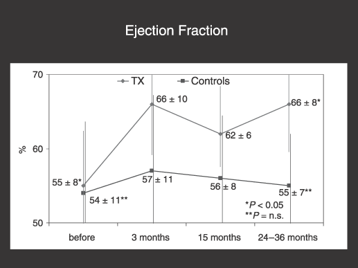
Long-term effect of intracoronary stem cell therapy (Tx) on ejection fraction. Note (i) the improvement of ejection immediately (3 months) after cell transplantation and (ii) the long-term effect, up to 3 years.
These long-term data demonstrate that negative remodeling of the left ventricle after myocardial infarction, which is expected under conventional therapy, can be completely stopped. Reversed remodeling can be realized, with the consequence of a longstanding improvement of ventricular function, in terms of improvement of shortening of cardiac output and of contractility. There have been no complications due to stem cell therapy.
Chronic infarction and heart failure
In patients with chronic infarction many years after the acute attack, intracoronary stem cell treatment is as effective as in the acute postinfarction period. In a randomized study, our group has performed three types of investigatory steps: (i) cardiac catheterization 9 months before stem cell therapy; (ii) cardiac catheterization at the time of stem cell therapy; and (iii) cardiac catheterization 3 months after stem cell therapy in both control and in the treatment groups (Strauer et al. 2005). These patients had suffered from myocardial infarction (mean time, 8 years) prior to the study. For those in the first protocol there was significant improvement in ejection fraction, in infarct size and in infarct wall movement velocity. Moreover, hypokinetic and akinetic zones decreased, and there was significant increase of maximum spiroergometric oxygen uptake and in FDG uptake by the injured and destroyed heart muscle. All together, ejection fraction increased significantly by 15%, contractility as directly determined by the wall movement velocity by 57%, infarct size decreased by 30%, and both overall oxygen uptake and myocardial glucose storage increased by 11% and 15%, respectively (Table 3). These data indicate that bone marrow cell therapy may also be beneficial in chronic coronary disease and may offer a further new therapeutic approach for the substantial numbers of coronary patients with heart failure (Menasche et al. 2001; Perin et al. 2003; Smits et al. 2003; Stamm et al. 2003).
| Before cell-therapy | After cell-therapy | Difference in % | P | |
|---|---|---|---|---|
| LV-ejection fraction, % | 52 ± 9 | 60 ± 7 | +15 | 0.0001 |
| Wall movement velocity in infarct area, cm/s | 1.86 ± 0.7 | 2.9 ± 0.9 | +57 | 0.0001 |
| Infarct area, % | 27 ± 8 | 19 ± 9 | –30 | 0.0001 |
| VO2, ml/min | 1602 ± 533 | 1776 ± 523 | +11 | 0.0001 |
| FDG-PET, % | 43.8 ± 8.0 | 50.5 ± 11.6 | +15 | 0.012 |
- Strauer et al. 2005.
Dilatative cardiomyopathy
Several preclinical as well as clinical trials have shown that transplantation of autologous bone marrow cells or precursor cells improves cardiac function after myocardial infarction and chronic heart disease. Seth et al. and our Düsseldorf group undertook the first studies of intracoronary stem cell implantation in patients with non-ischaemic dilated cardiomyopathy (Seth et al. 2006).
This first-in-man study of autologous bone marrow cells in dilated cardiomyopathy (First-in-Man ABCD) investigated 44 patients (24 randomly allocated to stem cell therapy and 20 as controls) and the Düsseldorf autologous bone marrow cells in dilated cardiomyopathy trial (Düsseldorfer ABCD Trial) investigated 20 patients (10 randomly allocated to the stem cell therapy and 10 as controls). In both studies none of the patients had coronary disease (excluded by angiography) or myocarditis (excluded by endomyocardial biopsy). In both trials cell transplantation was performed via the intracoronary administration route in either coronary artery.
All procedures should lead to an adequate prolonged contact time of the stem cells with arteries and capillaries and facilitates cell migration (Strauer et al. 2002). There has been a significant increase in New York Heart Association Functional Classification in treatment patients in both trials (Table 4). Ejection fraction improved by 5.4% in the First-in-Man ABCD trial and 8% in the Düsseldorfer ABCD Trial. In parallel with the Düsseldorfer ABCD trial, physical ability (functional capacity) rose from 25 watts to 75 watts. In addition, there has been improvement of maximum oxygen uptake under stress from 1236 ± 217 ml/min up to 1473 ± 198 ml/min (Table 4). Furthermore, reduction of arrhythmia was documented. Both trials found reduction in end-systolic volumes and no change in end-diastolic volumes. In the control groups no significant changes were documented. No side-effects of intracoronary autologous stem cell therapy were found, particularly no arrhythmias, no heart insufficiency, no dyspnoea and no palpitations.
| Before stem cell therapy (n = 10) | 3 months after stem cell therapy (n = 10) | Differences (%) | P | |
|---|---|---|---|---|
| Ejection fraction (%) | 18 ± 1 | 26 ± 3 | 44% | < 0.001 |
| LVEDD (mm) | 74 ± 4 | 68 ± 4 | 8% | < 0.01 |
| Average functional capacity (watt) | 25 | 75 | 200% | < 0.001 |
| Sense of well-being (%)(SF-36) | 10 | 70 | 600% | < 0.001 |
In the First-in-Man trial, endomyocardial biopsy was performed before cell transplantation as well as 3 months thereafter. Histopathology revealed no evidence of persisting stem cells after 3 months, no evidence of any new immature myocytes, and also no evidence of any inflammation, infarction or neovascularization, but the ratio of capillaries to myocytes showed a significant increase (Fig. 6) (Seth et al. 2006). There were some data to suggest cell proliferation (binucleate cells and Ki-67 positivity). Laboratory experiments in non-ischaemic dilated cardiomyopathy have suggested that benefit from stem cell therapy comes mainly from lower levels of fibrosis and increased vascularity. Data from the First-in-Man ABCD trial suggest that the benefit of stem cell therapy could be a paracrine effect with changes in vascularity. Results from the First-in-Man ABCD and the Düsseldorfer ABCD trials show that transplantation of autologous bone marrow cells, as well as the intracoronary approach, represent a novel and effective therapeutic procedure for dilated cardiomyopathy.
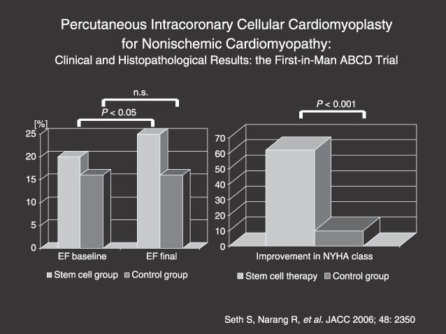
First-in-Man ABCD Trial. Note improvement in ejection fraction and New York Heart Association Functional Classification increase (Seth et al. 2006).
MODE OF ACTION OF STEM CELLS IN HEART DISEASE
The regenerative potential of bone marrow-derived stem cells may be explained by any of four mechanisms: direct cell differentiation from mononuclear cells to cardiac myocytes (Oh et al. 2003), cytokine-induced myocyte growth (Orlic et al. 2001) and increase of residual viable myocytes (especially in the border zone of the infarcted area), stimulation of intrinsic myocardial stem cells (endogenous stem cells) (Nadal-Ginard et al. 2003; Anversa et al. 2006), and induction of cell fusion between transplanted bone marrow cells and resident myocytes (Terada et al. 2002; Alvarez-Dolado et al. 2003; Murry et al. 2004, 2006; Balsam et al. 2004). Trans-differentiation has been described by previous investigators (Bittner et al. 1999); however, it has been questioned by recent experimental studies. The influence of cytokines has been shown to restore coronary blood vessels and muscle cells after experimental myocardial infarction. This regeneration is most pronounced in the border zone of ischaemic and/or infarcted tissue, demonstrating enhancement of mitotic and cycling cells up to fourfold compared to areas remote from necrotic myocardium. Moreover, mononuclear bone marrow stem cells express a bounty of cytokines (e.g. vascular endothelial growth factor, insulin-like growth factor, platelet-derived growth factor), thereby stimulating residual normal myocytes for regeneration and proliferation, and instrinsic myocardial stem cells (endogenous stem cells) for cell regeneration and for cell fusion. Mitotic indices are three to four times more frequent within the border zone of myocardial necrosis when compared with non-injured heart muscle. Also, 20% to 40% of intracoronarily transplanted bone marrow-derived stem cells may accumulate within the border zone of myocardial infarction (Anversa et al. 2006). No signs of microcirculation disturbances have been apparent because all patients had thrombolysis in myocardial Infarction flow grade 3. Thus, it is conceivable that mitotic border zone represents the optimum niche for exogenously transplanted stem cells, stimulating mitotic rates and heart muscle regeneration, preferably originating in and expanding from these areas. Cell fusion may also contribute to heart muscle regeneration, which takes its origin from the border zone, expanding gradually to the necrotic core of the infarcted area.
Clinical studies cannot determine which cell biological and molecular mechanims are responsible for heart muscle repair or which of the studied factors play the predominant role. However, the final functional outcome of this kind of cell therapy demonstrates three main target effects: improvement in muscle function (pumping ability and contractility), myocardial perfusion (SPECT), and myocardial glucose metabolism (PET), thus providing evidence that heart muscle repair must have taken place by this intracoronary bone marrow cell transplantation procedure.
SPECIAL ASPECTS OF STEM CELL THERAPY
Homing myocardium and preconditioning
For the involvement of injected progenitor cells with myocardial regeneration, the stem/progenitor cells must be directed in their migration by cytokines and chemotactic factors. Stem/progenitor cells can be mobilized by ischaemia-related inflammatory cytokines and chemotactic factors. The SDF-1/CXCR4 axis seems to be important in this homing, chemotaxis, engraftment and retention ischaemic tissue, as has been shown in recent experimental studies performed by Elmadbouh et al. (2007). Here, it was demonstrated that SDF-1 is a potent chemotactic factor for CXCR4 expressing cells and is markedly up-regulated locally in the myocardium under ischaemic conditions. This indicates that repetitive balloon occlusion represents a useful method for ischaemia production, thereby enhancing stem cell migration to the heart muscle. Damås et al. have reported significantly decreased concentrations of SDF-1 in patients with stable angina and particularly low levels in unstable angina compared with healthy control subjects. SDF-1 interacts with the single specific receptor, CXCR4, thus forming an SDF-1/CXCR4 axis, which is pivotal to mobilization, homing and survival of progenitor cells (CD34+) and endothelial progenitor cells (EPCs). In experiments on mice, it was shown that SDF-1-dependent migration of EPCs led to increased neovascularization and improved ischaemic tissue perfusion. With reference to the receptor for SDF-1, altered function of the SDF-1/CXCR4 axis has been described (Damas et al.). This study described alterations in patients with unstable angina, in whom low levels of SDF-1 coexisted with reduced cellular membrane expression of CXCR4 on peripheral blood mononuclear bone marrow cells (MNCs) despite overexpression of the corresponding gene, as evidenced by high CXCR4 mRNA levels in these cells. The importance of the SDF-1/CXCR4 axis in chemoattraction and homing of CXCR4+/CD34+ cells is evidenced by selective inhibition of CXCR4, which significantly reduces chemotaxis of these cells. It also seems that other chemoattractants must be involved, as blockade of CXCR4 does not completely prevent the chemotaxis. Because the gradient of SDF-1 concentration through endothelium is an important signal for bone/muscle progenitor cell homing, increased secretion of SDF-1 from the ischaemic myocardium can direct the flux of cells into the myocardium, thus facilitating tissue repair.
For clinical situations, five important methodological prerequisites have to be considered: the intracoronary approach of cell infusion directly into the artery, simultaneous ischaemic preconditioning by four intracoronary balloon insufflations for 2–4 min each (Strauer et al. 2001), a sufficient quantity of mononuclear bone marrow cells (more than 80 × 106 cells), the intracoronary application of macroalbumin aggregates before stem cell transplantation and low-dose dobutamine therapy (10 µg/kg BW/min) intravenously with the objective of raising the heart rate about 20 beats per minute and finally additionally fractionated administration of dipyridamol (Persantin®) (Heidland et al. 2000). The most successful way to transplant bone marrow cells into the heart is intracoronary delivery. However, if stem cells were only to be infused into the coronary arteries, without concomitant ischaemia, contact between them and myocardial cells is sparse. Simultaneous ischaemic preconditioning should include sufficient production of ischaemia (ST elevation in the ECG, precordial pain of the patient).
Border zone – regeneration area
Regeneration of blood vessels and muscle cells is most pronounced in the border zone of ischaemic and/or infarcted tissue, demonstrated by enhancement of mitotic cells and all cycling cells up to fourfold, when compared with areas remote from the necrotic myocardium. Furthermore, mononuclear bone marrow stem cells express a variety of cytokines, thereby stimulating residual normal myocytes for regeneration and proliferation and intrinsic myocardial stem cells (endogenous stem cells) for cell regeneration and for cell infusion. Mitotic indices are three to four times more frequent within the border zone of myocardial necrosis compared to non-injured heart muscle; 20–40% of intracoronarily transplanted bone marrow-derived stem cells may accumulate within the border zone of myocardial infarction. It is conceivable that in myocardial infarction the border zone represents the optimal area in for exogenously transplanted stem cells, stimulating mitotic rates and heart muscle regeneration, preferably originating in and expanding from these areas (Anversa et al. 2006). Cell fusion may also contribute to heart muscle regeneration, which takes its origin from the border zone, expanding gradually to the necrotic core of the infarcted area.
Repetition of intracoronary stem cell therapy
Repetition of intracoronary application of autologous stem cells in patients with chronic coronary artery disease and non-ischaemic dilated cardiomyopathy seems to have an accumulative beneficial effect. As yet, there are no published data on repetitive intracoronary stem cell therapy; however, already 21 patients with chronic ischaemic coronary artery disease and two patients with dilated cardiomyopathy have been treated in Düsseldorf for a second time (between 3 and 12 months after the first cell transplantation) with intracoronary autologous bone marrow cells. A significant improvement in ejection fraction was measured (first time: increase of 5.3%, second time: additional increase of 6%). Furthermore, 18 patients improved by at least one functional class. No side-effects of intracoronary autologous stem cell therapy were found. Of course, further experimental studies and controlled prospective clinical trials must follow.
Clinical safety
The procedure of intracoronary autologous bone marrow cell tranplantation in patients with acute myocardial infarction, chronic coronary artery disease and non-ischaemic dilated cardiomyopathy is effective and safe. No increase of malignant diseases or inadequate progression of coronary artery disease have been documented.
To assess any inflammatory response and myocardial reaction after intracoronary autologous stem cell transplantation, white blood cell count, serum levels of C-reactive protein and of creatine phosphokinase are measured before, during and after treatment, and these data collected revealed no evidence of inflammation. No procedural, no cell-induced complications and no side-effects of other nature have occurred in any patient.
Stem cell tracking for detection of cell fate
Visualization of the injected bone marrow-derived progenitor cells provides important information concerning cell fate in the myocardium. Homing of injected cells in the infarcted myocardium is a critical early event after intracoronary cell administration. In animal models reported by Kocher et al. (2001) and Aicher et al. (2003) myocardial homing of transplanted progenitor cells was monitored by fluorescence or radioactive labelling. Hofmann et al. (2005) assessed myocardial homing and biodistribution of injected bone marrow-derived cells. In this study, 2-[18F]-fluoro-2-deoxy-D-glucose (18F-FDG) labelled cells were used and a 3D PET imaging technique was performed. It was demonstrated that a small fraction, about 3%, of the transplanted mononuclear cells remained in the infarcted myocardium, while the majority homed to the liver and spleen within 1 h after intracoronary delivery. More precise investigation with labelled CD34+ progenitor cells showed enhanced engraftment of the cells (about 40%) in human infarcted myocardium and predominantly homed to the infarcted border zone. Thus, intracoronary BMC transplantation enhances left ventricular contractility, primarily in myocardial segments adjacent to the injured area.
Visualization of stem cells in chronic ischaemic heart disease
Bone marrow-derived progenitor cell homing to ischaemic or injured myocardium is necessary for local repair. Chronic ischaemia of the myocardium leads to tissue loss, remodelling, reduction in contractility and progressive ventricular failure. In recent studies, cell therapy in chronic ischaemic heart disease led to improvement of left ventricular function and enhanced myocardial perfusion in the border zone of the chronic infarction area. Caveliers et al. (2007) demonstrated that 111indium-labelled CD133+ cells are suitable for follow-up investigations of cell distribution during the first day after cell transplantation. This group found a significant quantity of CD133+ peripheral blood stem cells in the heart after intracoronary transplantation. Shortly after cell administration, 8% of the injectate (presumed to be cells) remained in the target zone, decreasing to 3% after 12 h. This decline in activity, presumably in cell number, could be related to technical problems such as efflux of 111indium from viable cells, or because of 111indium leakage. Three days after cell injection, 1% of injected CD133+ cells still resided within the post-infarction area. Exact localization of labelled cells within the infarcted zone was not possible due to technical limitations. Further studies with advanced molecular imaging techniques may be necessary for exact localization of the cells in the target region.
Dose-dependent contribution for cardiac recovery
Compelling evidence suggests that transplantation of bone marrow-derived progenitor cells contributes to recovery of left ventricular function after myocardial infarction through enhancing ischaemic neovascularization (Kawamoto et al. 2001, 2003; Kocher et al. 2001). The mechanism of this therapeutic effect was previously considered to be incorporation, differentiation, and proliferation of bone marrow-derived progenitor cells. Several working groups (Orlic et al. 2001; Yeh et al. 2003) have demonstrated trans-differentiation of bone marrow-derived progenitor cells into endothelial cells, cardiomyocytes and smooth muscle cells, while Balsam et al. (2004) and Murry et al. (2004) have reported that in animal models BMCs do not trans-differentiate into cardiomyocytes in infarcted myocardium. Further investigations are necessary to solve these controversial results. Iwasaki et al. (2006) investigated dose-dependent contributions of CD34+ progenitor cells (1 × 10³, 1 × 10⁵, 5 × 10⁵ cells/kg) in animal models after myocardial infarction. They found dose-dependent augmentation of cardiomyogenesis and vasculogenesis after transplantation of human CD34+ cells into rat infarcted myocardium. Enhanced capillary density, inhibition of left ventricular fibrosis and increased recovery of the left ventricular function was associated with higher numbers of transplanted CD34+ cells. These findings suggest that use of higher doses of CD34+ cells may be more potent for therapeutic application to the damaged myocardium than a lower dose. Iwasaki et al. (2006) in their study also found that there was no beneficial effect of CD34+ cells in their low-dose group (1 × 10³ cells/kg).
CLINICAL INDICATIONS FOR STEM CELL THERAPY
On the basis of numerous studies within the past few years, intracoronary stem cell therapy is considered to be a safe therapeutical procedure in heart disease, when destroyed and/or compromised heart muscle must be regenerated. This kind of cell therapy with autologous bone marrow cells is completely justified ethically, except for the small numbers of patients with direct or indirect bone marrow disease (e.g. myeloma, leukaemic infiltration) in whom there would be lesions of mononuclear bone marrow cells.
Best examined indications for stem cell therapy are previous myocardial infarction with large infarct area, aneurysm and depressed ejection fraction and heart failure due to chronic myocardial infarction (Table 5). The age of infarction seems to be irrelevant to regenerative potency of stem cells, since stem cell therapy in old infarcts (older than 8 years) is almost equally effective in comparison to previous infarcts. This regenerative phenomenon most probably may be related to persistence of the border zone, which is also present in chronic infarcts and which has an approximately fourfold mitotic rate. Further indications are ischaemic and non-ischaemic cardiomyopathy and heart failure due to hypertensive heart disease. Not yet established, but facultative indications, are refractory ventricular arrhythmia and pulmonary hypertension.
| • Previous myocardial infarction with heart failure (depressed ejection fraction, large akinesia and aneurysm) |
| • Heart failure due to cardiomyopathy |
| – ischaemic |
| – non-ischaemic |
| • Decompensated hypertensive heart disease |
| • Facultative indications: |
| – refractory ventricular arrhythmia |
| – pulmonary hypertension |
PERSPECTIVES CARDIAC STEM CELL THERAPY
Since the first description of the use of bone marrow-derived stem cells for treatment of heart disease in 2001, a large number of clinical studies has been published demonstrating the effectiveness of stem cells in various clinical conditions, but with very different bone marrow preparation techniques. The use of non-standardized cell transplantation procedures is common with large variation in (i) numbers of transplanted cells, (ii) intracoronary balloon insufflation time, and (iii) additive preconditioning measures. Therefore, important future studies must be performed in order to define the optimum technique of cell preparation, to discover the best cell type for myocardial regeneration, to pretherapeutically test the viability and migration potency of the cells, to analyse their homing characteristics to the cardiac niche and to other extracardiac organs, to characterize the mode of action of stem cells for cardiac regeneration, to improve cell delivery techniques, to label stem cells for determining stem cell fate, and to try to find clinically relevant and established indications for cell therapy in the future in various heart diseases (Strauer & Kornowski 2003). In realizing these perspectives, joint and cooperative studies between preclinical and clinical research are essential. More interest should be focused and given to adult stem cell projects that already have proven significant clinical efficacy without any ethical problems, as there are with hypothetical use of embryonic stem cells.
ETHICAL CONSIDERATIONS
The use of human autologous mononuclear bone marrow cells containing (progenitor) stem cells are clinically justified and ethically unquestionable because no side-effects have been reported, especially with regard to teratocarcinoma. Moreover, in contrast to differentiation of embryonic stem cells, there is no arrhythmogenic potential, and immunosuppressive therapy is unnecessary. Thus, the therapeutical advantage clearly prevails, and clinical use has already been realized. Further and intensified research using autologous human bone marrow stem cells is needed and is essential in order to promote stem cell therapy for the numerous cardiac diseases.
Clinical use of autologous bone marrow mononuclear cells has no ethical problems. Therapy is performed with usual cardiac catheterization techniques and PTCA (time ~30 min). In severe cardiac failure, physical activity increases twofold and spiroergometric VO2 by 10–15%. Ejection fraction increases by 20–30% (in non-ischaemic) and by 8–15% (in ischaemic) cardiomyopathy. Myocardial perfusion (scintigraphy) and myocardial glucose uptake (PET) increase considerably demonstrating neo-perfusion and neo-metabolism in formerly avital myocardial areas. No stem cell-related side-effects, especially no cardiac arrhythmia or inflammation have been reported.
This review has described the most important methodological prerequisites for bone marrow-derived transplantation into the myocardium. These are (i) careful mononuclear cell preparation (GMP facilities), (ii) strict intracoronary method of cell application, and (iii) procedures for enhancing cell transfer from the coronary vascular bed into the myocardium, for example by induced ischaemia (balloon dilatation, dipyridamole intracoronary dipyridamole application), by increase in contractility (dobutamine infusion), by promotion of cell migration (prolongation of cell contact time by i.c. albumine microsphere injection).


