A tactile approach to introduce the skin autoimmune disease psoriasis to the general public and the vision-impaired community
Abstract
Scientific outreach activities play an important role in disseminating knowledge, connecting the general public to research and breaking down scientific skepticism barriers. However, the vision-impaired community is often disadvantaged when the most common audio–visual approach of scientific communication is applied. Here we integrated tactile clues in the scientific communication of immune processes involved in the autoimmune skin disease psoriasis. We fostered the involvement of the vision-impaired community through interactive experiences, including tactile scientific origami art, a haptic poster and wood-carved molecular models. Readily accessible science communication that engages a number of senses is a critical step toward making science more inclusive and engaging for individuals with a wide range of sensory abilities. The approach of the 2023 Monash Sensory Science exhibition aligns with the principles of equity, diversity and inclusion and helps to empower a more informed and scientifically literate public.
CONCEPT AND INTRODUCTION
The goal of the 2023 Monash Sensory Science exhibition was to disseminate knowledge about autoimmune diseases with a particular focus on the vision-impaired community which is often left out through the conventional use of figures and graphs as prime methods of scientific communication. We work on the highly prevalent noncommunicable skin disease psoriasis that affects 2–3% of the global population. This condition is commonly misunderstood as contagious, leading to stigma in patients who often experience comorbidities such as anxiety and depression. It is therefore important to raise awareness and understanding of the disease. To familiarize exhibition visitors with the immune dysregulation in psoriasis, we decided to take our participants on a journey from the highly familiar perception of the skin to an exploration of skin layers by providing tactile insights into the cellular players and key molecular events of skin autoimmunity.
WHAT YOU FEEL IS HOW PSORIASIS FEELS
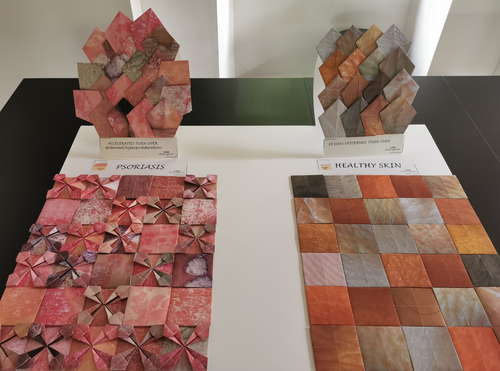
Everyone has had experiences when they were comfortable in their own skin versus periods of itch and discomfort. We built on this lived experience and initiated our engagement with the participants in an artistic way. PEAUrigami® (peau: French for skin) scientific art exhibits have an educational purpose at their core; PEAUrigami® seeks to communicate knowledge of the pathophysiology of skin to different audiences through origami artwork with skin pattern–printed papers.1, 2 Previously, PEAUrigami® educational–artistic compositions have been exhibited at several dermatologic congresses (European Society for Dermatological Research 2019 and 2022, European Academy of Dermatology and Venereology 2020 and International Society of Investigative Dermatology 2023) as well as in a private dermatology practice to sensibilize patients to melanoma prevention.3 Here, the PEAUrigami® medical art aimed to mobilize the sense of touch in a visually impaired audience. The international artist took great care to represent a wide array of skin of color in a steady state using close-up photography of the skin. The origami-folded surface was deliberately chosen to be very smooth, similar to stroking healthy skin (Figure 1, bottom right). As a contrasting exhibit, the psoriasis panel included origami pieces with a spiky nature that created a hindrance and interrupted the flow of the hand in the vision-impaired audience, leaving a somewhat uncomfortable feel (Figure 1, bottom left). The psoriasis panel was compiled of psoriatic plaque photographs that allowed us to show to visitors with some degree of vision that the lesions oscillate between a very red and a silvery white appearance. We could further explain that one of the altered processes during lesion formation is an increased turnover rate of keratinocytes with epidermal turnover in healthy skin being about 4 weeks versus one week in psoriasis, with a resulting thickening of the epidermal and cornified layers.4 This was represented by an orderly cornified layer in healthy skin versus an overcrowded arrangement during the shedding of epidermal cells in psoriasis (Figure 1, upper panels).

ANATOMY OF THE SKIN AND ALTERATIONS IN PSORIATIC LESIONS
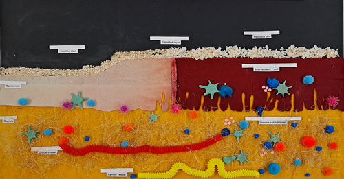
After bringing the attention of visitors to the disease at the first station, we encouraged them to explore the second station which was a tactile poster of the skin in cross section (Figure 2). It allowed us to again showcase both the nonlesional and the lesional skin side-by-side and delve deeper into the anatomical as well as cellular levels. The poster demonstrated the cornified top layer as smooth oats in a steady state or spiky coconut chips in psoriatic plaques. The depiction of the epidermis included epidermal thickening, redness and rete ridges on the lesional side, all hallmark features of psoriasis. The dermis contains blood vessels carrying immune cells that can immigrate into the skin, an extracellular matrix facilitating cell migration and lymphatics with perforations to facilitate cell exit from the skin. On the cellular level, the visitors could familiarize themselves with some key players of the immune response in psoriasis: melanocytes located in the basal epidermis served as an example of antigen-presenting cells, dendritic cells as professional facilitators of the immune response, CD8+ T cells (blue) were shown to be able to immigrate into the epidermis in higher numbers and CD4+ T cells (orange) were localized in the dermis. Some T cells were further shown to excrete cytokines to enable us to discuss the currently most successful treatment options for severe psoriasis: biologics targeting the T-cell cytokines tumor necrosis factor (TNF)-α, interleukin (IL)-17 and IL-12/23.5

The cellular players and anatomic structures were labeled with braille to aid accessibility. Prerecorded audio buttons were also mounted near the poster, but received little use as visitors preferred personal interaction and seemed to value the ability to lead the conversation with their own questions at their own pace.
THE CRITICAL MOLECULAR PLAYERS—INTRODUCING A SCIENTIFIC PROJECT
At the final station, we wanted to drill down to the molecular level and expose visitors to the scientific work we do. We specialize in the mass spectrometric study of peptides presented by the major histocompatibility complex (MHC) with the goal of elucidating peptide triggers of psoriasis that are presented by the main risk gene, the MHC class I molecule HLA-C*06:02. We are also investigating the T-cell receptor (TCR) specificity of skin-resident CD8+ T cells that are key recipients of HLA-C*06:02-mediated signals. In the periphery of the session room, our most recent work of the solved crystal structure of the TCR–melanocyte ADAMTSL5 peptide–HLA-C*06:02 complex and our multiphoton microscopy movies of immune cell migration were projected on big screens around the session room, thus forming the backdrop and providing some further instigation for discussion.6-8
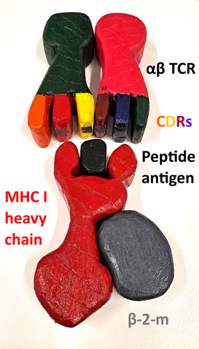
To explain our work at the third station, a local wood carving artist generated a 3D model of the TCR–peptide–MHC I complex (Figure 3). The individual pieces were molded to fit well into the palm, to tinker with and explore. The exploration phase of each individual piece (MHC heavy chain, beta-2-microglobulin, peptide, TCR alpha and beta) allowed us to explain the function of each molecule and elaborate on how they come together as a complex. The complementarity-determining regions of the TCR chains were shaped as “fingers” that probe each peptide–MHC molecule and convey antigen specificity. This resonated highly with the vision-impaired participants who were right at that moment using their own fingers to probe the exhibit. Once the visitors knew and understood more about the molecular players that convey antigen specificity in the immune system, they could arrange the puzzle pieces on the table and were guided by magnets integrated into the individual wooden molecules. This station was highly popular, but a drawback of the time investment for the puzzle and the often-ensuing broader scientific discussions was that one guide dog became increasingly bored and eventually continued to instigate that their owner moved on. A researcher's unanticipated note to self: Mind the attention span of the visitors, but also the patience of the guide dogs.

OUTCOMES OF THE PUBLIC ENGAGEMENT ACTIVITY
The relevance of conducting outreach activities such as the one presented here became very obvious when a visitor commented: “Mum has psoriasis, but I don't understand it” (Table 1). The feedback during the session was overwhelmingly positive with participants commenting that the means of the presentation was a “less scary” way of introducing science, “more accessible” and “a way in for people”. Another visitor mentioned that the exhibition helped them to “understand how it [psoriasis] impacts people”. Overall, we hope that we were able to not only increase the understanding of the autoimmune disease psoriasis, but also help visitors glean a deeper understanding of the skin and immune compartments within it. We welcome anyone to take inspiration from the Monash Sensory Science communication program as it is rewarding for visitors and scientists alike. This approach can be used to educate and raise awareness among the general public about various skin conditions and other diseases.
| “Helpful to look at it side by side.” (at the PEAUrigami® station) |
| “That's a great way to show the flakiness.” (at the PEAUrigami® station) |
| “Good to understand how it [psoriasis] impacts people.” |
| “It looks similar to other skin conditions. How is it different?” (Young visitor pointing to a lesional flareup area on their hand) |
| “Mum has psoriasis, but I don't understand it.” |
| “We had psoriasis. Is there a cure for it?”, “That's interesting, I hope you can find a cure” (Elderly couple at the tactile poster) |
| “illustrates very nicely the difference between psoriasis and healthy [skin]” (at the tactile poster) |
| “It's cool.”, “great” (at the pMHC–TCR model) |
| “less scary”, “more accessible”, “a way in for people” (Different quotes pertaining to the science exhibition) |
ACKNOWLEDGMENTS
We thank Dr Erica Tandori, Professor Jamie Rossjohn and the rest of the Monash Sensory Science team for their long-standing outreach engagement and for organizing the 2023 Monash Sensory Science exhibition, which we had the privilege to be a part of. We also acknowledge the LEO Foundation which supports our psoriasis antigen discovery work (LF-OC-22-001014). Open access publishing facilitated by Monash University, as part of the Wiley - Monash University agreement via the Council of Australian University Librarians.
AUTHOR CONTRIBUTIONS
Runqiu Song: Investigation; visualization. Jingran Ye: Investigation; visualization. Shanzou Chung: Investigation; visualization; writing – review and editing. C Chris Meyer: Methodology; visualization. Corinne Déchelette: Methodology; visualization; writing – review and editing. Anthony Purcell: Supervision; writing – review and editing. Asolina Braun: Conceptualization; investigation; supervision; writing – original draft; writing – review and editing.
CONFLICT OF INTEREST
The authors have no conflicts of interest to disclose.
Open Research
DATA AVAILABILITY STATEMENT
All data are available in the article.


