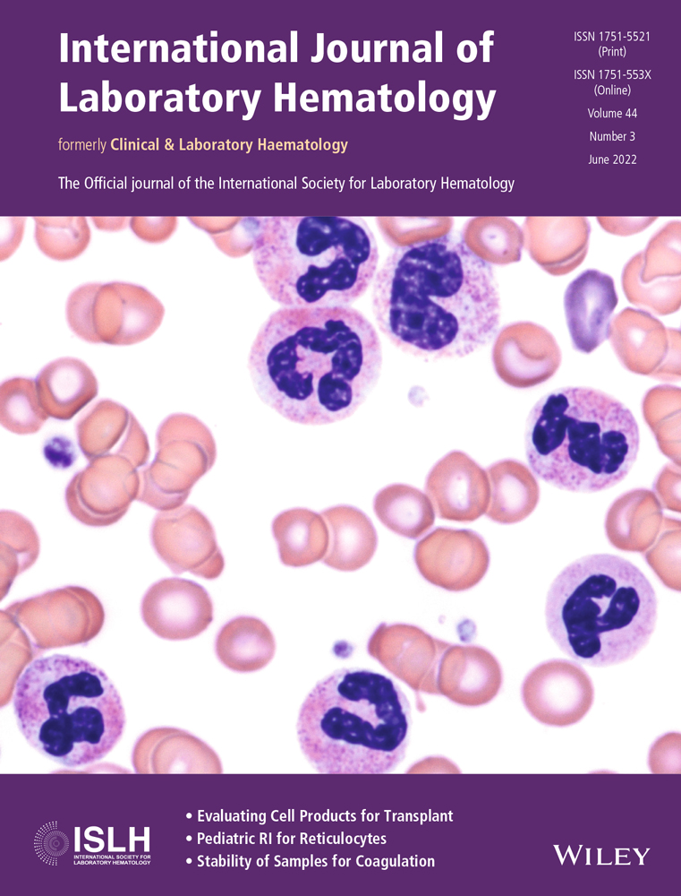
Changes in peripheral blood cellular morphology as diagnostic markers for COVID-19 infection
Abstract
Introduction
Real-time reverse-transcriptase polymerase chain reaction (RT-PCR) assays were established to detect severe acute respiratory syndrome-coronavirus 2 (SARS-CoV-2). However, due to the high rate of false negative results, additional tests as computed tomography (CT) scans of the chest and blood chemistry are required to properly diagnose COVID-19 infection. Abnormal morphological changes of peripheral blood cells as granulocytic dysmorphism and abnormal reactive lymphocytes have been described in some cases. The aim of the present study was to investigate the morphological changes affecting all peripheral blood cells of COVID-19 patients, in order to find any specific abnormalities that could help in the early diagnosis and/or prognosis.
Methods
Peripheral blood smears of 113 COVID-19 patients and 50 non-COVID-19 controls were examined for morphological changes in the period between October 2020 and January 2021 (second wave). We set a score value in which every morphological abnormality was given one point in each examined blood smear. Score, neurophil/lymphocyte (N/L) ratio, and blood chemistry were compared to the severity and outcome of the disease.
Results
Significant morphological changes were found when compared to control blood smears. Various abnormalities as pyknotic cells, broken cells, pseudo Pelger-Huët, abnormal lymphocytes, abnormal monocytes, and leukoerythroblastic reaction were found. Cases with higher scores had unfavorable outcomes (p = .005). High interleukin-6 (IL-6) levels were correlated to pyknotic cells (p = .003).
Conclusion
The blood picture of COVID-19 patients revealed various morphological changes that are not detected with the same frequency and variability in other viral infections. The prominent morphological changes can be predictive of an undesirable outcome of the disease.
CONFLICT OF INTEREST
The authors have no competing interests.
Open Research
DATA AVAILABILITY STATEMENT
The data that support the findings of this study are available from the corresponding author upon reasonable request.


