Overexpression of FOXC1 correlates with poor prognosis in gastric cancer patients
Abstract
Aims
The aim of this study was to determine FOXC1 expression in gastric tissues, and the clinical significance of FOXC1 in the development, progression and metastasis of gastric cancer (GC).
Methods and results
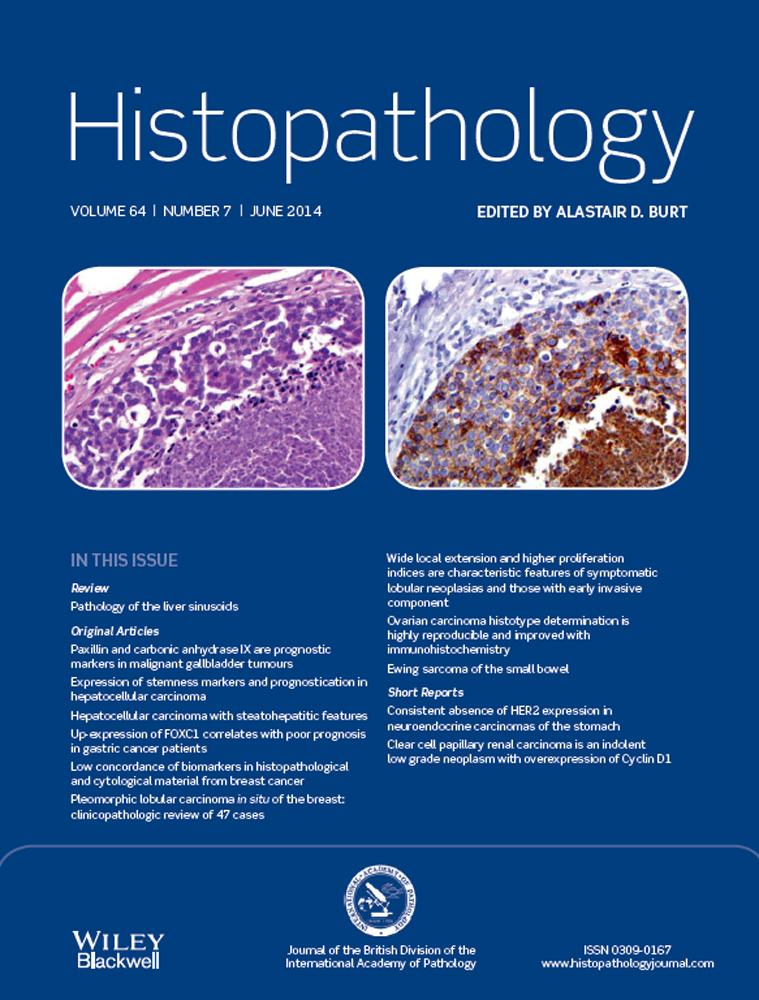
We screened GCs for the expression of FOXC1 using the Affymetrix U133 plus 2.0 Gene Chip Array, and found that expression was significantly higher in GC tissues than in controls. Furthermore, we validated the expression levels of FOXC1 using real-time quantitative RT-PCR (qRT-PCR), and of FOXC1 using immunohistochemistry (IHC). Our study showed that expression levels of FOXC1 mRNA and FOXC1 in GC tissues were significantly higher than those in corresponding non-tumour tissues. High FOXC1 expression correlated with the degree of histological differentiation (P < 0.01), TNM stage (P < 0.001), invasive depth (P < 0.05), lymph node metastasis (P < 0.05), and distant metastasis (P < 0.01). Survival analysis revealed that patients with high FOXC1 expression had shorter overall survival than those with low expression (P < 0.001). Multivariate analysis showed that high FOXC1 expression was an independent prognostic factor for GC patients (P < 0.01).
Conclusions
Overexpression of FOXC1 may play a key role in the progression of GC, and FOXC1 expression may serve as a useful marker for predicting the outcome of patients with GC.