Afferent loop syndrome following pancreatic head cancer surgery treated with metal stent placement using a short-type single-balloon enteroscope
Abstract
Watch a video of this article.
BRIEF EXPLANATION
Afferent loop syndrome is a rare complication that occurs following reconstructive intestinal tract surgery as a result of postoperative adhesions or peritoneal dissemination due to recurrence. Obstruction of the afferent loop can be fatal, and often requires surgical treatment. However, patients who develop afferent loop syndrome due to recurrence of malignancy are often in poor general health, making surgery invasive.1 With the development of balloon-assisted enteroscopy, there have been reports of these patients being treated endoscopically.2-5
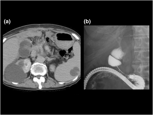
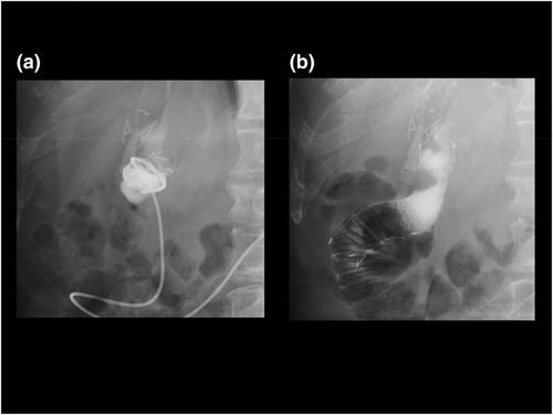
The patient was a 74-year-old woman who underwent subtotal stomach-preserving pancreaticoduodenectomy for pancreatic head cancer. She was found to have multiple liver metastases on contrast-enhanced computed tomography (CT) 3 years after surgery. While receiving chemotherapy for recurrence of pancreatic head cancer, she presented with fever and abdominal pain. Contrast-enhanced CT led to a diagnosis of afferent loop syndrome caused by peritoneal dissemination. Conservative treatment was unsuccessful (Fig. 1a). Therefore, we decided to treat the afferent loop syndrome by drainage using a short-type single-balloon enteroscope (s-SBE) with a working channel diameter of 3.2 mm (SIF-H290S; Olympus Medical, Tokyo, Japan). We advanced the s-SBE and identified the stenotic area in the afferent loop. We traversed the stenosis with a catheter and guidewire, advancing the guidewire into the dilated bowel (Fig. 1b). In view of elevated inflammatory markers, a nasobiliary drainage tube was placed in the afferent loop (Fig. 2a). When the patient's condition improved, we placed a metal stent at the stricture site using the s-SBE. The s-SBE was advanced to the site of the stricture via the nasobiliary drainage tube. A 22 mm × 15 cm duodenal metal stent with a caliber of 3.0 mm (uncovered Niti-S stent; Taewoong Medical, Seoul, South Korea) was placed in the stenotic area, and patency was confirmed with contrast medium (Fig. 2b, Video S1). There were no postprocedural complications.


Authors declare no conflict of interest for this article.


