Endocytoscopy for the diagnosis of marginal zone B-cell lymphoma of mucosa-associated lymphoid tissue type in the rectum: Report of two cases
Abstract
Watch a video of this article
Brief Explanation
Endocytoscopy enables in vivo microscopic imaging at 500-fold magnification, allowing real-time analysis of the gastrointestinal epithelium at the cellular level. Recently, the efficacy of endocytoscopy has been reported in optical diagnosis not only for epithelial neoplasms but also for subepithelial lesions such as gastric malignant lymphoma.1-4 However, little is known about typical endocytoscopic findings of colorectal malignant lymphoma. We herein report two cases of rectal marginal zone B-cell lymphoma of mucosa-associated lymphoid tissue (MALT) type (MALT lymphoma) that were seen using endocytoscopy.
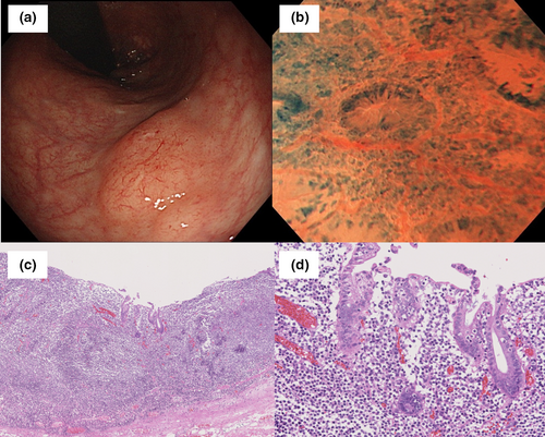
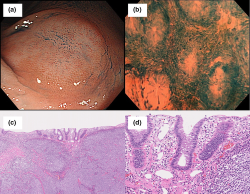
The first patient was a 68-year-old woman and the second patient was a 79-year-old woman. They were referred to our division for evaluation of submucosal tumors in the rectum. Colonoscopy showed a slightly elevated lesion covered with non-neoplastic epithelium (Figs 1a and 2a). Methylene blue staining of the endocytoscopic specimen showed diffuse interglandular infiltration of small atypical cells. Cytological atypia of infiltrated cells of case 1 were slightly more irregular than those of case 2 (Figs 1b, 2b and Video ).


These lesions were resected en bloc by endoscopic submucosal dissection, and pathology showed infiltration of small to medium-sized atypical lymphoid cells with irregular nuclei (Figs 1c and 2c). In both cases, the lymphoid cells had infiltrated the duct epithelium, but lymphoma cells of case 2 were not exposed to the luminal surface (Figs 1d and 2d). Immunohistochemical study of the lesions was positive for CD20, CD79a, and bcl-2, and negative for CD3, CD5, CD10, and cyclin D1. The final diagnosis was MALT lymphoma.
In the present cases, subtle endocytoscopic and pathological images could easily be seen. Given that the depth of endocytoscopic observation is approximately 50 μm from the mucosal surface,5 endocytoscopic findings may correspond to infiltration of atypical cells within the epithelium and glands. Similar reproducible endocytoscopic findings in the two cases suggested that these might be common endocytoscopic findings in MALT lymphoma. Endocytoscopy may aid in the diagnosis of colonic MALT lymphoma.
Authors declare no conflicts of interest for this article.


