A novel thin wire snare-assisted en bloc cold snare endoscopic mucosal resection of a colonic adenoma 10–14 mm in size
Abstract
Watch a video of this article
Brief Explanation
Cold snare polypectomy (CSP) is carried out as a standard technique for small colorectal polyps. However, CSP for 10–14-mm colorectal polyps was reported to have limitations in terms of the histological margin,1 and CSP is not indicated for polyps ≥10 mm.2 Recently, piecemeal cold snare endoscopic mucosal resection (CS-EMR), which combines submucosal injection and the CSP technique, has been used for colorectal polyps ≥10 mm.3 However, the feasibility of en bloc CS-EMR for 10–14-mm colorectal adenomas is unclear.
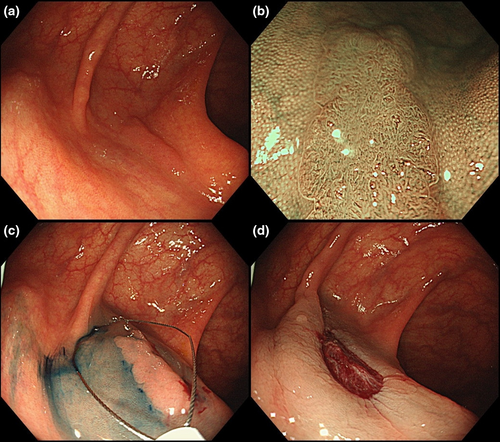
Colonoscopy detected a flat elevated 12-mm polyp. Endoscopic diagnosis was low-grade adenoma because magnifying narrow-band imaging (NBI) suggested type 2A in the Japan NBI Expert Team classification.4 An indigo carmine-tinged glycerol solution was injected into the submucosal layer. Then, using a 15-mm oval snare with a wire diameter of 0.30 mm (SnareMaster Plus; Olympus, Tokyo, Japan), this lesion was ensnared and guillotined without cautery. En bloc CS-EMR was achieved without complications (Fig. 1, Video S1). Histopathological examination showed a low-grade adenoma with negative resection margins and no submucosal tissue (Fig. 2).


This thin wire snare-assisted en bloc CS-EMR technique has two advantages. First, submucosal injection of indigo carmine-tinged solution may improve visibility of the margin and facilitate appropriate lesion capturing. Second, the thin wire snare may improve cutting, even for lesions with submucosal injection; one group reported improved complete removal by CSP for small polyps with a thin wire snare (0.30 mm) versus a thick wire snare (0.47 mm).5 We noted that the resected specimen did not contain submucosal tissue, suggesting insufficient pathological evaluation of the submucosal tissue. Therefore, submucosal invasive cancer should be ruled out before using CS-EMR.
In summary, CS-EMR using a 15-mm snare with a thin wire may be used for en bloc resection of 10–14-mm colorectal low-grade adenomas diagnosed by magnifying endoscopy.
Authors declare no conflicts of interest for this article.


