Volume 31, Issue 3 pp. e54-e55
DEN Video Article
Free Access

Tips for endoscopic retrieval of a large rectal tumor after endoscopic submucosal dissection by a forefinger-compression method

Lancharat Chuncharunee

Lancharat Chuncharunee

Department of Molecular Gastroenterology and Hepatology, Graduate School of Medical Science, Kyoto Prefectural University of Medicine, Kyoto, Japan

Search for more papers by this author
Naohisa Yoshida

Corresponding Author

Naohisa Yoshida

Department of Molecular Gastroenterology and Hepatology, Graduate School of Medical Science, Kyoto Prefectural University of Medicine, Kyoto, Japan

Corresponding: Naohisa Yoshida, Department of Molecular Gastroenterology and Hepatology, Kyoto Prefectural University of Medicine, Graduate School of Medical Science, 465 Kajii-cho, Kawaramachi-Hirokoji, Kamigyo-ku, Kyoto 602-8566, Japan. Email: [email protected]Search for more papers by this author
Ken Inoue

Ken Inoue

Department of Molecular Gastroenterology and Hepatology, Graduate School of Medical Science, Kyoto Prefectural University of Medicine, Kyoto, Japan

Search for more papers by this author
First published: 10 January 2019
Citations: 3

Abstract

Watch a video of this article

Brief Explanation

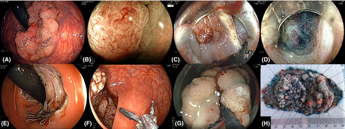
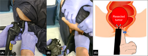
During endoscopic submucosal dissection (ESD) of large colorectal lesions, retrieval of specimens though the anal canal is sometimes challenging. We introduced a simple technique to solve this problem using a forefinger-compression method (FCM). A superficial tumor 80 mm in size showed nearly circumferential infiltration in the rectum (Fig. 1A). Magnified blue laser imaging showed an irregular surface and vessel patterns corresponding to Japanese NBI Expert Team (JNET) classification type 2B (Fig. 1B).1 We carried out ESD with the pocket-creation method using a scissor-shaped knife (ClutchCutter 3.5 mm; Fujifilm Medical Co., Tokyo, Japan), which could minimize perforation and was useful for stopping bleeding (Fig. 1C,D).2-4 The lesion was resected en bloc in 140 min (Fig. 1E). The endoscope was extracted from the patient's body to remove the hood and then inserted again. The dissected side of the resected tumor was then grasped using large forceps for removal (Fig. 1F,G). The patient was placed in the left-lateral position. The endoscopist's right hand was lubricated and then inserted into the anus parallel to the endoscope (Fig. 2A; Video S1). The body of the endoscope was held with two fingers of the left hand. While the scope was removed, along with the resected specimen, FCM was carried out. An endoscopist compressed the anal canal to the anterior side using the forefinger in order to distend the anal canal (Fig. 2B,C). A resected lesion (100 × 45 mm) was removed from the patient's body without any damage (Fig. 1H). Histopathological diagnosis was tubular adenocarcinoma with a negative margin. FCM is simple and requires no special devices, and large tumors can be retrieved from the anal canal without causing tissue damage. As a limitation, particularly large lesions might be better retrieved by the sliding overtube method or by the bowel preparation method if FCM fails.5

NaohisaYoshida received a research grant fromFujifilmCo. Other authors declare no conflict of interests for this article.
Details are in the caption following the image
A superficial tumor 80 mm in size resected by endoscopic submucosal dissection (ESD) and removed with a forfinger-compression method. (A) A superficial tumor (granular-type) 80 mm in size in the rectum. (B) Blue-laser imaging showed an irregular pattern. (C,D) The pocket-creation method was carried out with a grasping scissors knife (ClutchCutter; Fujifilm Medical Co., Tokyo, Japan). (E) The lesion was resected en bloc in 140 min. (F,G) The resected specimen was grasped using large forceps without a hood and retrieved by a forefinger-compression method. (H) The resected lesion (100 × 45 mm) was removed from the patient's body without any damage. Histopathological diagnosis was tubular adenocarcinoma with a negative margin.
Details are in the caption following the image
A forefinger-compression method for endoscopic retrieval of a large tumor. (A) The endoscopist's right hand was lubricated and inserted into the anus parallel to the endoscope. (B) An assistant pulled the patient's left hip upwards to distend the anus. The endoscope was removed along with the resected specimen. An endoscopist compressed the anal canal to the anterior side using the forefinger in order to distend the anal canal. (C) Illustration of the forefinger-compression method.

    The full text of this article hosted at iucr.org is unavailable due to technical difficulties.