Volume 29, Issue 5 pp. 626-633
Case Report

Usefulness of confocal laser endomicroscopy to diagnose ulcerative colitis-associated neoplasia

Naoki Ohmiya

Corresponding Author

Naoki Ohmiya

Department of Gastroenterology, Fujita Health University School of Medicine, Toyoake, Japan

Corresponding: Naoki Ohmiya, Department of Gastroenterology, Fujita Health University School of Medicine, 1-98 Kutsukake-cho, Toyoake, Aichi 470-1192, Japan. Email: [email protected]Search for more papers by this author
Noriyuki Horiguchi

Noriyuki Horiguchi

Department of Gastroenterology, Fujita Health University School of Medicine, Toyoake, Japan

Search for more papers by this author
Tomomitsu Tahara

Tomomitsu Tahara

Department of Gastroenterology, Fujita Health University School of Medicine, Toyoake, Japan

Search for more papers by this author
Dai Yoshida

Dai Yoshida

Department of Gastroenterology, Fujita Health University School of Medicine, Toyoake, Japan

Search for more papers by this author
Hyuga Yamada

Hyuga Yamada

Department of Gastroenterology, Fujita Health University School of Medicine, Toyoake, Japan

Search for more papers by this author
Mitsuo Nagasaka

Mitsuo Nagasaka

Department of Gastroenterology, Fujita Health University School of Medicine, Toyoake, Japan

Search for more papers by this author
Yoshihito Nakagawa

Yoshihito Nakagawa

Department of Gastroenterology, Fujita Health University School of Medicine, Toyoake, Japan

Search for more papers by this author
Tomoyuki Shibata

Tomoyuki Shibata

Department of Gastroenterology, Fujita Health University School of Medicine, Toyoake, Japan

Search for more papers by this author
Tetsuya Tsukamoto

Tetsuya Tsukamoto

Department of Diagnostic Pathology I, Fujita Health University School of Medicine, Toyoake, Japan

Search for more papers by this author
Makoto Kuroda

Makoto Kuroda

Department of Diagnostic Pathology I, Fujita Health University School of Medicine, Toyoake, Japan

Search for more papers by this author
First published: 28 February 2017
Citations: 10

Abstract

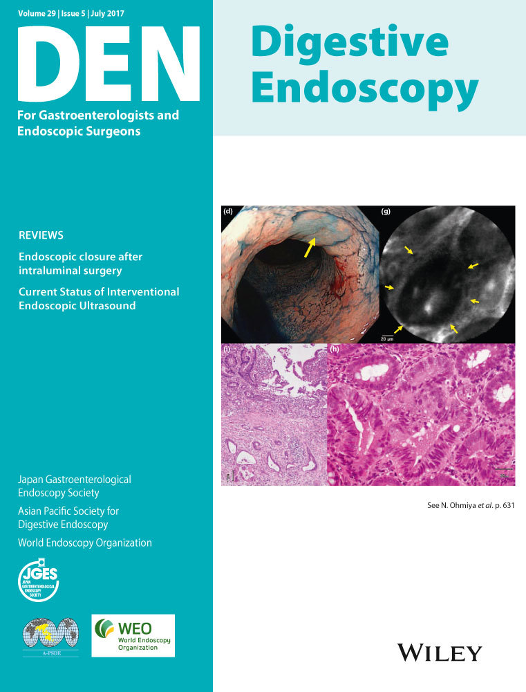
Chromoendoscopy, narrow-band imaging (NBI), and confocal laser endomicroscopy (CLE) have been introduced in ulcerative colitis (UC)-associated neoplasia surveillance. We aimed to determine the ability of CLE to differentiate among UC-associated neoplasia (differentiated type or undifferentiated type), sporadic adenoma, and circumscribed regenerative lesions. Of 665 patients with UC, we carried out probe-based CLE (pCLE) on 12 patients with suspected UC-associated neoplasia in addition to magnifying chromoendoscopy with crystal violet and NBI. We compared pCLE findings with pathological diagnoses. pCLE could differentiate UC-associated differentiated cancer from other pathologies such as solitary adenoma and non-neoplastic circumscribed regenerative lesions on the basis of back-to-back orientation of crypts (P = 0.048), and UC-associated undifferentiated cancer from other pathologies on the basis of dark trabecular architecture (P = 0.015). Sensitivity, specificity, and accuracy of combination of back-to-back orientation of crypts and dark trabecular architecture for carcinoma or dysplasia were 100%, 83%, and 92%, respectively. In vivo microscopic observation with pCLE was helpful to evaluate the suspected UC-associated neoplasia.

The full text of this article hosted at iucr.org is unavailable due to technical difficulties.