Volume 47, Issue 3 pp. 263-268
CASE REPORT

Metastatic pancreatic adenosquamous carcinoma to the scalp: A case report and review of the literature

Behzad Salari

Behzad Salari

Lauren V. Ackerman Laboratory of Surgical Pathology, School of Medicine, Washington University Medical Center, St. Louis, Missouri

Search for more papers by this author
David M. Sheinbein

David M. Sheinbein

Division of Dermatology, School of Medicine, Washington University Medical Center, St. Louis, Missouri

Search for more papers by this author
Ilana S. Rosman

Ilana S. Rosman

Lauren V. Ackerman Laboratory of Surgical Pathology, School of Medicine, Washington University Medical Center, St. Louis, Missouri

Division of Dermatology, School of Medicine, Washington University Medical Center, St. Louis, Missouri

Search for more papers by this author
Louis P. Dehner

Corresponding Author

Louis P. Dehner

Lauren V. Ackerman Laboratory of Surgical Pathology, School of Medicine, Washington University Medical Center, St. Louis, Missouri

Correspondence

Louis P. Dehner, Department of Pathology and Immunology, Washington University School of Medicine, 660 S Euclid Ave, Campus Box 8118, 3rd Floor, Rm 3421, Institute of Health Bldg. (IOH), St. Louis, MO 63110.

Email: [email protected]

Search for more papers by this author
First published: 06 October 2019
Citations: 3
Poster presentation at 55th ASDP Annual meeting, Chicago, IL, USA, 2018.

Abstract

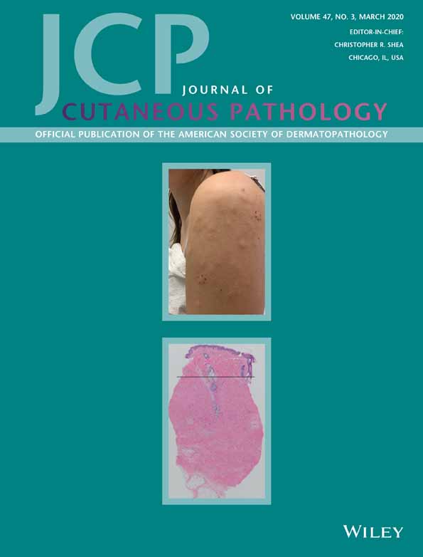
Metastatic carcinoma to the skin occurs in only a minority of patients with a visceral or internal malignancy, with breast, lung, and colorectum accounting for the majority of cases. We present the case of a 66-year-old man with a recent violaceous nodule of the left scalp (1.2 × 1.0 × 0.2 cm) that was a metastatic pancreatic adenosquamous carcinoma, representing a seemingly rare event. Two months prior, after complaining of right hip pain, an image revealed a right femoral lesion. A biopsy of that lesion showed moderately differentiated adenocarcinoma. Subsequent imaging showed a mass in the pancreatic tail and also markedly elevated serum tumor markers, CA 19-9 and carcinoembryonic antigen (5325 and 111.5 U/mL, respectively). Before the appearance of the scalp nodule, the patient received radiotherapy and was started on chemotherapy, which was continued after diagnosis and resection of the nodule. Subsequent metastases developed in the liver, lung and additional cutaneous lesions. He died 11 months after initial presentation with right hip pain. As this case shows, cutaneous metastases confer a poor prognosis, often with less than a year survival following their appearance.

CONFLICT OF INTEREST

None.

The full text of this article hosted at iucr.org is unavailable due to technical difficulties.