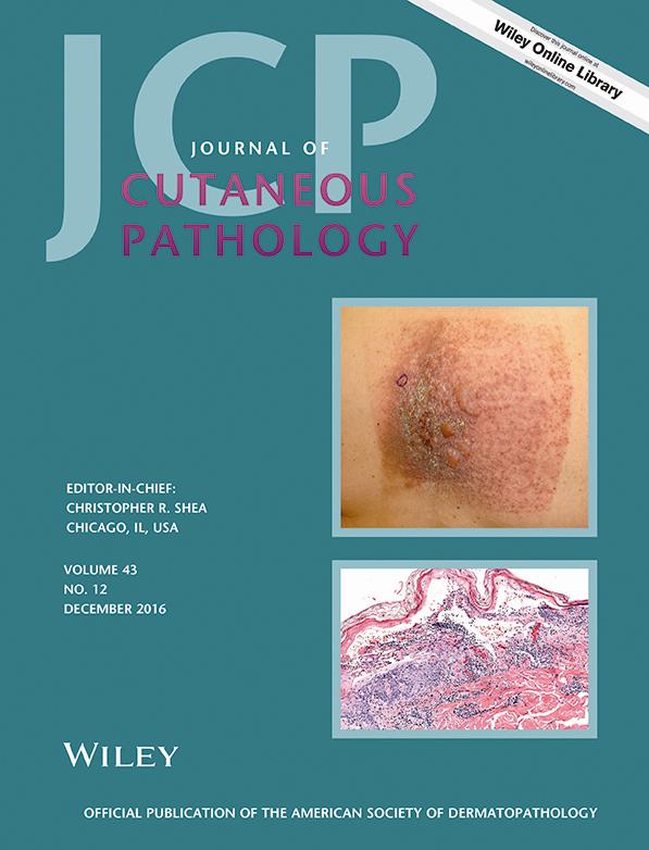
Cutaneous histoplasmosis with prominent parasitization of epidermal keratinocytes: report of a case
Hedieh H. Honarpisheh
Department of Pathology, University of Texas MD Anderson Cancer Center, Houston, TX, USA
Department of Pathology, Duke University School of Medicine, Durham, NC, USA
Search for more papers by this authorJonathan L. Curry
Department of Pathology, University of Texas MD Anderson Cancer Center, Houston, TX, USA
Department of Dermatology, University of Texas MD Anderson Cancer Center, Houston, TX, USA
Search for more papers by this authorKristen Richards
Department of Dermatology, University of Texas MD Anderson Cancer Center, Houston, TX, USA
Search for more papers by this authorPriyadharsini Nagarajan
Department of Pathology, University of Texas MD Anderson Cancer Center, Houston, TX, USA
Search for more papers by this authorPhyu P. Aung
Department of Pathology, University of Texas MD Anderson Cancer Center, Houston, TX, USA
Search for more papers by this authorCarlos A. Torres-Cabala
Department of Pathology, University of Texas MD Anderson Cancer Center, Houston, TX, USA
Department of Dermatology, University of Texas MD Anderson Cancer Center, Houston, TX, USA
Search for more papers by this authorDoina Ivan
Department of Pathology, University of Texas MD Anderson Cancer Center, Houston, TX, USA
Department of Dermatology, University of Texas MD Anderson Cancer Center, Houston, TX, USA
Search for more papers by this authorCarol R. Drucker
Department of Translational and Molecular Pathology, University of Texas MD Anderson Cancer Center, Houston, TX, USA
Search for more papers by this authorRichard Cartun
Department of Pathology, Hartford Hospital, Hartford, CT, USA
Search for more papers by this authorVictor G. Prieto
Department of Pathology, University of Texas MD Anderson Cancer Center, Houston, TX, USA
Department of Dermatology, University of Texas MD Anderson Cancer Center, Houston, TX, USA
Department of Translational and Molecular Pathology, University of Texas MD Anderson Cancer Center, Houston, TX, USA
Search for more papers by this authorCorresponding Author
Michael T. Tetzlaff
Department of Pathology, University of Texas MD Anderson Cancer Center, Houston, TX, USA
Department of Translational and Molecular Pathology, University of Texas MD Anderson Cancer Center, Houston, TX, USA
Michael T. Tetzlaff
Department of Pathology, University of Texas MD Anderson Cancer Center, 1515 Holcombe Blvd, Houston, TX 77030, USA
Tel: (713)-792-2585
Fax: (713)-745-0778
e-mail: [email protected]
Search for more papers by this authorHedieh H. Honarpisheh
Department of Pathology, University of Texas MD Anderson Cancer Center, Houston, TX, USA
Department of Pathology, Duke University School of Medicine, Durham, NC, USA
Search for more papers by this authorJonathan L. Curry
Department of Pathology, University of Texas MD Anderson Cancer Center, Houston, TX, USA
Department of Dermatology, University of Texas MD Anderson Cancer Center, Houston, TX, USA
Search for more papers by this authorKristen Richards
Department of Dermatology, University of Texas MD Anderson Cancer Center, Houston, TX, USA
Search for more papers by this authorPriyadharsini Nagarajan
Department of Pathology, University of Texas MD Anderson Cancer Center, Houston, TX, USA
Search for more papers by this authorPhyu P. Aung
Department of Pathology, University of Texas MD Anderson Cancer Center, Houston, TX, USA
Search for more papers by this authorCarlos A. Torres-Cabala
Department of Pathology, University of Texas MD Anderson Cancer Center, Houston, TX, USA
Department of Dermatology, University of Texas MD Anderson Cancer Center, Houston, TX, USA
Search for more papers by this authorDoina Ivan
Department of Pathology, University of Texas MD Anderson Cancer Center, Houston, TX, USA
Department of Dermatology, University of Texas MD Anderson Cancer Center, Houston, TX, USA
Search for more papers by this authorCarol R. Drucker
Department of Translational and Molecular Pathology, University of Texas MD Anderson Cancer Center, Houston, TX, USA
Search for more papers by this authorRichard Cartun
Department of Pathology, Hartford Hospital, Hartford, CT, USA
Search for more papers by this authorVictor G. Prieto
Department of Pathology, University of Texas MD Anderson Cancer Center, Houston, TX, USA
Department of Dermatology, University of Texas MD Anderson Cancer Center, Houston, TX, USA
Department of Translational and Molecular Pathology, University of Texas MD Anderson Cancer Center, Houston, TX, USA
Search for more papers by this authorCorresponding Author
Michael T. Tetzlaff
Department of Pathology, University of Texas MD Anderson Cancer Center, Houston, TX, USA
Department of Translational and Molecular Pathology, University of Texas MD Anderson Cancer Center, Houston, TX, USA
Michael T. Tetzlaff
Department of Pathology, University of Texas MD Anderson Cancer Center, 1515 Holcombe Blvd, Houston, TX 77030, USA
Tel: (713)-792-2585
Fax: (713)-745-0778
e-mail: [email protected]
Search for more papers by this authorAbstract
Disseminated histoplasmosis most commonly occurs in immunosuppressed individuals and involves the skin in approximately 6% of patients. Cutaneous histoplasmosis with an intraepithelial-predominant distribution has not been described.
A 47-year-old man was admitted to our institution with fever and vancomycin-resistant enterococcal bacteremia. He had been diagnosed with T-cell prolymphocytic leukemia 4 years earlier and had undergone matched-unrelated-donor stem cell transplant 2 years earlier; on admission, he had relapsed disease. His medical history was significant for disseminated histoplasmosis 6 months before admission, controlled with multiple antifungal regimens. During this final hospitalization, the patient developed multiple 2–5 mm erythematous papules, a hemorrhagic crust across the chest, shoulders, forearms, dorsal aspect of the fingers, abdomen and thighs. Skin biopsy revealed clusters of oval yeast forms mostly confined to the cytoplasm of keratinocytes and within the stratum corneum; scattered organisms were present in the underlying superficial dermis without any significant associated inflammatory infiltrate. Special stains and immunohistochemical studies confirmed these to be Histoplasma organisms.
We highlight this previously unrecognized pattern of cutaneous histoplasmosis to ensure its prompt recognition and appropriate antifungal therapy.
References
- 1Chu JH, Feudtner C, Heydon K, Walsh TJ, Zaoutis TE. Hospitalizations for endemic mycoses: a population-based national study. Clin Infect Dis 2006; 42: 822.
- 2Goodwin RA Jr, Des Prez RM. Pathogenesis and clinical spectrum of histoplasmosis. South Med J 1973; 66: 13.
- 3Chang P, Rodas C. Skin lesions in histoplasmosis. Clin Dermatol 2012; 30: 592.
- 4Tuon FF, Gomes V, Pagliari C, et al. Isolated lymphadenitis due to Histoplasma capsulatum diagnosed by fine-needle aspiration biopsy and immunohistochemistry. Rev Iberoam Micol 2008; 25: 50.
- 5Bauder B, Kubber-Heiss A, Steineck T, Kuttin ES, Kaufman L. Granulomatous skin lesions due to histoplasmosis in a badger (Meles meles) in Austria. Med Mycol 2000; 38: 249.
- 6Jensen HE, Schonheyder HC, Hotchi M, Kaufman L. Diagnosis of systemic mycoses by specific immunohistochemical tests. APMIS 1996; 104: 241.
- 7Knox KS, Hage CA. Histoplasmosis. Proc Am Thorac Soc 2010; 7: 169.
- 8Wheat LJ, Azar MM, Bahr NC, Spec A, Relich RF, Hage C. Histoplasmosis. Infect Dis Clin North Am 2016; 30: 207.
- 9Wheat LJ, Slama TG, Norton JA, et al. Risk factors for disseminated or fatal histoplasmosis. Analysis of a large urban outbreak. Ann Intern Med 1982; 96: 159.
- 10Kauffman CA, Israel KS, Smith JW, White AC, Schwarz J, Brooks GF. Histoplasmosis in immunosuppressed patients. Am J Med 1978; 64: 923.
- 11Hong JH, Stetsenko GY, Pottinger PS, George E. Cutaneous presentation of disseminated histoplasmosis as a solitary peri-anal ulcer. J Cutan Pathol. 2016; 43: 438.
- 12Eidbo J, Sanchez RL, Tschen JA, Ellner KM. Cutaneous manifestations of histoplasmosis in the acquired immune deficiency syndrome. Am J Surg Pathol 1993; 17: 110.
- 13Karimi K, Wheat LJ, Connolly P, et al. Differences in histoplasmosis in patients with acquired immunodeficiency syndrome in the United States and Brazil. J Infect Dis 2002; 186: 1655.
- 14P KR, Mosam A, Dlova NC, N BS, Aboobaker J, Singh SM. Disseminated cutaneous histoplasmosis in patients infected with human immunodeficiency virus. J Cutan Pathol 2002; 29: 215.
- 15Wheat LJ, Freifeld AG, Kleiman MB, et al. Clinical practice guidelines for the management of patients with histoplasmosis: 2007 update by the Infectious Diseases Society of America. Clin Infect Dis 2007; 45: 807.
- 16Theel ES, Harring JA, Dababneh AS, Rollins LO, Bestrom JE, Jespersen DJ. Reevaluation of commercial reagents for detection of Histoplasma capsulatum antigen in urine. J Clin Microbiol 2015; 53: 1198.
- 17de Vries HJ, Reedijk SH, Schallig HD. Cutaneous leishmaniasis: recent developments in diagnosis and management. Am J Clin Dermatol 2015; 16: 99.
- 18Kenner JR, Aronson NE, Bratthauer GL, et al. Immunohistochemistry to identify Leishmania parasites in fixed tissues. J Cutan Pathol 1999; 26: 130.
- 19Rand AJ, Buck AB, Love PB, Prose NS, Selim MA. Cutaneous acquired toxoplasmosis in a child: a case report and review of the literature. Am J Dermatopathol 2015; 37: 305.
- 20Meissner EG, Bennett JE, Qvarnstrom Y, et al. Disseminated microsporidiosis in an immunosuppressed patient. Emerg Infect Dis 2012; 18: 1155.
- 21Das A, Bar C, Patra A. Norwegian scabies: rare cause of erythroderma. Indian Dermatol Online J 2015; 6: 52.
- 22Ghosh SK, Bandyopadhyay D, Biswas SK, Mandal RK. Generalized scaling and redness in a 2-month-old boy. Crusted (Norwegian) scabies (CS). Pediatr Dermatol 2010; 27: 525.
- 23Mehta V, Balachandran C, Monga P, Rao R, Rao L. Images in clinical practice. Norwegian scabies presenting as erythroderma. Indian J Dermatol Venereol Leprol 2009; 75: 609.
- 24Tirado-Sanchez A, Bonifaz A, Montes de Oca-Sanchez G, Araiza-Santibanez J, Ponce-Olivera RM. Crusted scabies in HIV/AIDS infected patients. Report of 15 cases. Rev Med Inst Mex Seguro Soc 2016; 54: 397.
- 25Bilan P, Colin-Gorski AM, Chapelon E, Sigal ML, Mahe E. Crusted scabies induced by topical corticosteroids: a case report. Arch Pediatr 2015; 22: 1292.
- 26http://www.immy.com/wp-content/uploads/2014/08/HAG102-Histo-Ag-EIA-PI.pdf (accessed on 19 August 2016).


