Impact of intratumoural CD96 expression on clinical outcome and therapeutic benefit in gastric cancer
Chang Xu, Hanji Fang, Yun Gu and Kuan Yu contributed equally to this work.
Abstract
CD96 was identified as a novel immune checkpoint. However, the role of CD96 in the gastric cancer (GC) microenvironment remains fragmentary. This study aimed to probe the clinical significance of CD96 to predict prognosis and therapeutic responsiveness, and to reveal the immune contexture and genomic features correlated to CD96 in GC patients. We enrolled 496 tumor microarray specimens of GC patients from Zhongshan Hospital (ZSHS) for immunohistochemical analyses. Four hundred and twelve GC patients from the Cancer Genome Atlas (TCGA) and 61 GC patients treated with pembrolizumab from ERP107734 published in the European Nucleotide Archive (ENA) were gathered for further analysis of the association between CD96+ cell infiltration and immune contexture, molecular characteristics, and genomic features by CIBERSORT and gene set enrichment analysis. Clinical outcomes were analyzed by Kaplan–Meier curves, the Cox model, interaction testing, and receiver operating characteristic analysis. High CD96+ cell infiltration predicted poor prognosis and inferior survival benefits from fluorouracil-based adjuvant chemotherapy in the ZSHS cohort whereas superior therapeutic responsiveness to pembrolizumab was shown in the ENA cohort. CD96-enriched tumors showed an immunosuppressive tumor microenvironment featured by exhausted CD8+ T-cell infiltration in both the ZSHS and TCGA cohorts. Moreover, in silico analysis for the TCGA cohort revealed that several biomarker-targeted pathways displayed significantly elevated enrichment levels in the CD96 high subgroup. This study elucidated that CD96 might drive an immunosuppressive contexture with CD8+ T-cell exhaustion and represent an independent adverse prognosticator in GC. CD96 could potentially be a novel biomarker for precision medicine of adjuvant chemotherapy, immunotherapy, and targeted therapies in GC.
Abbreviations
-
- TCGA
-
- The Cancer Genome Atlas
-
- ENA
-
- European Nucleotide Archive
-
- GSEA
-
- Gene Set Enrichment Analysis
-
- ROC
-
- Receiver Operating Characteristic
-
- AUC
-
- area under curve
-
- ZSHS
-
- Zhongshan Hospital
-
- ACT
-
- adjuvant chemotherapy
-
- PD-1
-
- programmed cell death protein 1
-
- ICB
-
- Immune checkpoint blocking
-
- PD-L1
-
- programmed cell death ligand 1
-
- TILs
-
- tumor-infiltrating lymphocytes
-
- NK
-
- natural killer
-
- TIGIT
-
- T cell immunoreceptor with Ig and ITIM domains
-
- ITIM
-
- immunoreceptor tyrosine-based inhibitory motif
-
- ICOS
-
- inducible T cell costimulator
-
- mRNA
-
- messenger RNA
-
- HCC
-
- hepatocellular carcinoma
-
- FFPE
-
- formalin-fixed and paraffin-embedded
-
- TMA
-
- tissue microarray
-
- TNM
-
- tumor-node-metastasis
-
- OS
-
- overall survival
-
- DFS
-
- disease-free survival
-
- IHC
-
- immunohistochemistry
-
- GSVA
-
- Gene Set Variation Analysis
-
- GC
-
- gastric cancer
-
- CPS
-
- combined positive score
-
- MSI
-
- microsatellite instability
-
- IFN-γ
-
- interferon-γ
-
- LAG-3
-
- lymphocyte-activation gene 3
-
- ARID1A
-
- AT-Rich Interaction Domain 1A
-
- PIK3CA
-
- phosphatidylinositol-4,5-bisphosphate 3-kinase catalytic subunit alpha
-
- TP53
-
- tumor protein p53
-
- SYNE1
-
- spectrin repeat containing nuclear envelope protein 1
-
- EBV
-
- Epstein- Barr virus
-
- CIN
-
- chromosomal instable
-
- HER2
-
- human epidermal growth factor receptor 2
-
- FGFR2
-
- fibroblast growth factor receptor 2
-
- EGFR
-
- epidermal growth factor receptor
-
- MET
-
- MET proto-oncogene receptor tyrosine kinase
-
- HHR
-
- homologous recombination repair
-
- CLDN18.2
-
- claudin18.2
-
- TME
-
- tumor microenvironment
-
- PC
-
- pancreatic cancer
-
- CTLs
-
- cytotoxic T lymphocytes
-
- CTLA-4
-
- cytotoxic T-lymphocyte-associated protein 4
-
- PI3K
-
- phosphatidylinositol 3-kinase
-
- G/GEJ
-
- gastric and gastro-esophageal junction
1 INTRODUCTION
Gastric cancer (GC) is the fifth most commonly diagnosed cancer and the fourth leading cause of cancer death worldwide, with 1,089,103 new cases and 768,793 new deaths globally in 2020.1 To date, surgical intervention remains the optimal treatment and the only curative approach for patients in the early stages, while the addition of adjuvant chemotherapy (ACT) has brought survival benefits for advanced GC patients.2 Therefore, in addition to new treatment regimens, it is also necessary to find novel biomarkers that can predict survival outcomes and therapeutic responsiveness to facilitate the appropriate use of existing treatment measures.3
The previous clinical successes of immune checkpoint blockade marked the beginning of a new era in cancer immunotherapy. According to the results of the KEYNOTE-059 Phase II clinical trial, the programmed cell death protein 1 (PD-1) inhibitor pembrolizumab (Keytruda) has been approved for the third-line treatment of GC.4 Recently, nivolumab plus chemotherapy has been approved for the first-line treatment of patients with advanced GC by the US Food and Drug Administration on the basis of CheckMate 649.5 Nevertheless, various response rates to immune checkpoint blocking (ICB)6, 7 could be due to differences in individual patients, tumor types, biomarker selection, treatment regimens, and so on5. Additionally, multiple promising factors, including PD-L1/PD-1 level,8 number of tumor-infiltrating lymphocytes (TILs),9 interferon signaling,10 mutational burden,10, 11 mismatch repair deficiency,11, 12 and intestinal microbiota,13 are still unable to yield accurate prediction for survival and therapeutic responsiveness. Of note, the prognostic values of PD-L1 expression have been debated for the discrepancy that some patients with PD-L1 positive expression cannot gain clinical benefit from PD-1 blockade.6 Hence, a pressing unmet need is to seek precise biomarkers of response to immunotherapeutic agents for identifying which patients are more sensitive to ICB and may derive better clinical benefits from specific treatments.14
CD96 (TACTILE) is a member of the extended nectin/NECL family.15 Expression of CD96 is confined to immune cells, primarily presented on T cells, NK cells, and NKT cells.15 Akin to TIGIT and CD226 (DNAM), the main ligand CD96 binding to is CD155 (necl-5, PVR) associated with tumor proliferation and migration.16 Human CD96 cytoplasmic domains include a short basic/proline rich motif and a single ITIM-like domain indicating potential inhibitory function, and also contain a YXXM motif which can be found in the activating receptors CD28 and ICOS,17 implying an activating receptor in certain contexts.18 Recent studies have revealed that CD96 functions as an intrinsic inhibitory receptor on CD8+ T cells and NK cells, and that anti-CD96 enhances antitumor immunity19; however, other studies have shown the opposite, i.e. that CD96 acts as a co-stimulatory receptor to enhance CD8+ T-cell activation and effector responses.20
Two studies reported contrasting observations of the correlation between CD96 expression and clinical outcomes in cancer patients.21 Peng et al. reported that the decrease in the frequency of CD96+ and CD226+ NK cells was correlated with lymph-node metastasis of pancreatic cancer.22 In contrast, Sun et al. found that elevated expression of CD96 predicted poor clinical outcomes in hepatocellular carcinoma (HCC) patients.23 Hence, the role of CD96 in GC still needs to be clarified.
The current study aimed to discuss the clinical significance and functional characteristics of CD96, and summarize its therapeutic potential and genomic features in GC.
2 MATERIALS AND METHODS
2.1 Study population
The research consisted of three independent patient cohorts. Cohort 1 included 496 patients from Zhongshan Hospital (ZSHS), Fudan University (Shanghai, China), the ZSHS cohort, with 59 patients excluded because of missing data, dot loss or because they suffered from metastatic diseases. The remaining 437 patients underwent radical gastrectomy and standard D2 lymphadenectomy between August 2007 and December 2008. All tissue samples from the ZSHS cohort were formalin-fixed and paraffin-embedded (FFPE). Patient clinicopathological characteristics, including age, gender, tumor size, tumor grade, Lauren's classification, tumor-node-metastasis (TNM) stage, and application of fluorouracil-based ACT were collected retrospectively. The T, N classification and TNM stage were assessed according to the 2010 International Union Against Cancer TNM staging system. Postoperative routine fluorouracil-based ACT was primarily given to patients with TNM II/III advanced tumors. No radiotherapy was administered to enrolled patients in ZSHS cohort. The endpoints of interest were overall survival (OS) and disease-free survival (DFS), computed from the date of gastrectomy to the date of death or disease recurrence, or the last follow-up. Subsequently, cohort 1 was randomly assigned into two independent data sets (discovery set, n = 219; validation set, n = 218). Our research had approval from the Clinical Research Ethics Committee of Zhongshan Hospital, Fudan University. All enrolled patients were informed of the usage of resected gastric tissue samples. Written consent was acquired from each patient. Cohort 2 was derived from the Cancer Genome Atlas (TCGA) with 412 GC patients in all, but 37 were excluded because of missing data or because they suffered from metastatic diseases. All patient characteristics and mRNA data were downloaded from https://xenabrowser.net/datapages/ on September 9, 2020. Cohort 3 was derived from ERP107734 published on the European Nucleotide Archive (ENA) with 61 patients treated with pembrolizumab. Due to the integrity of the mRNA expression matrix uploaded, we included 39 patients with certain characteristics in our research. An illustration of patients enrolled and study design is presented in Figure S1.
2.2 Immunohistochemistry
Immunohistochemistry (IHC) was applied to detect the expression of CD96 and the infiltration of immune cells on each TMA slide. The TMAs were constructed by Shanghai Outdo Biotech Co, Ltd. The protocol details of TMA construction and IHC staining have been described elsewhere.24, 25 Blinded to the clinical data, two pathologists evaluated the IHC score of CD96 independently according to the number of stained cells in three randomly selected 200× fields of view. The mean score of their evaluation was adopted. The cut-off value for the classification of high CD96 and low CD96 subgroups was the median value. On the basis of TCGA data, CIBERSORT was constructed to calculate the relative proportion of 22 immune cell types recognized as LM22. In addition, we selected CD8+ T cells, Treg cells (Foxp3+), and M2 macrophages (CD163+) as significant immune contexture in GC with high CD96+ cell infiltration. The associated antibodies are listed in Table S1.
2.3 Statistical analysis
SPSS 21.0 (SPSS Inc.) was applied for statistical analysis. The cut-off value for CD96 expression was the median value. Pearson's χ2 test and Fisher's exact test were applied for categorical variables, and Student's t-test, the Mann Whitney U test, and one-way ANOVA test were applied for continuous variables. Kaplan–Meier curves, the log-rank test, and univariate and multivariate analyses were applied for survival outcomes. Receiver operating characteristic (ROC) analysis was used to compare the accuracy of the prediction of clinical outcome by the parameters. All analyses mentioned above were visualized by R (4.1.1). All statistical analyses were two-sided and p < 0.05 was regarded as statistically significant.
2.4 In silico analysis
The gene expression profile data in the TCGA cohort were used to quantify the infiltration of immune cells in tumor tissues by single-sample gene set enrichment analysis (ssGSEA)26 in the R Bioconductor package Gene Set Variation Analysis (GSVA), and the infiltration of immune cells was obtained. The ssGSEA algorithm is a rank-based method that computes an enrichment score representing the degree to which genes in a particular gene set are coordinately up/downregulated in a single sample. The source and details of the gene signature are listed in Table S2.
3 RESULTS
3.1 CD96+ cells are enriched in GC tissues and associated with tumor progression
To discover the clinical roles of CD96 in the tumor progression of GC, clinical information for all GC patients enrolled in the ZSHS cohort was retrospectively collected and IHC staining of CD96 in gastric tissues is shown in Figure S2A. Interestingly, the intratumoral tissues showed significantly higher infiltration of CD96+ cells compared with peritumoral tissues (p < 0.001; Figure S2B), therefore the role of intratumoral CD96+ cells was the main focus of our following study. The clinicopathological characteristics of GC patients are listed in Table 1. Notably, the infiltration of CD96+ cells was significantly associated with tumor stages in GC and elevated in TNM stage III tumors, indicating that CD96+ cells might be correlated with the progression of GC (Figure S2C). Taken together, these findings suggest that CD96+ cells are enriched in GC tissues and associated with tumor progression.
| Characteristics | Patients | Discovery set | Validation set | Combined set | |||||||
|---|---|---|---|---|---|---|---|---|---|---|---|
| NO. | % | low CD96 | high CD96 | p value | low CD96 | high CD96 | p value | low CD96 | high CD96 | p value | |
| All patients | 437 | 113 | 106 | 113 | 105 | 226 | 211 | ||||
| Age (years) | |||||||||||
| Median (IQR) | 60 (53–69) | 58 (52–69) | 64 (54–73) | 59 (52–66) | 61 (54–70) | 59 (53–69) | 61 (53–69) | ||||
| Gender | |||||||||||
| Male | 309 | 70.7 | 83 | 73 | 0.460 | 77 | 76 | 0.554 | 160 | 149 | 0.967 |
| Female | 128 | 29.3 | 30 | 33 | 36 | 29 | 66 | 62 | |||
| Localization | |||||||||||
| Proximal | 109 | 24.9 | 29 | 26 | 0.837 | 25 | 29 | 0.403 | 54 | 55 | 0.804 |
| Middle | 64 | 14.6 | 12 | 14 | 23 | 15 | 35 | 29 | |||
| Distal | 264 | 60.4 | 72 | 66 | 65 | 61 | 137 | 127 | |||
| Tumor size (cm) | |||||||||||
| Median (IQR) | 3.80 (2–5) | 3.00 (2–5) | 3.75 (2–5) | 3.50 (2–5) | 3.50 (2–5) | 3.78 (2–5) | 3.82 (2–5) | ||||
| Differentiation | |||||||||||
| Well + moderately | 121 | 27.7 | 32 | 34 | 0.559 | 31 | 24 | 0.533 | 63 | 58 | 0.928 |
| Poorly | 316 | 72.3 | 81 | 72 | 82 | 81 | 163 | 153 | |||
| Lauren classification | |||||||||||
| Intestinal type | 273 | 62.5 | 72 | 69 | 0.888 | 71 | 61 | 0.491 | 143 | 130 | 0.72 |
| Diffuse type | 164 | 37.5 | 41 | 37 | 42 | 44 | 83 | 81 | |||
| T stage | |||||||||||
| T1 | 80 | 18.3 | 17 | 17 | 0.873 | 25 | 21 | 0.002 | 42 | 38 | 0.427 |
| T2 | 63 | 14.4 | 16 | 14 | 18 | 15 | 34 | 29 | |||
| T3 | 82 | 18.8 | 26 | 20 | 48 | 14 | 48 | 34 | |||
| T4 | 212 | 48.5 | 54 | 55 | 48 | 55 | 102 | 110 | |||
| N stage | |||||||||||
| N0 | 170 | 38.9 | 43 | 40 | 0.005 | 52 | 35 | 0.029 | 95 | 75 | 0.007 |
| N1 | 50 | 11.4 | 18 | 3 | 13 | 16 | 31 | 19 | |||
| N2 | 83 | 19.0 | 21 | 19 | 26 | 17 | 47 | 36 | |||
| N3 | 134 | 30.7 | 31 | 44 | 22 | 37 | 53 | 81 | |||
| TNM stage | |||||||||||
| I | 107 | 24.5 | 24 | 23 | 0.331 | 35 | 25 | 0.070 | 59 | 48 | 0.036 |
| II | 106 | 24.3 | 33 | 22 | 31 | 20 | 64 | 42 | |||
| III | 224 | 51.3 | 56 | 61 | 47 | 60 | 103 | 121 | |||
- Note: p < 0.05 marked in bold font shows statistical significance.
- After surgery, 5-fluorouracil-based chemotherapy was primarily given to patients with advanced tumors (stage II/III). Patients with adjuvant chemotherapy received at least one cycle of 5-fluorouracil-based chemotherapy. No radiotherapy was administered to anyone of the patients recruited.
- Abbreviations: IQR, interquartile range; NA, not available; TNM, tumor-node-metastasis.
3.2 High infiltration of CD96+ cells predicts poor prognosis in GC patients
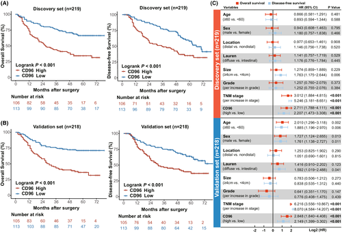
To investigate the prognostic significance of CD96 in GC, clinical outcomes of GC patients with high CD96+ cells infiltration were compared with those of patients with low CD96+ cell infiltration. In both the discovery and validation sets, the high CD96+ cell subgroup patients had significantly inferior OS and DFS (p < 0.001, p < 0.001, p < 0.001, and p < 0.001; Figure 1A,B). Since CD96+ cell infiltration appears to be a potential prognosticator for GC, multivariate analysis based on clinicopathological characteristics was constructed to demonstrate whether CD96+ cells could serve as a potential independent prognostic factor for the survival outcomes. Notably, CD96+ cell infiltration was identified as an independent adverse prognostic factor for OS and DFS in both the discovery (hazard ratio [HR] 2.711, 95% confidence interval [CI] 1.788–4.111, p < 0.001 and HR 2.207, 95% CI 1.473–3.308, p < 0.001; Figure 1C) and validation (HR 2.848, 95% CI 1.841–4.408, p < 0.001 and HR 2.149, 95% CI 1.399–3.302, p < 0.001; Figure 1C) sets. Hence, high infiltration of CD96+ cells could predict inferior prognosis in GC patients.
3.3 CD96 indicates therapeutic responsiveness in GC patients
We investigated whether CD96+ cell infiltration could predict therapeutic responsiveness in GC patients. Since we found that receiving fluorouracil-based postoperative adjuvant chemotherapy could indicate superior overall survival for TNM stage II/III patients (p < 0.001; Figure 2A), we sought to explore the interaction between different CD96+ cell infiltration subgroups and the therapeutic responsiveness to ACT in stage II/III patients. Interestingly, ACT could lead to significantly better OS in low CD96+ cell subgroup patients, contrary to high CD96+ cell subgroup patients(p < 0.001 and p = 0.340, respectively; Figure 2A). This might indicate that low CD96+ cell subgroup patients received better survival benefits after ACT (p for interaction = 0.001; Figure 2A).
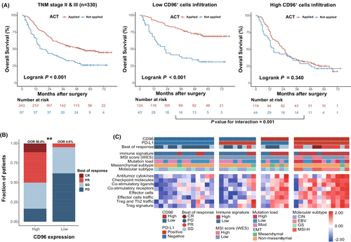
In addition to the ZSHS cohort, we enrolled a cohort of GC patients treated with pembrolizumab published as ERP107734 on the ENA to examine the potential value of CD96 in indicating immunotherapeutic responsiveness to PD-1 inhibition in GC. The baseline characteristics of GC patients are listed in Table S3.27 Notably, response rates were significantly associated with CD96 mRNA expression (p = 0.002; Figure 2B), with objective response rates (ORRs) of 50.0% and 4.8% in the high CD96 subgroup and the low CD96 subgroup, respectively. Since the PD-L1 combined positive score (CPS) has been proven to be an indicator of response to pembrolizumab and is widely used as a clinical guideline,28 we conducted a combined analysis of PD-L1 CPS and CD96 expression to more accurately identify patients who respond well to pembrolizumab. Interestingly, PD-L1-positive CD96 high patients showed the best responsiveness to pembrolizumab (Figure 2C). Additionally, several predictive indicators and characteristics of responsiveness to pembrolizumab in terms of immune signature, microsatellite instability, mutational load, mesenchymal subtype, and molecular subtype were grouped. Notably, the patients with better responsiveness showed an enrichment of biomarkers, including antitumor cytokines, checkpoint molecules, co-stimulatory ligands, co-stimulatory receptors, effector cells and effector cells traffic, Treg and Th2 traffic, and Treg signature, indicating a high-infiltration immune microenvironment (Figure 2C).
Accumulatively, our results suggest that CD96 could serve as a potential predictive biomarker for ACT and ICB treatment, as well as a promising therapeutic target for individualized precision medicine treatment of GC patients.
3.4 CD96 is associated with an immunosuppressive contexture characterized by an exhausted CD8+ T-cell phenotype in GC
Given that CD96 is regarded as an immune checkpoint,18 we next focused on the potential impact of CD96 on immune contexture. CIBERSORT was performed to investigate the association between CD96 mRNA expression and the typical immune cells infiltration in the TCGA cohort. Notably, high expression of CD96 mRNA expression was associated with high infiltration of memory B cells, CD8+ T cells, activated memory CD4+ T cells, follicular helper T cells, and M1 macrophages while low expression of CD96 mRNA was associated with resting memory CD4+ T cells, M0 macrophages, resting mast cells, and neutrophils (Figure 3A). To depict the functional states of immune cells more precisely, we found that CD96 mRNA expression was significantly associated with upregulation of both immune checkpoints and effector molecule mRNA level, which might indicate an inflamed but exhausted immune microenvironment (Figure 3A). To validate the results derived from the TCGA database, we further conducted IHC staining of immune cells, immune checkpoints, and effector molecules on the TMAs from the ZSHS cohort. Remarkably, high infiltration of CD96+ cells was significantly associated with high infiltration of CD8+ T cells, regulatory T cells, and M2 macrophages, and upregulation of interferon-γ, PD-L1, and lymphocyte-activation gene 3 IHC score (p = 0.006, p < 0.001, p < 0.001, p = 0.001, p < 0.001, p = 0.001; Figure 3B), which is consistent with the deduction that CD96 could potentially facilitate the immunosuppressive contexture. We further explored the association of these immune markers with the survival of GC patients and found that only PD-L1 expression significantly correlated with the poor prognosis in the ZSHS cohort (HR 1.619, 95% CI 1.213–2.162, p < 0.001; Figure S3). However, ROC analysis indicated that CD96 was associated with significantly higher prognostic accuracy compared with PD-L1 (CD96: AUC = 0.686; PD-L1: AUC = 0.583; Figure S4).
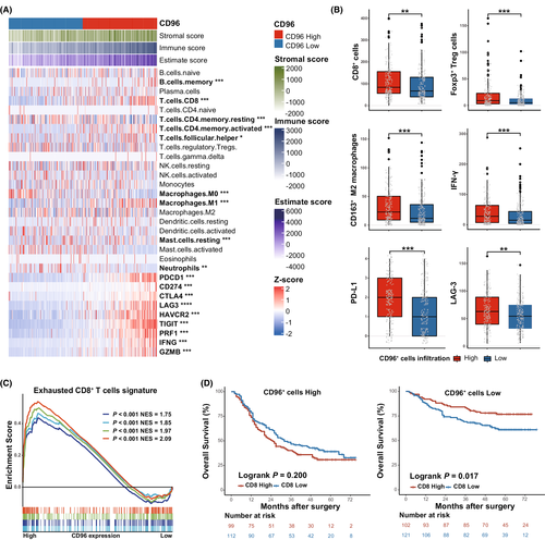
High infiltration of CD8+ T cells was regarded as a positive prognosticator in most cancer types.29 However, recent studies have indicated that the induction of T-cell dysfunction in tumors with high infiltration of CD8+ T cells could correlate with tumor immune evasion.30 To verify if CD8+ T cells displayed an exhausted phenotype, we further performed GSEA analysis. Notably, the exhausted CD8+ T cell signatures were significantly enriched in the high CD96 subgroup (Figure 3C). Furthermore, we combined CD96 and CD8 levels for survival analysis to discover the clinical significance of CD96-associated cytotoxic T-cell dysfunction. The analysis demonstrated that high CD8+ T cell infiltration could only identify prolonged OS in the high CD96+ cell subgroup, rather than the low CD96+ cell subgroup (Figure 3D). These results indicate that high CD96 expression is associated with immunosuppressive contexture characterized by exhausted a CD8+ T-cell phenotype, leading to poor prognosis.
3.5 Features of gene mutations and molecular subtypes based on CD96 expression in GC
Considering that progressive accumulation of gene alterations could facilitate tumorigenesis,31 we intended to profile detailed associations between CD96 and genomic features by delineating a holistic gene alteration landscape of GC from the TCGA cohort (Figure 4A). As a result, we identified the top 20 variant mutated genes, and AT-rich interaction domain 1A (ARID1A) and phosphatidylinositol-4,5-bisphosphate 3-kinase catalytic subunit alpha (PIK3CA) gene alterations were frequently found in the high CD96 subgroup while tumor protein p53 (TP53) and spectrin repeat containing nuclear envelope protein 1 (SYNE1) were in the low CD96 subgroup (p = 0.003, p < 0.001, p < 0.001, p = 0.003, p = 0.029; Figure 4B).
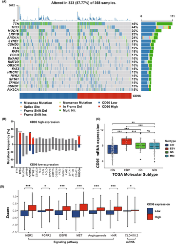
Further research has revealed that molecular subtypes of GC have provided novel perspectives for patient stratification and individualized therapy.32, 33 Notably, CD96 mRNA expression was highest in Epstein–Barr virus (EBV)-positive subtype GC and lowest in chromosomal instable (CIN) subtype GC in the TCGA cohort (Figure 4C).
Recently, molecular targeted therapies, which target specific molecular deviations in signal transduction pathways and/or cancer proteins, including trastuzumab, bemarituzumab, savolitinib and zolbetuximab, have attracted growing attention in GC precision medicine. Thus, we wondered whether there was an association between CD96 mRNA expression and activation of the corresponding signaling pathways. Known as promising therapeutic targets in GC, human epidermal growth factor receptor 2, fibroblast growth factor receptor 2, epidermal growth factor receptor, MET, angiogenesis, homologous recombination repair signaling score, and CLDN18.2 mRNA expression were all elevated in the high CD96 subgroup (p < 0.001, p = 0.040, p < 0.001, p < 0.001, p < 0.001, p < 0.001, p = 0.018; Figure 4D). Taken together, these results suggest that CD96 is associated with unique genomic characteristics and specific molecular subtypes, and might guide the application of targeted therapies in GC.
4 DISCUSSION
In this research, we analyzed 851 GC samples and summarized the clinical significance of CD96 in GC. Remarkably, our study was the first to identify CD96 as a useful prognostic factor in GC. We also revealed the predictive value of CD96 in response to ACT and pembrolizumab immunotherapy. High CD96 expression or CD96+ cell infiltration was associated with an immunosuppressive tumor microenvironment (TME) characterized by the exhausted T-cell phenotype. These findings highlighted the critical role of CD96 as a feasible stratification biomarker and as a strong candidate for immunotherapy target in GC.
Recent research has reported observations of distinct correlations between CD96 expression and clinical outcomes in patients with different types of cancer.21 In our study, we found that patients with high infiltration of CD96+ cells had poorer prognosis in GC, consistent in HCC but reversed in pancreatic cancer.22, 23 The reason for these two opposite outcomes may be that CD96 acts as a stimulatory receptor in GC and HCC but as an inhibitory receptor in pancreatic cancer. However, both of these receptors can eventually cause dysfunction of cytotoxic T lymphocytes (CTLs). To confirm the role of CD96, it is necessary to further study the inhibitory function and regulatory mechanism of CD96 on CTLs in GC. Additionally, fluorouracil-based ACT is considered as a first-line adjuvant therapy regimen for stage II/III patients.34, 35 Remarkably, low infiltration of CD96+ cells showed superior survival benefits after fluorouracil-based ACT in GC while high infiltration of CD96+ cells was associated with an exhausted T-cell phenotype and failed to show survival benefits after ACT, which was useful for appropriate treatment choices. The reason for this phenomenon was unclear, but we propose a hypothesis. The chemotherapy drugs may kill tumor cells by an immunogenic cell death pathway, which induces robust antitumor immune responses and has the potential to greatly increase the efficacy of chemotherapy.36 However, the enhanced antitumor effect may be partially offset by exhausted CTLs, which are possibly associated with high infiltration of CD96+ cells. Our findings demonstrated that CD96 expression on TILs and its correlation with prognostic assessment is a crucial indicator for the role of CD96 in tumor controls and precision medicine.
Immunotherapies, especially ICB (anti-PD-1/PD-L137 or anti-cytotoxic T lymphocyte associated protein 438), that aim to reverse immune cell exhaustion have demonstrated profound clinical benefit in several cancers; however, some patients with clinical and molecular discordance cannot respond to ICB as predicted. As PD-L1-positive patients were predicted to respond to pembrolizumab but not all were responders,6 it is important to identify additional biomarkers for patient selection. Remarkably, CD96 low expression could eliminate a subset of nonresponders from PD-L1-positive patients. It is of clinical significance that accurate screening for patients and rational use of PD-1 inhibitor via CD96 stratification can improve drug efficacy and reduce toxic side effects. Consequently, our findings suggest that CD96 has the potential to be a companion immunotherapeutic biomarker for optimized efficacy and classification in GC.
CTLs in the TME were considered a pivotal prognosticator for most cancer types as they were required to fight tumorigenesis and tumor progression as frontline cells.39, 40 However, due to persistent antigen exposure and/or inflammation, CTLs change into an exhausted state with suboptimal functions failing to eradicate tumors.30 Interestingly, our study revealed that the TME of high CD96 subgroups exhibited high expression of both effector and checkpoint molecules, consistent with the findings in the immunotherapy cohort. This TME combining the dual characteristics of effect and exhaustion state, which might be associated with dual functions of the huCD96 cytoplasmic domains, indicates that these exhausted CTLs might not be inert or terminally exhausted.30, 41 This finding may also partly explain why high CD96 patients could experience durable and efficient responses on ICB therapies.
Since we have found that CD96 might be involved in the tumor progression of GC, we subsequently attempted to describe the genomic profiling of alterations associated with CD96 in GC, which might be the driving forces causing genetic intratumoral heterogeneities and changes in cellular states and the TME, and finally impact the prognostic value of CD96.42, 43 In our study, genetic alterations of ARID1A and PIK3CA were frequently found in the high CD96 subgroup, correlated with tumor progression and tumorigenesis.44-46 As the molecular classification of GC proposed by TCGA was useful to reveal tumor biological properties and help the selection of targeted therapies for individual patients,47, 48 we found CD96 mRNA expression was elevated in EBV-positive GC characterized by frequent PIK3CA and ARID1A mutations.32 It is possible that EBV-positive patients are likely to gain clinical benefits from phosphatidylinositol 3-kinase inhibitors.45, 49 Furthermore, we compared the enrichment level of biomarker-targeted pathways between high and low CD96 levels.47 Intriguingly, those pathways of biomarker-targeted therapies for advanced-stage gastric and gastro-esophageal junction cancers might be activated in the high CD96 subgroup, implying that CD96 has the potential to be a companion biomarker of sensitive patient selection.
On the other hand, we realize that the underlying mechanism of CD96-associated tumorigenesis remains to be explored and further investigation is needed in subsequent studies. Hence, we advocate further confirmation of our findings within the framework of larger, multicentered, and randomized clinical trials to validate the clinical significance of CD96 in GC.
In conclusion, our study demonstrates that CD96 is an independent prognosticator. It is also a biomarker of precise patient selection for fluorouracil-based ACT and immunotherapies (pembrolizumab) in GC. The infiltration of CTLs exhibited an exhausted phenotype in high CD96+ cell infiltration tumors. Thus, CD96 may be a promising biomarker for individual-based treatment in GC.
AUTHOR CONTRIBUTIONS
C. Xu, H. Fang, Y. Gu, and K. Yu: acquisition, analysis and interpretation of data, statistical analysis, and drafting of the manuscript. J. Wang, C. Lin, H. Zhang, H. Li, and H. He: technical and material support. H. Liu and R. Li: study concept and design, analysis and interpretation of data, drafting of the manuscript, obtained funding, and study supervision. All authors read and approved the final manuscript.
ACKNOWLEDGMENTS
We thank Dr. Lingli Chen (Department of Pathology, Zhongshan Hospital, Fudan University, Shanghai, China) and Dr. Yunyi Kong (Department of Pathology, Shanghai Cancer Center, Fudan University, Shanghai, China) for their excellent pathological technology help.
FUNDING INFORMATION
This study was funded by grants from the National Natural Science Foundation of China (81,871,930, 81,902,402, 81,902,901, 81,972,219, 82,003,019, 82,103,313), the Shanghai Rising-Star Program (22QA1401700), and the Shanghai Sailing Program (18YF1404600, 19YF1407500, 21YF1407600). All the sponsors have no roles in the study design or the collection, analysis, and interpretation of data.
DISCLOSURE
The authors have no conflicts of interest.
ETHICS STATEMENTS
Approval of the research protocol by an Institutional Reviewer Board: The study was approved by the institutional review board and ethics committee of Zhongshan Hospital, Fudan Universtiy.
Informed Consent: Written informed consent was obtained from each patient.
Registry and the Registration No. of the study/trial: N/A.
Animal Studies: N/A.


