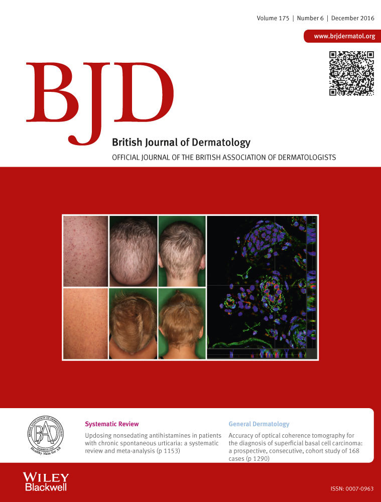
Accuracy of optical coherence tomography for the diagnosis of superficial basal cell carcinoma: a prospective, consecutive, cohort study of 168 cases
Summary
Background
Superficial basal cell carcinoma (sBCC) can be safely treated topically. Potentially noninvasive imaging techniques, such as optical coherence tomography (OCT), may be useful to diagnose and manage patients with sBCC and obviate the need for biopsy.
Objectives
To evaluate in OCT (i) the sensitivity and specificity for sBCC diagnosis, (ii) the accuracy in determining BCC depth and (iii) the role in management of sBCC mimickers.
Methods
A prospective, consecutive cohort of lesions for which sBCC was considered in the differential diagnosis. These lesions underwent clinical, dermoscopic and OCT assessment. Diagnosis and its confidence were recorded for each modality and were correlated with the histopathological diagnosis (punch biopsy). Interpretation of the OCT images and assessment of individual features were performed blinded to the biopsy results.
Results
In total, 168 lesions were recruited: 52% were sBCC, 26% were other BCC variants and the remaining lesions were actinic keratosis, squamous cell carcinoma in situ, other benign inflammatory processes and two other malignant tumours. The sensitivity and specificity of OCT for diagnosis of sBCC were 0·87 and 0·80, respectively. There was excellent correlation between OCT and biopsy for tumour depth amongst tumours ≤ 0·4 mm (Pearson correlation r = 0·86, P < 0·001), but the correlation was less as depth increased (Pearson correlation r = 0·71, P < 0·001 for all tumours < 1·0 mm).
Conclusions
OCT has good diagnostic accuracy for diagnosing sBCC and measuring depth in tumours ≤ 0·4 mm. Potentially OCT can reduce the need for biopsy in clinically suspected sBCCs. However, careful follow-up is required in such cases as there is a small risk (5%) of misdiagnosis.