Scanning electron microscopy analysis of the developmental, inter- and intraspecific variation in the otoliths of Anguilla anguilla (Linnaeus, 1758) collected from Bafa Lake in Western Anatolia, Türkiye
Abstract
This study aimed to identify unique traits, grounded in a thorough description of the sagitta otolith of Anguilla anguilla, to characterize its ontogenetic stages between juvenile and preadult individuals. Twenty-one characteristics were designated to investigate the ontogenetic (when comparing the otolith characteristics of fishes of different sizes, interspecific variation (comparing the otolith characteristics of the studied species with those of related species), and intraspecific variation (comparing the otolith characteristics of the studied species with those of the same species from different localities, i.e. geographical variation). This investigation concluded that the sagittal otoliths exhibited ontogenetic disparities based on the shapes recorded by different age groups. Otoliths of the young individuals belonging to fish total length groups GI and GII have shown differences in shape from those of the older individuals in the other seven length groups surveyed. An assessment of previously available otolith descriptions of A. anguilla collected from other areas revealed differences between species across larger geographic areas, making some features unsuitable for further investigation. However, other otolith traits were found to be reliable across different length groups and could be considered species-specific features for identification. These traits include otolith width, otolith thickness, the shape of the mesial and lateral surfaces, the shape of the sulcus acusticus, the presence or absence of ostio-caudal differentiation, and the shape, size, and thickness of the rostrum.
1 INTRODUCTION
1.1 General
The European eel (Anguilla anguilla Linnaeus, 1758) is a catadromous fish species with a complicated life history. It is found in many countries along the coasts of North Africa and Europe (Moriarty & Dekker, 1997). This eel species inhabits the Sargasso Sea, where the leptocephali larvae hatch. After drifting passively for about 5000 to 6000 km of passive drift driven by ocean currents, the larvae cross the Atlantic Ocean to reach the continental slopes of Europe and Africa (Zydlewski & Wilkie, 2012). As they mature, yellow eels undergo a transformation into migratory silver eels, which return to the sea to reproduce and eventually die (Tesch, 2003). The concept of European eel panmixia is typically recognized and has been considered for a long time (Bianchini et al., 2009; Wirth & Bernatchez, 2003). The choice of the species' spawning areas is based on early data obtained in 1923. In the investigation at hand, however, more emphasis has been placed on migration timings and routes, feeding characteristics, habitats, and bio-ecological features (Durif et al., 2020; Jensen et al., 2018). All these studies contribute synergistically to the conservation of the species.
Anguilla anguilla, which has lived in the waters of the European continent for at least 800,000 years (Kettle et al., 2008), is currently facing many impacts, and there has been a significant decline in production over the years. The considerable reduction observed in natural stocks has attracted international attention, resulting in the European eel being listed as a critically endangered species under the protection of the IUCN (International Union for Conservation of Nature) Red List (Pike et al., 2020). Consequently, international trade is also conducted under the scope of CITES (Convention on International Trade in Endangered Species of Wild Fauna and Flora) (Diekmann et al., 2019). Under CITES, the export of European eel, which is guided by quotas, was announced by the Ministry of Agriculture and Forestry in Türkiye with an export quota of 100 tonnes for the year 2020. In this regard, beyond its ecological significance, the survival of the species is also crucial from an economic perspective.
Numerous features have been applied to study the taxonomy and phylogeny of fish species, including morphometric and meristic data (e.g. Bektas & Belduz, 2009), morphological descriptions (e.g. Jawad, 2008), coloration (e.g. Karcher et al., 2012), and genetic/molecular data (Bani et al., 2013). Some of these methods, such as molecular and genetic analyses, require time and specific laboratory setup and can be expensive to run (Anderson & Thompson, 2002). The morphology of the solid parts of the fish body, such as otoliths, scales, and bones, are also valuable traits for classifying fish species (Battaglia et al., 2015; Jawad, 2005, 2008; Kishipour et al., 2023; Teimori et al., 2014, 2023; Tuset et al., 2003, 2006).
1.2 Otolith
Teleost fishes have three pairs of acellular solid calcium carbonate structures in their inner ear. These structures, known as otoliths or ear stones, are of three types: the lapillus (otolith of the utricle), sagitta (otolith of the saccule), and asteriscus (otolith of the lagena) (Nolf, 1985; Schulz-Mirbach et al., 2019). Otoliths exhibit many forms in diverse species and populations and consequently can demonstrate species-specific morphology (Teimori, Iranmanesh, et al., 2021; Teimori, Motamedi, et al., 2021). Although three types of otoliths exist on each side of the teleost fish head, many investigations and inspections have been conducted on the largest one (in many teleost fishes), which is termed the sagitta or saccular otolith (e.g. Echreshavi et al., 2021; Mehraban et al., 2021). These investigations are valuable in studies related to age and growth (e.g. Campana & Neilson, 1985; Green et al., 2009), migration (e.g. Hanson et al., 2022), identification of larval phases (e.g. Lord et al., 2010), palaeobiologic and systematic studies on fishes (e.g. Echreshavi et al., 2021; Jawad, 2007, 2008; Jawad et al., 2021; Jawad, Hoedemakers, et al., 2018; Jawad, Sabatino, et al., 2018; Lord et al., 2010; Mehraban et al., 2021; Nolf, 1985, 2013; Teimori et al., 2011; Tuset et al., 2008, 2012), habitat (e.g. Avigliano et al., 2014; Lombarte & Lleonart, 1993), or evaluation of fish populations (e.g. Campana & Thorrold, 2001).
Numerous investigations have been performed to study the ontogeny of otoliths (Jawad et al., 2008, 2023; Jawad, Hoedemakers, et al., 2018; Jawad, Sabatino, et al., 2018; Teimori, Motamedi, & Zeinali, 2021). The outcomes show that habitat and genetics both impact otolith shape and size (Hüssy, 2008; Lombarte et al., 2003; Lombarte & Lleonart, 1993). Additionally, it has been declared that otoliths obtained from juvenile fish are not valuable for species identification because the developed otolith features are present only in adult individuals (Hüssy, 2008; Jawad, Hoedemakers, et al., 2018; Jawad, Sabatino, et al., 2018; Lombarte et al., 2003; Lombarte & Lleonart, 1993). Also, ontogenetic explorations can enable the palaeontological appraisal of fossil otoliths (Nolf, 1985, 2013).
The aim of this study was to identify unique traits, grounded on a thorough description of the sagittal otolith of A. anguilla, to characterize ontogenetic stages between juvenile and preadult individuals. This study will benefit European eel taxonomy and palaeoichthyology. Otoliths of A. anguilla collected for this study have barely been studied, and otoliths of some individuals at different developmental stages are dealt with and figured for the first time.
2 MATERIALS AND METHODS
2.1 Study area
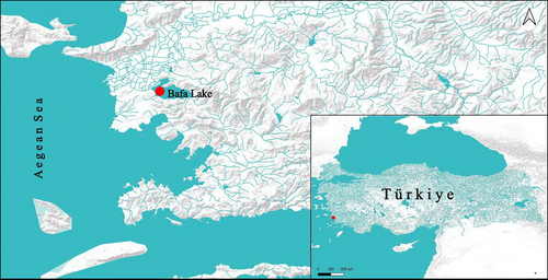
The study was conducted in Lake Bafa, located in Aydın Province, which ranks first in European eel production in Türkiye. Lake Bafa is situated within the boundaries of the Söke District of Aydın Province in Western Anatolia and the Milas District of Muğla Province. The lake extends east west and measures 16 km in length and 6 km in width. It has an average elevation of 2 m above sea level, a maximum depth of 21 m, and a surface area of 6708 hectares. Serçin Lake, located to the north of Lake Bafa and completely separated from it during dry periods, has a depth of 2 m and a surface area of 673 hectares. In some studies, it has been reported that the northern part of the lake, referred to as Serçin Lake, has completely dried up (Tosunoğlu et al., 2017). Fish samples were taken from Lake Bafa (37°30′11.14″ N; 27°24′10.28″ E) (Figure 1). The map (Figure 1) was generated using QGIS version 3.34.11-Pi, accessible at https://www.qgis.org.
2.2 Sampling collection
To collect European eel specimens (76), fishing attempts were conducted during the 2023 fishing season using 260 modified and 20 traditional eel fyke nets with poor size selectivity, rigged with a 14 mm stretched mesh size, to ensure a wide size range. These fyke nets, connected by the codend ropes, were transformed into a combined fishing gear. Four fishing attempts were performed at 10 different points in the lake, with 28 fyke nets used at each station. The mean soak time of the fyke nets was 7 days. The weights of captured fish were recorded using a portable digital scale with a precision of 0.01 g, and their total lengths were determined with a measurement board with a precision of 1 mm and recorded on forms. The fish specimens were grouped by total length (Table 1). It is imperative to note that the species studied can attain larger lengths than the specimens speared (Verreycken et al., 2011), but no fish of the maximum stated size were caught during this study. The data obtained are used to draw initial inferences, and looking for the ideal situation where specimens of all sizes become available for study is not validated. Otoliths were placed in their respective total length groups so that species-specific ontogenetic changes in otolith shape could be tracked. Specimens of A. anguilla were divided into seven groups (Group I, 200–300 mm; Group II, 310–400 mm; Group III, 410–500 mm; Group IV, 510–600 mm; Group V, 610–700 mm; Group VI, 710–800 mm; Group VII, 810–900 mm).
| CHARACTERS | A | B | C | D | E | F | G |
|---|---|---|---|---|---|---|---|
| Otolith shape | Elliptical-rectangular | Oval –rectangular | Rectangular | Rectangular | Rectangular | Rectangular | Rectangular |
| Otolith width | Wide | Wide | Wide | Wide | Wide | Wide | Wide |
| Otolith thickness | Thick | Deep | Deep | Deep | Deep | Deep | Thick |
| Mesial surface | Slightly concave | Slightly concave | Slightly concave | Slightly concave | Slightly concave | Slightly concave | Slightly concave |
| Lateral surface | Slightly convex | Slightly convex | Slightly convex | Slightly convex | Slightly convex | Slightly convex | Slightly convex |
| Shape of the dorsal margin | Irregular-entire | Waved or irregular | Irregular | Flat, irregular, or rounded, with indentation at the middle | Slightly irregular | Smooth - irregular | Irregular, elevated over the junction between ostium and cauda |
| Shape of the ventral margin | Lobate | Waved or irregular | Irregular | Flat | Straight posteriorly and coarsely irregular anteriorly | Smooth irregular | Smooth |
| Shape of the posterior margin | Lobate–slightly crenate | Entire, broadly pointed, lobate | Rounded or slightly irregular | Lobate or produced rounded | Rounded, bulging posteriorly | Straight or pointed | Broadly pointed |
| Sulcus acusticus | Heterosulcoid, ostial | Heterosulcoid, ostial | Heterosulcoid, ostial | Heterosulcoid, ostial | Heterosulcoid, ostial | Heterosulcoid, ostial | Heterosulcoid, ostial |
| Ostio-caudal differentiation | Absent | Absent | Absent | Absent | Absent | Absent | Absent |
| Ostium | Funnel-like, shorter than cauda | Funnel-like, shorter than cauda, shallow or deep | Funnel-like, shorter than cauda, deep | Funnel-like, shorter than cauda, shallow | Funnel-like, shorter than cauda, deep | Funnel-like, shorter than cauda, shallow | Funnel-like, shorter than cauda, shallow |
| Cauda | Tubular, short | Tubular, short, shallow, or deep | Tubular, short, shallow, or deep | Tubular, short, shallow, or deep | Tubular, short, shallow, or deep | Tubular, short, shallow | Tubular, short, shallow |
| Crista superior | Poorly or well-developed | Poor to well-developed | Poorly developed | Absent | Poorly developed | Absent | Moderately developed |
| Crista inferior | Poorly developed or moderately developed | Absent | Absent | Absent | Poorly developed | Absent | Poorly developed |
| Shape of the dorsal depression | Shallow or absent | Absent to moderately developed | Absent or moderately developed | Absent | Absent | Absent | Absent |
| Shape of the ventral depression | Poorly developed or absent | Absent to moderately developed | Absent or moderately developed | Absent | Absent | Absent | Absent |
| Shape of the rostrum | Broadly pointed | Broadly pointed | Broadly pointed | Broadly pointed | Broadly pointed | Broadly pointed | Broadly pointed |
| Size of the rostrum | Large | Large | Large | Large | Large | Large | Large |
| Thickness of the rostrum | Thick | Thick | Thick | Thick | Thick | Thick | Thick |
| Antirostrum | Poorly or well-developed | Poorly or well-developed | Small, poorly developed | Small and rounded | Small and rounded | Small and rounded | Large, broad, or small and straight |
| Excisura | Very wide, with a shallow angle | Absent or moderately developed, with a narrow angle | Small or large, shallow | Large or medium, shallow | Medium and shallow | Wide and shallow | Wide and shallow |
- Note: A—GI (200–300 mm TL), n = 2; B—GII (310–400 mm TL), n = 5; C—GIII (410–500 mm TL), n = 3; D—GIV (510–600 mm TL), n = 2; E—GV (610–700 mm TL), n = 1; F—GVI (710–800 mm TL), n = 4; G—GVII, (810–900 mm TL), n = 2; TL, fish total length.
2.3 Otolith sampling
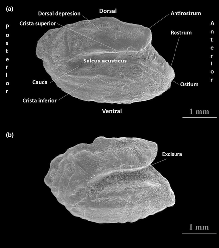
Sagittae were extracted, cleaned, dried, and stored in numbered Eppendorf tubes for subsequent inspection. The otic capsules were removed, and the otoliths were detached with a pair of fine forceps. They were cleaned in 70% ethanol and stored dry. Otoliths from each size group were examined with scanning electron microscopy (SEM), which allowed observation of morphological characteristics on the mesial face of the otoliths. In preparation for SEM, otoliths were air-cleaned and mounted on an aluminium stub using double-sided carbon tape. Otoliths were viewed in an SEM device, model FEI Quanta FEG 250. No coating was applied to the sample surfaces to ensure conductivity. Instead, conductivity was achieved using the device's low vacuum mode, and the chamber pressure was set to 60 pascals. The terminology of otolith morphology follows Smale et al. (1995) (Figure 2). In this study, we examined and compared 21 otolith characteristics among seven length groups of Anguilla anguilla (Linnaeus 1758) collected from the Bafa Lake in Western Anatolia, Türkiye (Table 1).
3 RESULTS
This section will provide a detailed description of the surface morphology of the otoliths for each fish length group will be designated distinctly. The SEM images show in detail the surface of otoliths, allowing the documentation of morphological features on the surface of the otoliths.
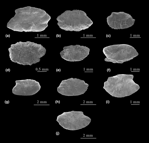
We observed variations in the surface morphology of the lateral side of the otoliths of the European eel examined. Specifically, the otolith surfaces of individuals in groups GII (310–400 mm), GIII (410–500 mm), and GIV (510–600 mm) exhibited varying levels of irregularities, predominantly around the margins. In contrast, the lateral surfaces of otoliths belonging to individuals in groups GV (610–700 mm), GVI (710–800 mm), and GVII (810–900 mm) were less irregular. Notably, the lateral surface of the otoliths in group GVII was relatively smooth (see Figure 3).
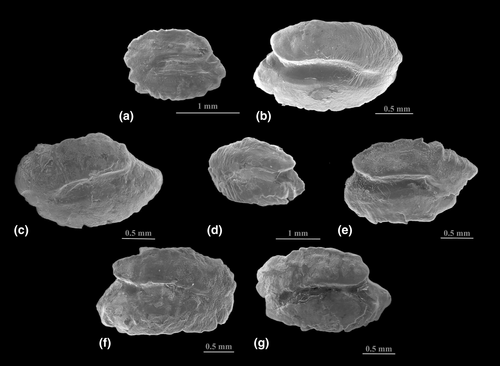
An ontogenetic divergence in the morphology of the otoliths of the European eel was discovered. The following otolith characteristics are sustained in young and adult fish: Otolith width, otolith thickness, the shape of both mesial and the lateral surfaces, the shape of the sulcus acusticus, the presence or absence of the ostio-caudal differentiation, and shape, size, and the thickness of the rostrum (Figures 4-6).

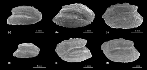
Juvenile otoliths of A. anguilla in Group I (GI) differ from adult ones in 8 out of 21 characters explored. The shape descriptions of the otoliths in seven size classes are given in Table 1. The shape of the otoliths of the different fish length groups exhibits slight change. Except for GI and GII, where the shape of the otolith was either elliptical or oval, the rectangular shape is the normal shape of the otolith observed in the remaining fish length groups.
The lateral and mesial surfaces are conservative in their shape through the growth phases of the fish. These characteristics are convex and concave, respectively.
Generally, the shape of the dorsal margin of the otoliths varied in irregularity across the seven fish length groups examined. In some otoliths of individuals of GIV (51–60 mm), this dorsal margin appeared flat or elevated at the junction between the ostium and cauda, while otoliths from individuals in group GVII (81–90 mm) showed different characteristics (Figures 4-6). The ventral margin of the otoliths also varied among the different fish length groups between smooth and irregular. In the otolith of the individuals of GI (20–30 mm), GII (31–40 mm), GIII (41–50 mm), and GIV (51–60 mm) this margin is lobate, waved to irregular, irregular, and flat respectively. For individuals in groups GI (20–30 mm), GII (31–40 mm), GIII (41–50 mm), and GIV (51–60 mm), the ventral margin was lobate, waved to irregular, irregular, and flat, respectively (Figures 4-6).
The posterior margin of the otoliths in various fish length classes primarily exhibited lobed and rounded shapes. In the otoliths of individuals from length classes GVI (71–80 mm) and GVII (81–90 mm), which included the largest fish sizes, the posterior margin was either straight or pointed, or broadly pointed, respectively. In the otoliths of individuals from length classes GI (20–30 mm), GII (31–40 mm), GIII (41–50 mm), GIV (51–60 mm), and GV (61–70 mm), the posterior margin appeared with the following characteristics: GI has a lobate–slightly crenate shape; GII has an entire shape; GIII was broadly pointed; GIV was lobate; GV was rounded or slightly irregular, and bulges posteriorly. In the otoliths of individuals from length classes GVI (71–80 mm) and GVII (81–90 mm), which included the largest fish sizes, the posterior margin was either straight or pointed, or broadly pointed, respectively.
Notably, ostio-caudal differentiation was absent in the otoliths of all the fish length classes examined.
The shape of the ostium and cauda in the otoliths was generally consistent; however, their depths varied among the individuals from the seven fish classes studied. For the ostium, there was inconsistency in depth, ranging from shallow to deep in the otoliths of both young and adult fish. The cauda depth varied between shallow and deep in the otoliths of individuals from groups GI (20–30 mm), GII (31–40 mm), GIII (41–50 mm), and GIV (51–60 mm). However, in the larger fish length groups, GVI (71–80 mm) and GVII (81–90 mm), the cauda was consistently shallow (Figures 4-6).
The shape and presence or absence of the crista superior and crista inferior showed variation, ranging from absent to poorly developed and moderately developed (Figures 4-6).
The dorsal and ventral depressions present in the otoliths of individuals in groups GI (20–30 mm), GII (31–40 mm), and GIII (41–50 mm) may be absent, poorly developed, or moderately developed. In contrast, these features were absent in the otoliths of individuals from the larger fish length groups: GIV (51–60 mm), GV (61–70 mm), GVI (71–80 mm), and GVII (81–90 mm) (Figures 4-6).
The shape and size of the antirostrum in the otoliths of all length groups examined varied from small and poorly developed to larger forms. In the case of the largest fish length group, GVII (81–90 mm), the antirostrum can be large, broad, or small and straight (see Figures 4-6).
The excisura and its angle or notch also varied among the otoliths of fish across the seven size groups. In the otoliths of GI (20–30mm), the excisura was very wide, and the angle was shallow. In group GII (31–40 mm), these features were either absent or moderately developed. For fish length groups GIII (41–50 mm), GIV (51–60 mm), and GV (61–70 mm), the excisura and its angle vary in size, with a generally wide angle. For fish length groups GIII (41–50 mm), GIV (51–60 mm), and GV (61–70 mm), the excisura and its angle vary in size, with a generally wide angle (Figures 4-6).
4 DISCUSSION
4.1 General
The phylogenetic relationship of the European eel A. anguilla is an intriguing topic. The genus Anguilla embraces several eel species, which share common ancestry and exhibit similar morphological features (Tsukamoto & Aoyama, 1998). Molecular studies suggested that the European eel and the Japanese eel share a more recent common ancestor than does with the American eel (Rozenfeld et al., 2019).
The external features of the species of the genus Anguilla are very alike, and it can be hard to see dissimilarities between them upon first seeing them. However, certain features are crucial for identifying and differentiating species. These include the presence or absence of parti-coloured body patterns, the types of maxillary band arrangement of the teeth, the locations of the dorsal fins origin, and the number of vertebrae.
Several morphological features are used to identify the European eel. These features include specific external body proportions, such as preanal length, predorsal length, the distance between the vertical lines through the anus, and the origin of the dorsal fin Ege (1939). In addition to these features, variations in colour and tooth patterns are also utilized for identification (Aoyama et al., 2001; Watanabe et al., 2005; Watanabe & Bischof, 2004). Therefore, additional morphological characteristics are needed to recognize the European eel from other anguillid species.
4.2 Developmental variation
During the early life stages, otoliths are small with a relatively high accretion rate and can be significantly reshaped by environmental issues (Hüssy, 2008; Motamedi et al., 2021). The ontogenetic growth from the circular larval otolith to the oblong adult otolith has been identified in a wide range of species (see Hüssy, 2008). Capoccioni et al. (2010) on eels and Vignon (2012) on Lutjanus kasmira indicated that otoliths remain relatively unchanged in shape at smaller sizes compared to larger ones. These results are consistent with those of Lombarte et al. (2003) on 11 species of the genus Merluccius, pointing out a noteworthy habitat effect on otolith growth upsurge in undeveloped fish, compared with the adults. Usually, the growing of the otolith shape is an ontogenetic procedure outside the earliest life stages that can reshape the overall otolith outline, notwithstanding small-scale environmental issues. The reason for such early steadiness would be a biological boundary modelled by otolith function as a sound transducer (Gauldie & Nelson, 1990). Proteins within the endolymphatic epithelium continue as a noteworthy component of the mechanism controlling the shape of the otolith and endolymph physiological issues allowing bio-mineralization are under genetic regulator and guideline (Borelli et al., 2003; Morales-Nin, 2000). This may validate why the early shape growth displays as an ontogenetically resolute procedure, with otolith growing following the build-up of either calcium carbonate or protein.
In theory, habitat disparateness may enable the link between organisms and their environment. Nonetheless, most of the investigations were made at large geographic scales, such as latitudinal gradient and marine ecological regions (e.g. Lombarte & Lleonart, 1993). On the contrary, surveys dealing with small-scale variations (i.e. over a few hundred metres) are inadequate (Vignon et al., 2008) and frequently aimed at local pollution or anthropogenic variabilities. The absence of findings at a small-scale may specify the shortage of expectation that local niche divergency would influence otolith shape over short distances in the absence of genetic differences. Furthermore, shape divergence improves with age/size, highlighting that the environment can reconcile the developmental ontogeny of otolith in a substantial way and that could be through growth rate.
Numerous traits of the sagittal otolith can contribute to taxonomic research (Figure 3), with some acknowledged as early as the start of the twentieth century (Nolf, 1985, 2013). The teleost sagittal otolith is the most extremely used anatomical reliability in comparative taxonomic investigations, due to its large size and degree of interspecific variations (Jawad, 2007, 2018). In the study at hand, we inspect a diversity of otolith traits, of which some are taxonomically valuable for identification at the species level. Such features are the otolith width, otolith thickness, shape of both mesial and the lateral surfaces, shape of the sulcus acusticus, the presence or absence of the ostio-caudal differentiation, shape, size, and the thickness of the rostrum.
The surface morphology of the lateral side of the otolith of the European eel showed ontogenetic variation. This variation can be seen as irregularities and lobulations mainly around the different edges of the otolith. Irregularities and the different levels of lobulations are more obvious in the otolith of individuals of groups GII (310–400 mm), GIII (410–500 mm), and GIV (510–600 mm), while otoliths of individuals belonging to groups GV (610–700 mm), GVI (710–800 mm), and GVII, (810–900 mm) showed fewer irregularities and lobulations. The difference in the surface irregularities of the lateral side of the otolith of the European eel suggests that this characteristic can be clearly used to separate the otoliths of groups GII–GIV from the otolith of the larger fish sizes GV–GVII, where the lateral surface of the otolith of G VII is entirely smooth. This trait can be considered a taxonomic value to recognize the European eel from the rest of the other anguillid species.
In the present study, the shape of the otolith of the individuals of A. anguilla GI (20–30 mm) and GII (31–40 mm) is elliptical-rectangular and oval-rectangular, respectively, which differs from the rectangular shape of the otolith in the remaining five fish length groups. Such differences represent important ontogenetic variations and can mislead the process of separating the young from the adult individuals.
Juvenile otoliths of A. anguilla in Group I (GI) differ from adult ones in 8 out of 21 characters explored (Table 1). These differences include otolith shape, the shape of the dorsal, ventral, and posterior margins, the shape of crista inferior, the shape of the dorsal and ventral depressions, and the shape of the excisura. These features represent an ontogenetic change. Similar outcomes were obtained by Jawad (2007) on triplefin fish species, Jawad (2018) on some members of the family Scaridae collected from the Solomon Islands, Jawad et al. (2023) on some members of the family Lethrinidae from the waters of the Republic of Yemen, and Reis et al. (2023) on the mullet Chelon auratus (Risso, 1810) collected from Köyceğiz Lagoon, Aegean Sea, Türkiye. Such inconsistency in these features makes these traits unusable in identifying individuals of the nine fish length groups examined.
The individuals of different length groups of A. anguilla are considered to share 13 otolith constant traits from smaller to larger individuals (Table 1). However, these characteristics can be beneficial as a means to recognize individuals of A. anguilla explored and could be an indicative characteristic of this species. With the aid of these features, A. japonica can be separated from A. anguilla by having an elliptic otolith shape, the posterior and the ventral margins are entire, the posterior margin is tapering, and the cauda is directed downward posteriorly (Lin & Chang, 2012). Anguilla marmorata has the following set of characteristics that separate it from A. anguilla, the elliptic shape of the otolith, the dorsal and the ventral margins however are entire, the cauda is directed downward posteriorly, and the rostrum is short. The following features can separate A. obscura from A. anguilla, oblong otolith shape, crista superior and inferior are well-developed, the rostrum is very long, the ventral margin is distinguished in having three spines projected downward, and the dorsal depression is very developed (Rivaton & Bourret, 1999). Finally, the following set of characters can separate the otolith of A. rostrata from those of A. anguilla. The crista superior and inferior are very well-developed, the sulcus acusticus is curved downward posteriorly, and the dorsal depression is very well-developed (Campana, 2004).
The outcome of the present study showed consistency in the shape of the sulcus acusticus, which is heterosulcoid and ostial, in both juvenile and adult individuals of A. anguilla. Some earlier investigations on fossil and extant otoliths have revealed that the morphology of this otolith feature has a uniform shape within a single genus (Jawad et al., 2023; Nolf, 1985) and that such similarity is likely genetically controlled (Gauldie, 1988). Reichenbacher et al. (2007) were capable of classifying species of the genus Aphanius (Cyprinodontiformes) into groups, based on distinctive and typical sulcus acusticus morphologies.
4.3 Intraspecific variation
Comparing the shape of the sulcus acusticus of A. anguilla with that of other members of the genus Anguilla like A. japonica, A. marmorata, and A. obscura, the shape of the sulcus acusticus of A. anguilla is wide, straight, and short. These features differ significantly from the sulcus acusticus of A. japonica, A. marmorata, A. obscura, and A. rostrata described by Lin and Chang (2012), Rivaton and Bourret (1999), and Campana (2004). Therefore, it is possible to consider the shape of the sulcus acusticus as taxonomic measure to distinguish individuals of A. anguilla from other anguillid fish species.
The data on the developmental account of the surface features of the otoliths of A. anguilla is deficient. As far as the authors are aware, the work of Tuset et al. (2008) is considered the only descriptive work on the otolith of A. anguilla from the Western Mediterranean, north and central Eastern Atlantic regions. Tuset et al. (2008) examined and described the otolith of three fish specimens with a total length of 120, 400, and 857 mm. These three fish specimens are smaller in total length than those of the individuals of GI (200–300 mm), Group GII (310–400 mm) group, and Group GVII (810 − 900 mm) of the specimens examined in the present study respectively. To compare these three otoliths with those described herein, it appears that the smallest otolith (120 mm) of Tuset et al. (2008) is shown to have an entire dorsal and ventral margin, and the posterior margin has a pointed extension at the midpoint. The sulcus acusticus is very shallow, and the dorsal and ventral depressions, the crista superior, and crista inferior are not developed. The antirostrum is absent. Comparing the otolith of the 400 mm total length of Tuset et al. (2008) with the otoliths from the specimens in the present study with a nearest total length of 398 mm, the dorsal, ventral, the posterior margins of this specimen appear less waved and irregular than our specimen. Additionally, the sulcus acusticus is shallow, with the dorsal and ventral depressions, crista superior, and crista inferior were not developed. However, the antirostrum was quite developed and produced. Comparing the surface morphology of the third otolith of Tuset et al. (2008) taken from fish specimen of 857 mm with our specimen from G VII (810–900 mm) group, it seems that the dorsal, ventral, and the posterior margins are wavier and more irregular than the otolith described in the present study. Moreover, the antirostrum is poorly developed compared with our specimens well-developed antirostrum. Finally, the sulcus acusticus is shallow, with the dorsal, ventral depressions, crista superior, and crista inferior not developed in the Tuset et al. (2008) specimen.
4.4 Interspecific variation
Some investigators have observed changes, where some of the otolith surface ornamentations can vary between species; accordingly, these researchers determined that the discrepancy in these features can be linked with zoogeography and phylogeny (Torres et al., 2000). Hence, the traits of the otolith that presented geographical dissimilarity cannot be dependably used to recognize A. anguilla from the Bafa Lake in Western Anatolia, Türkiye, as there seems to be too much intraspecific geographic inconsistency in the anguillid otoliths.
The outcome of the present study focuses on a specific level of ontogenetic inconsistency in the shape of the otolith for A. anguilla, aimed at distinguishing small-juvenile from large-adult specimens, assuming the species has been correctly identified when collected. The specific characteristics of the otoliths observed in this study can be added to the other detailed features of A. anguilla. Similar results have been reported by Reichenbacher et al. (2007), Echreshavi et al. (2021), and Mehraban et al. (2021) in studies on fish species other than anguillid. Further research is needed, including a comparative analysis of the shape and geometry of the sagittal otolith, to contribute to additional taxonomic characteristics for the identification of this species.
AUTHOR CONTRIBUTIONS
Salim Serkan Güçlü: Conceptualization; data curation; investigation; methodology; project administration; resources. Habil Uğur Koca: Data curation; investigation; methodology; resources; validation; writing review and editing. Mehmet Cilbiz: formal analysis; investigation; methodology; supervision; validation; visualization; review and editing. Laith A. Jawad: Conceptualization; formal analysis; investigation; methodology; project administration; supervision; validation; visualization; writing original draft; writing review and editing.
ACKNOWLEDGEMENTS
The authors thank TUBITAK for their support. We would especially like to thank Dr. Salih AKYÜREKLİ (Isparta), SEM responsible of Süleyman Demirel University YETEM-Innovative Technologies Application and Research Centre.
FUNDING INFORMATION
This study was supported by the Scientific and Technological Research Council of Turkey (TUBITAK) under the Grant Number 222O145.
CONFLICT OF INTEREST STATEMENT
The authors declare that they have no known competing financial interests or personal relationships that could have appeared to influence the work reported in this article.


