Comparison of different organic selenium supplementations on selenium status and serum biomarkers in dairy cows
Abstract
The objective of this study was to investigate the effects of two different organic selenium (Se) supplements, selenomethionine (Se-Met) and selenohomolanthionine (Se-Hlan), on the serum biochemical parameters and Se status of dairy cows. Different dietary Se supplementation treatments were set as follows: a control group (CON, adding sodium selenite at 0.3 mg Se/kg dry matter [DM]), 0.3 and 0.5 Se-Met (adding Se-Met at 0.3 and 0.5 mg Se/kg DM, respectively), as well as 0.3 and 0.5 Se-Hlan (adding Se-Hlan at 0.3 and 0.5 mg Se/kg DM, respectively). The experiment lasted 8 weeks. The serum measurements showed that both organic Se treatments resulted in higher uric acid than CON. Se-Met produced higher aspartate aminotransferase, glucose, urea, low-density lipoprotein cholesterol, and lactate dehydrogenase than Se-Hlan. Regarding the Se status, the highest milk Se values appeared in 0.5 Se-Met, with intermediate values in 0.3 Se-Met and 0.5 Se-Hlan, whereas the highest and lowest serum Se levels were presented in 0.5 Se-Met and 0.3 Se-Hlan, respectively. Our results suggest that Se-Hlan was not as efficient in boosting serum or milk Se as Se-Met and differences in serum biomarkers between Se-Met and Se-Hlan may be associated with distinct metabolic pathways for different forms of organic Se.
1 INTRODUCTION
Selenium (Se) plays an important role in human nutrition, not only via direct influence on the maintenance of immune and hormonal systems, protection against heavy metals and xenobiotics, and prevention of chronic diseases, but also in the key balance of benefits and competition between host and gut microbial flora (Ferreira et al., 2021). For Chinese residents, a reduction in grain consumption was correlated with a decrease in hair Se measurements (Li et al., 2014), a factor that may have contributed to adverse health impacts in Se-deficient areas. Moreover, a previous study demonstrated that 39–61% of Chinese residents consumed less Se than the WHO/FAO recommended level (26–34 μg/d) (Dinh et al., 2018). Therefore, as suggested by Givens et al. (2004), increasing Se-enriched milk consumption could relieve the Se deficiency in China.
A meta-analysis of Se intake by dairy cows showed that oral Se supplementation resulted in an increase in milk Se level by 9.24–16.34 μg/l (95% confidence interval) (Ceballos et al., 2009). The source and dosage of Se and their interaction explained approximately 70% of the variance. Compared with inorganic Se, organic Se was more efficient in increasing milk Se content using either selenized yeast or Se-fertilized forage (Séboussi et al., 2016). As the most common organic Se supplementation for dairy cattle, selenized yeast contains approximately 60–70% selenomethionine (Se-Met) (Calamari et al., 2010). A novel selenoamino acid, selenohomolanthionine (Se-Hlan), has been identified in Japanese pungent radishes (Ogra et al., 2007). Se-Met is the Se analog of methionine, and as such, it is similar to methionine in chemical structure and metabolic pathways. Se-Hlan is the common name of 4,4′-selenobis [2-aminobutanoic acid], and it is utilized only in the trans-selenation pathway for selenoprotein synthesis (Tsuji et al., 2010). Se-Hlan has been used in broilers to evaluate growth performance, nutrient utilization, oxidative stress, and Se tissue concentrations, and comparisons between Se-Met and Se-Hlan have also been made (Celi et al., 2013). However, to date, there are no comparable data available for the two organic Se supplements in dairy animals.
We hypothesized that the addition of Se-Hlan in dairy cattle feed would be as effective as Se-Met as a substitute for inorganic Se. This study aimed to evaluate the effects of different organic Se supplements, Se-Hlan and Se-Met, on milk production and composition, serum biochemical parameters, antioxidant indicators, immune parameters, and the Se concentration in milk and blood of dairy cows. According to the specification of feed additives issued by the Ministry of Agriculture of China (Ministry of Agriculture of China, 2009), the recommended and maximum additive amounts for Se are 0.1–0.3 and 0.5 mg/kg in total mixed rations for ruminants, respectively. To evaluate the potential milk performance and health issues affected by Se intake, low (0.3 mg/kg) and high (0.5 mg/kg) levels of Se were set in each organic Se treatment.
2 MATERIALS AND METHODS
2.1 Animals and experimental diets
This experiment complied with the Chinese regulations for experimental animals (in Chinese, http://www.gov.cn/gongbao/content/2017/content_5219148.htm). The experimental protocol was approved (Q/MN-CW/GL-2020-7) and supervised by the dairy cattle institute of the Mengniu Dairy Group. Eighty pre- or multi-parous Holstein dairy cows with days in milk (DIM) of 148 ± 29, parity 1.84 ± 0.96, and 26.8 ± 3.3 kg of milk/day were housed in a freestall barn at the Ziang Dairy farm (Hohhot, China). Fresh water was provided ad libitum. The animals were randomly assigned to one of five blocks that were balanced for DIM, parity, and milk yield. The Se concentration in the basal diet without Se supplementation was 0.02 mg/kg DM. Five treatments were designed as follows: a control group (CON, supplemented with sodium selenite at 0.3 mg of Se/kg DM), a low Se-Met group (0.3 Se-Met, supplemented with organic Se from Se yeast I [SelenoSourceAFTM2000, about 80% of total Se as Se-Met, provided by Diamond V, Mills Inc., USA] at 0.3 mg of Se/kg DM), a high Se-Met group (0.5 Se-Met, supplemented with organic Se from Se yeast I at 0.5 mg of Se/kg DM), a low Se-Hlan group (0.3 Se-Hlan, supplemented with organic Se from Se yeast II [AB Tor-Sel 4000, 80–85% of total Se as Se-Hlan, provided by AB Agri, Bio Processing Australia Pty Ltd., Australia] at 0.3 mg of Se/kg DM), and a high Se-Hlan group (0.5 Se-Hlan, supplemented with organic Se from Se yeast II at 0.5 mg of Se/kg DM). Cows were fed twice at 7:00 and 14:00 each day, and feed refusals were removed every morning. The pretrial period lasted one week, and the experimental period lasted eight weeks.
2.2 Sampling and analysis
The average daily feed intake of dairy cows was reported by subtracting the refusals from the total fresh TMR weekly. Feed samples were collected monthly and combined, then oven-dried at 65°C for 48 h, and ground into powders with particle sizes less than 1 mm. DM was measured by oven drying at 105°C for 4 h to a constant weight. The CP and ether extract of feed samples were determined using the Kjeldahl method and Soxhlet extraction (Thiex et al., 2003), respectively. The NDF and ADF contents were measured according to procedures described by Van Soest et al. (1991). Calcium and phosphorus contents were reported by atomic absorption spectroscopy and spectrophotometry, respectively (Chemists & Horwitz, 1990). The Se content in feed samples was detected using atomic fluorescence spectroscopy after microwave digestion (Ministry of Agriculture of China, 2010). Measurements of Se contents for CON, 0.3 Se-Met, 0.5 Se-Met, 0.3 Se-Hlan, and 0.5 Se-Hlan diets were 0.32, 0.33, 0.49, 0.29, and 0.50 mg/kg DM, respectively. The other detected nutrients are listed in Table 1.
| Composition | Content |
|---|---|
| Ingredient, g/kg DM | |
| Corn silage | 326.8 |
| Alfalfa hay | 118.0 |
| Flaked corn | 49.7 |
| Whole cottonseed | 68.3 |
| Ground corn | 119.8 |
| Rapeseed meal | 23.0 |
| Soybean meal | 129.8 |
| Soybean hull | 84.1 |
| Distillers dried grains with solubles | 22.0 |
| CMS 14041631 | 8.5 |
| Sodium bicarbonate | 6.1 |
| Magnesium oxide | 6.1 |
| Zeolite meal | 4.1 |
| Mineral meal | 25.5 |
| Rice dusk | 1.8 |
| Premix2 | 6.4 |
| Chemical composition, g/kg DM | |
| NEL (MJ/kg DM)3 | 629 |
| CP | 154.2 |
| Ether extract | 35.3 |
| NDF | 352.5 |
| ADF | 199.5 |
| Ca | 9.1 |
| P | 2.7 |
| Ash | 37.6 |
| Se (mg/kg DM) | 0.02 |
- 1 A syrup supplement (Vedan, Yantai, Shandong Province, China).
- 2 Contained (DM basis) 400,000 IU/kg of vitamin A; 50,000 IU/kg of vitamin D; 5,000 IU/kg of vitamin E; 5,000 mg/kg of Fe; 1,600 mg/kg of Cu; 6,500 mg/kg of Zn; 2,000 mg/kg of Mn; 60 mg/kg of I; and 45 mg/kg of Co. Se was added in accordance with the experimental design.
- 3 Calculated based on China NY/T 34, 2004.
Three milking for experimental cows was arranged at 06:00, 14:00, and 22:00 h, and individual milk production was reported at each milking. Milk samples were collected at three successive milking periods and combined in the proportion of 4:3:3 every Monday during the trial. The mixed milk samples were divided into two parts; one was stored at 4°C for milk composition (fat, protein, and lactose) analysis using Mid-infrared spectroscopy (Foss MilkoScan 2000, Foss Food Technology Corp., Eden Prairie, MN, USA), and the other was frozen at −20°C for Se detection. For the determination of Se contents, milk samples (approximately 1 g) were digested in the presence of 5 ml of HNO3 in a closed-vessel heating block. The diluted digested solution was analyzed using ICP-MS according to standard protocols (Ministry of Agriculture of China, 2016).
Twelve cows chosen at random in each group were selected to sample blood. Near the end of the experiment, whole blood samples were collected from the coccygeal vein into vacutainer tubes (AOSAITE Medical Equipment Co., Ltd., Heze, Shandong Province, China) with or without EDTAK2. The latter tubes with blood were stored at 4°C for 30 min and centrifugated at 4000 rpm for 20 min to separate serum samples. Blood samples were stored at 4°C for Se detection, and serum samples were frozen at −20°C for subsequent biochemical parameters, antioxidant capacity, immunologic indexes, and Se determination. Approximately 0.5 g of blood sample and 1 g of serum sample were prepared to determine Se contents according to standard protocols (Ministry of Agriculture of China, 2016), and similarly for analysis of milk samples. An automated biochemical analyzer (HITACHI 7080; Hitachi Valve Ltd., Tokyo, Japan) was used to detect biochemical parameters of serum samples, including aspartate aminotransferase (AST), alanine aminotransferase (ALT), alkaline phosphatase (ALP), cholinesterase (CHE), creatine kinase (CK), glucose (GLU), urea, creatinine (CREA), uric acid (UA), cholesterol (CHO), triglycerides (TG), high-density lipoprotein cholesterol (HDL-C), low-density lipoprotein cholesterol (LDL-C), lactate dehydrogenase (LDH), and total bile acids (TBA). Indexes reflecting antioxidant capacity (total antioxidant capacity (T-AOC), GSH-Px, lipid peroxide (LPO), and malondialdehyde (MDA)) and immunologic indexes (IL-6, IL-2, IgG, and TNF-α) in serum were measured using commercial assay kits (Nanjing Jian Cheng Bioengineering Institute, Nanjing, China) in this study.
2.3 Statistical analysis
All data were analyzed using R version 3.6.1. The Shapiro–Wilk and Bartlett tests were used to examine the normality and homoscedasticity of the data, respectively. The linear model for data on feed intake, milk composition, milk yield, and milk Se had fixed effects of the week, treatment, and the interaction of week and treatment and a random effect (cow). Data on serum biochemical parameters, antioxidant indexes, antibodies and cytokines, and blood and serum Se levels between treatments were analyzed using ANOVA. Tukey's multiple range test was used for the evaluation of differences between the groups. Orthogonal contrasts were constructed to compare (1) Se-Met vs. Se-Hlan: selenomethionine supplemented diets (0.3 Se-Met and 0.5 Se-Met) versus selenohomolanthionine supplemented diets (0.3 Se-Hlan and 0.5 Se-Hlan), (2) CON vs. Se-Met: CON versus selenomethionine supplemented diets (0.3 Se-Met and 0.5 Se-Met), (3) CON vs. Se-Hlan: CON versus selenohomolanthionine supplemented diets (0.3 Se-Hlan and 0.5 Se-Hlan), and (4) Organic Se level: organic Se level at 0.3 (0.3 Se-Met and 0.3 Se-Hlan) versus 0.5 (0.5 Se-Met and 0.5 Se-Hlan) mg of Se/DM. Data were presented as the least squares mean and standard error of the mean. Statistical significance was set at P < 0.05, and trends were reported when 0.05 ≤ P < 0.10. Measurements affected by treatments, Se sources, or significant levels (P < 0.05) were screened for principal component analysis (PCA), and data standardization before PCA was completed by centering (subtracting average values) and normalization (dividing by the standard deviation).
3 RESULTS
3.1 Milk production and composition
The results of Se supplementation on milk composition and production are shown in Table 2. Data for milk performance were recorded every week, and a significant sampling week effect on milk composition was observed in the primary analysis. Therefore, the final model contained reports from weeks 5 to 8 to exclude the transitory effect of Se supplementation on milk performance. No significant effect of sampling week was observed in the final model. As shown in Table 2, a trend toward an effect (P = 0.09) on the milk fat content of treatments was observed. A trend toward an effect (P = 0.06) on milk yield was presented in the comparison between Se-Met supplementation and CON.
| Item | Treatment mean1 | SEM2 | P-value | ||||||||
|---|---|---|---|---|---|---|---|---|---|---|---|
| CON | 0.3Se-Met | 0.5Se-Met | 0.3Se-Hlan | 0.5Se-Hlan | trt3 | Se-Met vs. Se-Hlan4 | CON vs. Se-Met5 | CON vs. Se-Hlan6 | Organic Se level7 | ||
| Total DMI, kg/d | 20.2 | 19.6 | 19.9 | 20.4 | 19.8 | 0.14 | 0.65 | 0.46 | 0.53 | 0.78 | 0.58 |
| Fat, g/kg | 36.4 | 36.2 | 36.3 | 35.1 | 36.3 | 0.24 | 0.09 | 0.15 | 0.82 | 0.17 | 0.10 |
| Protein, g/kg | 34.1 | 34.5 | 34.5 | 33.2 | 35.3 | 0.77 | 0.14 | 0.49 | 0.69 | 0.83 | 0.18 |
| Lactose, g/kg | 47.8 | 47.5 | 48.2 | 48.6 | 48.1 | 0.19 | 0.18 | 0.17 | 0.93 | 0.11 | 0.88 |
| Milk yield, kg/d | 23.4 | 22.5 | 22.6 | 22.2 | 22.3 | 0.47 | 0.11 | 0.38 | 0.06 | 0.22 | 0.16 |
- a,b Means with different superscripts in this row are significantly different (P < 0.05).
- 1 Treatment mean: CON is basal diet supplemented with sodium selenite at 0.3 mg of Se/kg DM; 0.3Se-Met and 0.5Se-Met are basal diets supplemented with selenized yeast I (mainly selenomethionine, SelenoSourceAFTM2000; Diamond V, Mills Inc., USA) at 0.3 and 0.5 mg of Se/kg DM, respectively; 0.3Se-Hlan and 0.5Se-Hlan are basal diets supplemented with selenized yeast II (mainly selenohomolanthionine, AB Tor-Sel 4000; AB Agri, Bio processing Australia Pty Ltd., Australia) at 0.3 and 0.5 mg of Se/kg DM, respectively.
- 2 SEM: standard error of mean.
- 3 trt: comparison of all treatments, including CON, 0.3 Se-Met, 0.5 Se-Met, 0.3 Se-Hlan, and 0.5 Se-Hlan.
- 4 Se-Met vs. Se-Hlan: Orthogonal contrasts used to compare selenomethionine supplemented diets (0.3 Se-Met and 0.5 Se-Met) versus selenohomoalanthionine supplemented diets (0.3 Se-Hlan and 0.5 Se-Hlan).
- 5 CON vs. Se-Met: Orthogonal contrasts used to compare CON versus selenomethionine supplemented diets (0.3 Se-Met and 0.5 Se-Met).
- 6 CON vs. Se-Hlan: Orthogonal contrasts used to compare CON versus selenohomoalanthionine supplemented diets (0.3 Se-Hlan and 0.5 Se-Hlan).
- 7 Organic Se level: Orthogonal contrasts of organic Se level at 0.3 (0.3 Se-Met & 0.3 Se-Hlan) versus 0.5 (0.5 Se-Met & 0.5 Se-Hlan) mg of Se/DM.
3.2 Biochemical parameters, antioxidant indicators, and immune parameters in the serum
The effects of different Se supplements on biochemical parameters, antioxidant indicators, and immune parameters in the serum are shown in Tables 3, 4, and 5, respectively. As shown in Table 3, the Se-Hlan supplementation group had lower ALT and AST than CON (P < 0.05) as well as lower AST than the Se-Met supplementation group (P = 0.04). Compared with Se-Hlan supplementation, both the Se-Met group and CON showed similar higher GLU values (P < 0.01). Urea concentrations in the Se-Met groups were higher than those in the Se-Hlan groups and CON (P < 0.01). Se-Met and Se-Hlan supplementation produced similar UA contents that were higher than the CON value (P < 0.01). Meanwhile, serum UA decreased with increasing organic Se intake (P = 0.02), but CREA showed opposite changes (P = 0.05). Se-Met groups showed higher TG concentrations than CON (P = 0.03). Although no significant differences between Se-Met or Se-Hlan groups and CON were observed in LDL-C or LDH, Se-Met groups presented higher LDL-C (P = 0.04) and LDH (P = 0.03) than Se-Hlan groups. For the antioxidant indexes shown in Table 4, Se-Hlan supplementation presented higher trends of GSH-Px and LPO than Se-Met groups (P = 0.08). Data for serum antibodies and cytokine levels listed in Table 5 presented no remarkable difference among Se supplements.
| Item1 | Treatment mean2 | SEM3 | P-value | ||||||||
|---|---|---|---|---|---|---|---|---|---|---|---|
| CON | 0.3Se-Met | 0.5Se-Met | 0.3Se-Hlan | 0.5Se-Hlan | trt4 | Se-Met vs. Se-Hlan5 | CON vs. Se-Met6 | CON vs. Se-Hlan7 | Organic Se level8 | ||
| ALT, U/L | 36.45 | 35.50 | 35.08 | 32.83 | 33.42 | 0.670 | 0.30 | 0.11 | 0.42 | 0.02 | 0.95 |
| AST, U/L | 103.36b | 104.33ab | 87.00ab | 80.50a | 83.17a | 5.079 | 0.02 | 0.04 | 0.28 | <0.01 | 0.28 |
| TP, g/L | 76.16 | 77.41 | 77.78 | 77.44 | 78.68 | 0.405 | 0.88 | 0.80 | 0.42 | 0.24 | 0.65 |
| ALB, g/L | 33.23 | 32.15 | 32.98 | 32.33 | 33.59 | 0.272 | 0.78 | 0.68 | 0.55 | 0.79 | 0.26 |
| TBIL, umol/L | 2.47 | 2.58 | 2.58 | 2.52 | 2.42 | 0.032 | 0.85 | 0.39 | 0.44 | 0.98 | 0.65 |
| ALP, U/L | 32.50 | 40.91 | 33.67 | 34.42 | 31.00 | 1.703 | 0.27 | 0.20 | 0.22 | 0.95 | 0.13 |
| GGT, U/L | 32.78 | 29.99 | 31.21 | 27.52 | 32.35 | 0.944 | 0.50 | 0.78 | 0.36 | 0.28 | 0.20 |
| GLU, mmol/L | 3.90b | 3.81ab | 3.85b | 3.60ab | 3.52a | 0.074 | <0.01 | <0.01 | 0.48 | <0.01 | 0.80 |
| UREA, mmol/L | 6.93a | 8.14bc | 8.30c | 7.02a | 7.10ab | 0.296 | <0.01 | <0.01 | <0.01 | 0.64 | 0.71 |
| CREA, umol/L | 84.67 | 77.25 | 83.00 | 79.82 | 82.75 | 1.318 | 0.18 | 0.60 | 0.14 | 0.31 | 0.05 |
| UA, umol/L | 40.83a | 63.73b | 50.08ab | 59.25b | 54.67ab | 3.943 | <0.01 | 0.93 | <0.01 | <0.01 | 0.02 |
| CHE, U/L | 351.42 | 342.58 | 350.18 | 347.25 | 341.67 | 1.961 | 0.68 | 0.77 | 0.43 | 0.28 | 0.89 |
| CHO, mmol/L | 5.26 | 4.98 | 5.26 | 4.75 | 4.58 | 0.136 | 0.57 | 0.18 | 0.77 | 0.22 | 0.87 |
| TG, mmol/L | 0.15a | 0.19b | 0.17ab | 0.17ab | 0.17ab | 0.007 | 0.05 | 0.15 | 0.03 | 0.19 | 0.17 |
| HDL-C, mmol/L | 2.81 | 2.66 | 2.83 | 2.86 | 2.81 | 0.035 | 0.82 | 0.48 | 0.65 | 0.83 | 0.66 |
| LDL-C, mmol/L | 0.58 | 0.60 | 0.56 | 0.51 | 0.47 | 0.023 | 0.31 | 0.04 | 0.92 | 0.20 | 0.41 |
| CK, U/L | 199.17 | 249.60 | 191.50 | 171.75 | 208.00 | 12.874 | 0.26 | 0.28 | 0.48 | 0.60 | 0.78 |
| LDH, U/L | 1073.33ab | 1122.08b | 1048.42ab | 929.25a | 1039.67ab | 31.743 | 0.05 | 0.03 | 0.84 | 0.14 | 0.70 |
| TBA, umol/L | 24.02 | 20.39 | 25.11 | 19.96 | 22.18 | 0.999 | 0.87 | 0.67 | 0.78 | 0.52 | 0.40 |
- a–c Means with different superscripts in this row are significantly different (P < 0.05).
- 1 Item: AST, aspartate aminotransferase; ALT, alanine aminotransferase; ALP, alkaline phosphatase; CHE: cholinesterase; CK, creatine kinase; GLU, glucose; CREA, creatinine; UA, uric acid; CHO, cholesterol; TG, triglycerides; HDL-C, high-density lipoprotein cholesterol; LDL-C, low-density lipoprotein cholesterol; LDH, lactate dehydrogenase; and TBA, total bile acids.
- 2 Treatment mean: CON is a basal diet supplemented with sodium selenite at 0.3 mg of Se/kg DM; 0.3 Se-Met and 0.5 Se-Met are basal diets supplemented with selenized yeast I at 0.3 and 0.5 mg of Se/kg DM, respectively; and 0.3 Se-Hlan and 0.5 Se-Hlan are basal diets supplemented with selenized yeast II at 0.3 and 0.5 mg of Se/kg DM, respectively.
- 3 SEM: standard error of mean.
- 4 trt: comparison of all treatments, including CON, 0.3 Se-Met, 0.5 Se-Met, 0.3 Se-Hlan, and 0.5 Se-Hlan.
- 5 Se-Met vs. Se-Hlan: Orthogonal contrasts used to compare selenomethionine supplemented diets (0.3 Se-Met and 0.5 Se-Met) versus selenohomoalanthionine supplemented diets (0.3 Se-Hlan and 0.5 Se-Hlan).
- 6 CON vs. Se-Met: Orthogonal contrasts used to compare CON versus selenomethionine supplemented diets (0.3 Se-Met and 0.5 Se-Met).
- 7 CON vs. Se-Hlan: Orthogonal contrasts used to compare CON versus selenohomoalanthionine supplemented diets (0.3 Se-Hlan and 0.5 Se-Hlan).
- 8 Organic Se level: Orthogonal contrasts of organic Se level at 0.3 (0.3 Se-Met and 0.3 Se-Hlan) versus 0.5 (0.5 Se-Met and 0.5 Se-Hlan) mg of Se/DM.
| Item1 | Treatment mean2 | SEM3 | P value | ||||||||
|---|---|---|---|---|---|---|---|---|---|---|---|
| CON | 0.3 Se-Met | 0.5 Se-Met | 0.3 Se-Hlan | 0.5 Se-Hlan | Trt4 | Se-Met vs. Se-Hlan5 | CON vs. Se-Met6 | CON vs. Se-Hlan7 | Organic Se level8 | ||
| MDA, nmol/mL | 12.60 | 11.77 | 12.88 | 13.15 | 11.41 | 0.331 | 0.62 | 0.96 | 0.82 | 0.79 | 0.73 |
| GSH-Px, U/mL | 69.20 | 63.38 | 68.00 | 69.57 | 73.79 | 1.668 | 0.26 | 0.08 | 0.31 | 0.47 | 0.20 |
| T-AOC, mM | 0.36 | 0.42 | 0.37 | 0.40 | 0.45 | 0.017 | 0.72 | 0.57 | 0.60 | 0.36 | 0.91 |
| LPO, umol/L | 7.48 | 6.89 | 6.28 | 7.01 | 7.73 | 0.251 | 0.18 | 0.08 | 0.11 | 0.84 | 0.90 |
- 1 Item: MDA, malonaldehyde; GSH-Px, glutathione peroxidase; T-AOC, total antioxidant capacity; LPO: lactoperoxidase.
- 2 Treatment mean: CON is basal diet supplemented with sodium selenite at 0.3 mg of Se/kg DM; 0.3Se-Met and 0.5Se-Met are basal diets supplemented with selenized yeast I (mainly selenomethionine, SelenoSourceAFTM2000; Diamond V, Mills Inc., USA) at 0.3 and 0.5 mg of Se/kg DM, respectively; 0.3Se-Hlan and 0.5Se-Hlan are basal diets supplemented with selenized yeast II (mainly selenohomolanthionine, AB Tor-Sel 4000; AB Agri, Bio processing Australia Pty Ltd., Australia) at 0.3 and 0.5 mg of Se/kg DM, respectively.
- 3 SEM: standard error of mean.
- 4 trt: comparison of all treatments, including CON, 0.3 Se-Met, 0.5 Se-Met, 0.3 Se-Hlan, and 0.5 Se-Hlan.
- 5 Se-Met vs. Se-Hlan: Orthogonal contrasts used to compare selenomethionine supplemented diets (0.3 Se-Met and 0.5 Se-Met) versus selenohomoalanthionine supplemented diets (0.3 Se-Hlan and 0.5 Se-Hlan).
- 6 CON vs. Se-Met: Orthogonal contrasts used to compare CON versus selenomethionine supplemented diets (0.3 Se-Met and 0.5 Se-Met).
- 7 CON vs. Se-Hlan: Orthogonal contrasts used to compare CON versus selenohomoalanthionine supplemented diets (0.3 Se-Hlan and 0.5 Se-Hlan).
- 8 Organic Se level: Orthogonal contrasts of organic Se level at 0.3 (0.3 Se-Met and 0.3 Se-Hlan) versus 0.5 (0.5 Se-Met and 0.5 Se-Hlan) mg of Se/DM.
| Item1 | Treatment mean2 | SEM3 | P-value | ||||||||
|---|---|---|---|---|---|---|---|---|---|---|---|
| CON | 0.3 Se-Met | 0.5 Se-Met | 0.3 Se-Hlan | 0.5 Se-Hlan | trt4 | Se-Met vs. Se-Hlan5 | CON vs. Se-Met6 | CON vs. Se-Hlan7 | Organic Se level8 | ||
| IL-6, ng/L | 92.92 | 96.23 | 94.63 | 91.69 | 92.36 | 0.826 | 0.97 | 0.50 | 0.72 | 0.90 | 0.93 |
| IL-2, ng/L | 67.12 | 66.73 | 61.68 | 63.49 | 60.89 | 1.274 | 0.71 | 0.61 | 0.53 | 0.28 | 0.34 |
| IgG, mg/mL | 9.38 | 9.38 | 10.18 | 9.89 | 9.68 | 0.155 | 0.61 | 0.98 | 0.46 | 0.46 | 0.47 |
| TNF-α, ng/L | 1.58 | 1.43 | 1.65 | 1.63 | 1.48 | 0.043 | 0.51 | 0.88 | 0.77 | 0.85 | 0.76 |
- 1 Item: IL, interleukin; Ig, immunoglobulin; TNF-α, tumour necrosis factor-α.
- 2 Treatment mean: CON is basal diet supplemented with sodium selenite at 0.3 mg of Se/kg DM; 0.3Se-Met and 0.5Se-Met are basal diets supplemented with selenized yeast I (mainly selenomethionine, SelenoSourceAFTM2000; Diamond V, Mills Inc., USA) at 0.3 and 0.5 mg of Se/kg DM, respectively; 0.3Se-Hlan and 0.5Se-Hlan are basal diets supplemented with selenized yeast II (mainly selenohomolanthionine, AB Tor-Sel 4000; AB Agri, Bioprocessing Australia Pty Ltd., Australia) at 0.3 and 0.5 mg of Se/kg DM, respectively.
- 3 SEM: standard error of the mean.
- 4 trt: comparison of all treatments, including CON, 0.3 Se-Met, 0.5 Se-Met, 0.3 Se-Hlan, and 0.5 Se-Hlan.
- 5 Se-Met vs. Se-Hlan: Orthogonal contrasts used to compare selenomethionine supplemented diets (0.3 Se-Met and 0.5 Se-Met) versus selenohomoalanthionine supplemented diets (0.3 Se-Hlan and 0.5 Se-Hlan).
- 6 CON vs. Se-Met: Orthogonal contrasts used to compare CON versus selenomethionine supplemented diets (0.3 Se-Met and 0.5 Se-Met).
- 7 CON vs. Se-Hlan: Orthogonal contrasts used to compare CON versus selenohomoalanthionine supplemented diets (0.3 Se-Hlan and 0.5 Se-Hlan).
- 8 Organic Se level: Orthogonal contrasts of organic Se level at 0.3 (0.3 Se-Met and 0.3 Se-Hlan) versus 0.5 (0.5 Se-Met and 0.5 Se-Hlan) mg of Se/DM.
3.3 Total se concentration in milk and blood samples
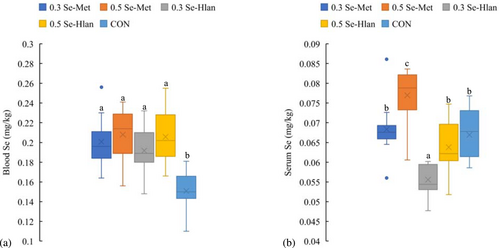
The effect of Se supplementation on milk Se is shown in Figure 1. Extreme impacts (P < 0.001) of Se supplementation, week, and Se supplementation × week interaction on milk Se level were shown in the statistical model (data not shown). During the experimental period, the highest milk Se values appeared in the 0.5 Se-Met group, and the lowest milk Se presented in CON and 0.3 Se-Hlan, with intermediate levels in the 0.5 Se-Hlan and 0.3 Se-Met groups. Total Se concentrations in blood and serum are summarized in Figure 2. In blood (Figure 2A), similar total Se values appeared in Se-Met and Se-Hlan supplementation groups, higher than that in CON (P < 0.01). Meanwhile, the highest serum Se appeared in the 0.5 Se-Met group, and the lowest value was in the 0.3 Se-Hlan group, with the other three groups having no significant difference (Figure 2B).


3.4 PCA results
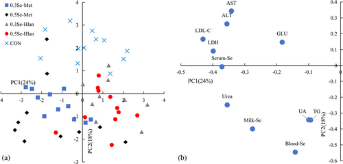
The PCA plots shown in Figure 3 illustrate the variance of measurements affected by Se supplementation (P < 0.05). The first two PCs explained 43% of the total variance. All the samples tended to cluster into three groups (Figure 3A): PC1 reflected the difference between Se-Met and Se-Hlan samples, while PC2 separated CON samples from organic Se samples. The corresponding loading plot lists the explained measurements (Figure 3B). AST, ALT, LDL-C, LDH, and GLU in serum were negatively correlated with PC1 and positively correlated with PC2, while urea, UA, TG in serum, and Se in milk and blood were negatively correlated with PC1 and PC2.

4 DISCUSSION
The influence of Se supplementation in dairy cow diets on milk composition and production in the present study was in line with the results from previous studies (Gong et al., 2014; Juniper et al., 2006), which found that neither organic nor inorganic Se supplementation affected milk composition or production. However, in other reports, fat (Sun et al., 2017), protein (Séboussi et al., 2016), and milk yields were affected by Se sources and dosages.
Most reports do not show a remarkable influence of Se supplementation on the blood biochemical parameters of dairy cows (Phipps et al., 2008), while some researchers have found that the source and amount of Se intake affected plasma metabolites such as BHBA, NEFA, and Na+ (Calamari et al., 2010). As indicators of liver function, lower ALT and AST than CON as well as lower AST than Se-Met supplementation appeared in Se-Hlan groups, a result that may reflect a potentially lower metabolic burden of Se-Hlan for the liver (Shaniko et al., 2015; Tsuji et al., 2010). In our study, more GLU and urea appeared in the Se-Met groups than in the Se-Hlan groups. Differences in serum urea among treatments in our study agree with the blood urea nitrogen (BUN) comparison of rats sampled at 1 h when the animals had received an injection of Se-HLan or Se-Met at 1.0 mg Se/kg body weight (Tsuji et al., 2010). The elevated GLU and urea levels for Se-Met intake seem to result from high basic metabolic functioning of the pancreas and liver, as explained by Shi et al. (2017). Although the addition of organic Se (5 g/head/day) for Tabapuã steers reduced UA excretion in urine samples (de Paiva Ferreira et al., 2020), the blood UA of dairy cows was not affected by Se source or dosage (Sun et al., 2019, 2021). Different from the above studies, we found that intake of selenized yeast by cows resulted in higher serum UA than with inorganic Se. Moreover, serum UA tended to decrease with increasing selenized yeast intake in our study. The increasing CREA trend with organic Se level also appeared in our results, and similar CREA levels were presented in all Se treatments, a result that was comparable with a study on mice using different Se sources injected at 0.1 and 1 mg Se/kg body weight. However, an overdose (10 mg Se/kg body weight) of Se-Met caused more serum CREA than that of Se-Hlan (Anan & Ogra, 2013). Considering the comparison of TG between treatments, Se-Met groups had similar TG values with Se-Hlan groups but had higher levels than CON, in accordance with observations in lactating ewes. The addition of inorganic Se for these animals caused lower blood TG than organic Se (Novoselec et al., 2018). Although not reported by other researchers, higher serum LDL-C in Se-Met groups than in Se-Hlan groups occurred in our study. High LDL-C may be associated with high serum Se levels because Se is bound to LDL-C in the blood (Schrauzer, 2000). Regarding LDH, serum measurements of mice injected with organic Se at 1 h observation showed that higher LDH values were presented in the Se-Met group compared to the Se-Hlan group (Tsuji et al., 2010), in line with our observations.
The influence of Se addition for dairy cows on plasma or serum antioxidant indexes was not consistent in previous reports. Some researchers have found that organic Se groups had higher GSH-Px and T-AOC as well as lower MDA compared with inorganic Se groups (Gong et al., 2014; Sun et al., 2017), while other researchers have observed no difference in these indexes between Se treatments (Séboussi et al., 2016). The addition of Se-Hlan in diets for dairy cows showed an MDA drop and a T-AOC increase in one farm observation (Li et al., 2020). In our study, serum GSH-Px and LPO levels showed a trend toward an effect affected by Se-Met and Se-Hlan supplementation. This was not in agreement with a report on broiler chickens that showed similar plasma GSH-Px levels between the two corresponding groups (Celi et al., 2013). As indicators of immune status, antibody IgG, the pro-inflammatory cytokines IL-6, TNF-α, and anti-inflammatory cytokines IL-2 in serum were not influenced by Se treatments in our study, similar to previous reports (Gong et al., 2014).
In our experiment, milk Se level surged rapidly after two weeks of Se-Met supplementation and increased slowly from week 3 to week 8 with some fluctuation, while inorganic Se treatment in CON showed little variation. This was similar to a previous report where the duration of transition for stable milk Se level was 18 days on average after dietary Se supplementation (Calamari et al., 2010). The stable milk Se concentration ranged from 37–53.6, 47–59.7, and 19.7–25.4 μg/kg from week 3 to week 8 for 0.3 Se-Met, 0.5 Se-Met, and CON, respectively, also corresponding to the results of the above research. In our results, the milk Se level for 0.3 Se-Hlan was higher than or similar to that of CON, and the milk Se level for 0.5 Se-Hlan was lower than or similar to that for 0.3 Se-Met. There are two potential reasons for the variation in milk Se among treatments. In ruminants, the absorption and digestibility of organic Se are higher than that of inorganic Se (Shaniko et al., 2015). From the aspect of metabolic pathways, Se-Met can be incorporated into protein synthesis as methionine or converted to selenohomocysteine via biosynthesis of Se-adenosylselenomethionine and then metabolized in a similar trans-selenation pathway as with Se-Hlan (Shaniko et al., 2015; Tsuji et al., 2010). Inorganic Se can be reduced and converted to selenoprotein (Shaniko et al., 2015). Therefore, the intake of Se-Met would lead to the enhancement of protein synthesis and an increase in the levels of selenoprotein, while the intake of Se-Hlan and inorganic Se would only lead to an increase in the levels of selenoprotein. Compared to inorganic Se, intake of organic Se (containing 63% Se-Met) increased the levels of Se-Met and selenocysteine (Se-Cys) in milk to a greater degree, with the Se level in the form of Se-Met being 5–6 times higher than that in the form of Se-Cys (Calamari et al., 2010). Based on our results, 0.5 Se-Met was more effective for the production of Se-enriched milk compared to other treatments.
In a comparison of dairy cows fed varied Se sources, organic Se from yeast or silage increased blood Se more than inorganic Se; different organic Se sources induced similar blood Se levels, while Se sources significantly affected serum Se levels (Séboussi et al., 2016). This trend was in agreement with our study, and our values for blood and serum Se were close to previously reported data (Li et al., 2020; Séboussi et al., 2016). However, there was no difference in blood Se levels between different level organic Se groups in this study, a result that was not in line with previous reports (Calamari et al., 2010). Serum Se was more sensitive to dietary Se supplementation than blood Se, because most of the blood Se is incorporated into red cells during erythropoiesis, and the lifespan of red blood cells for cows is 90–120 days (Calamari et al., 2010). In our observations, serum Se for cows was likely to be affected by the amount of organic Se intake, since the high Se treatment induced higher serum Se than the low Se treatment (Figure 2B). Unlike Se-Hlan, Se-Met from dietary sources is directly involved in serum albumin synthesis as methionine (Cheng et al., 2019), which may explain the higher serum Se level in the Se-Met groups compared to the Se-Hlan groups. Similar serum Se levels in CON and 0.3 Se-Met were presented in our study (Figure 2B), and this was in contrast to previous research (Séboussi et al., 2016). In addition, the lower serum Se in 0.3 Se-Hlan than in 0.3 Se-Met and CON may indicate a lesser impact of Se-Hlan intake on the serum Se fraction. The quantitative differences in serum Se caused by Se sources should be confirmed in further research. Regarding the difference in milk Se and serum Se in response to Se supplementation, milk Se was more sensitive to Se-Met intake for dairy cows (Figures 1, 2B, and 3). This may be associated with the higher Se proportion of Se-Met in milk (about 40–60%) than in serum (about 10–30%) (Awadeh et al., 1998; Calamari et al., 2010). The primary serum Se was incorporated into GSH and selenoprotein P, constant with the response to Se sources (Burk et al., 2001).
Overall, for the efficiency of feed Se sources, Se yeast was more efficient for increasing cows' milk and serum or plasma Se levels than inorganic Se (Youcef & Isabelle, 2016). For the comparison of different types of Se yeast, especially Se-Met vs. Se-Hlan, research on broilers (Celi et al., 2013) and mice (Anan & Ogra, 2013; Tsuji et al., 2010) has been performed, but there have been no previous studies on dairy cows. In our observations, Se-Met resulted in higher milk and serum Se levels than Se-Hlan. This was not in agreement with observations in mice made one hour after administration by bolus of a dose of 1 mg Se/kg body weight, where serum and blood Se levels using Se-Hlan were higher than with Se-Met (Tsuji et al., 2010). Regarding the distribution preference, Se-Met is more efficiently accumulated in the liver and pancreas than Se-Hlan (Anan & Ogra, 2013). A large dose (1 mg Se/kg body weight) bolus administration of Se-Met in mice caused higher levels of LDH, AST, and BUN as well as higher levels of amylase at 1 h compared to controls (without any treatments), while Se-Hlan did not have such effects, suggesting that a large dose of Se-Met induced liver and pancreatic damage (Tsuji et al., 2010). The accumulation of Se-Met for 8 weeks in these organs may affect basic metabolism, and this would cause higher AST, GLU, urea, LDL-C, and LDH than the values presented in the Se-Hlan groups (Table 3 and Figure 3).
In conclusion, our observations suggested that milk and serum Se levels were not affected equally by Se-Met and Se-Hlan. Compared with Se-Hlan, Se-Met was more effective in improving dairy cow serum or milk Se, and it is more likely to be used in Se-enriched milk production. The higher serum AST, GLU, urea, LDL, and LDH observed in Se-Met compared to Se-Hlan seemed to be correlated with a higher hepatic burden for Se-Met metabolization. The exact influence of Se-Met and Se-Hlan on cattle health needs further study.
ACKNOWLEDGMENTS
China Mengniu Dairy Company Limited is acknowledged for funding this study (grant number Q/MN-CW/GL-2020-7). We are grateful to all staff of the Ziang dairy farm for their support during animal care and sample collection. Gratitude is expressed to the Pony Test international group for sample analysis. We thank LetPub (www.letpub.com) for its linguistic assistance during the preparation of this manuscript.
CONFLICT OF INTEREST STATEMENT
We confirm that there is no conflict of interest with any financial organization in this paper.


