Computed tomography and cross-sectional anatomy of the head in the giant anteater (Myrmecophaga tridactyla)
Summary
The aim of this study was to identify the anatomical structures in the head region of the giant anteater (Myrmecophaga tridactyla) using computed tomography. For this, three giant anteater corpses, adult and female, were used. The tomographic analysis was performed in sequential order in rostrocaudal direction. Subsequently, the heads of these animals were sectioned in cross sections of approximately 1.5 cm each. Tomographic images were compared with anatomical sections. The association between computed tomography and anatomical sections allowed the visualization of anatomical structures found in the nasal, oral, cranial, pharyngeal and ear cavities. An elongated oral cavity delimited by the elongated mandible was observed. The oral cavity was mainly occupied by a thin and elongated tongue formed mainly by the union of the sternoglossal muscles. The nasopharynx and oropharynx extended up to the neck at the level of the fifth cervical vertebra. The findings of this study suggest that the giant anteater has a head with structures morphologically adapted to its feeding habits, such as an elongated skull, tongue, styloid bones and mandible. In addition, based on the results of this study, it is possible to suggest that computed tomography is a pivotal tool for the veterinary routine of wild animals, since it allowed the identification of anatomical structures found in the head of the giant anteater.
1 INTRODUCTION
The giant anteater (Myrmecophaga tridactyla) is the largest representative of the Myrmecophagidae family (Borges et al., 2017), with wide geographic distribution and that appears in all Brazilian biomes, from the Amazon to the southern fields (Miranda, 2012). Besides that, it is the largest species among neotropical vermilinguans (Gaudin et al., 2018). They are myrmecophagous since their diet consisting basically of ants and termites (Naples, 1999). This feeding habit affects digestive structures, metabolic rate, and animal behaviour and locomotion (Naples, 1999).
According to the International Union for Conservation of Nature (IUCN), the giant anteater is considered a vulnerable species of Brazilian fauna (Miranda et al., 2014). The specificity of the diet, low reproductive rates and its large size, associated with habitat degradation, have contributed to the threat of this species (Miranda et al., 2014). The destruction of its habitat, hunting, burning, dog attacks and run over have represented the main threats to the giant anteater, causing it to disappear in several Brazilian regions such as Santa Catarina (Cherem et al., 2004; Miranda, 2012) and Rio de Janeiro (Miranda et al., 2014).
The knowledge of anatomy is extremely important for the diagnosis and prognosis of wild animals, with imaging diagnostic being an important tool in diagnosis and treatment since it allows observing the topography of animals in a non-invasive way (Borges et al., 2017; Cunha et al., 2018; Costa et al., 2019). The computed tomography (CT) is adequate to measure, study and distinguish different types of tissues and structures, in addition to providing more accurate diagnoses in a shorter time. Another advantage of CT is the non-overlapping of structures, including for skull assessment (Mackey et al., 2008; Cunha et al., 2018).
Trauma resulting from being run over or predatory attacks is very common in wild animals, and the little anatomical knowledge of these species can compromise the entire recovery of these animals (Santos et al., 2020). Thus, knowledge of the anatomy of the giant anteater would facilitate the clinical-surgical approach of the animal, which has been increasingly the victim of trauma.
There are several studies associating computed tomography and anatomical sections describing the nasal, oral, pharyngeal and cranial cavities in the mammals such as in one-humped camel (Blanco et al., 2014), donkey (El-Gendy et al., 2014), Egyptian sheep (Awaad et al., 2019) and red fox (Mahdy & Zayed, 2020). The anatomy of the head and neck regions in the giant anteater been studied in some aspects, such as, for example, the anatomy of the masticatory system (Reiss, 1997; Naples, 1999; Endo et al., 2007), the shape and relationships of the hyoid apparatus (Borges et al., 2017), and morphology of the tongue (Casali et al., 2017). However, to our knowledge, there are no descriptions of the head in the giant anteater using CT. Thus, the aim of this study was to identify and describe the anatomical structures of the head of the giant anteater (Myrmecophaga tridactyla) using CT and to verify if the CT is a tool that can be used in the veterinary routine to identify anatomical structures of the giant anteater head.
2 MATERIAL AND METHODS
2.1 Animals
Three specimens of giant anteater (Myrmecophaga tridactyla) were used, adults and females, with an average weight of 40.95 kg. The corpses of these animals were provided by the Center for Medicine and Research on Wild Animals (CEMPAS), School of Veterinary Medicine and Animal Sciences, UNESP, Botucatu. The main cause of death was road vehicle crash-related injuries and the injuries did not involve the head of the animals. The corpses were frozen in a freezer at −20°C at the Laboratory of Wildlife Anatomy, UNESP, Botucatu. This study was authorized by the Committee on the Use of Animals of the School of Veterinary Medicine and Animal Science, UNESP (CEUA 1200/2019) and by the National Environmental and Wildlife Bureau (SISBIO – 77145-1).
2.2 Computed tomography
The specimens were previously thawed and taken to the Diagnostic Imaging Service, School of Veterinary Medicine and Animal Science, where the CT scans were performed, using the Single Slice CT scanner (Shimadzu SCT-7800 CT). The scanning parameters were 120 kVp, 190mA, 3-mm slice thickness and 1 s of rotation, using the soft tissue window and bone window. Tomographic images were taken in the rostrocaudal direction from the rostral end of the head to the level of the fifth cervical vertebra (C5) on transverse plane. Scans were also done on sagittal and dorsal planes to contribute to the findings. The images were reconstructed in MPR and subsequently in three dimensions (3D) using the Voxar Workstation 6.3 software (Barco®). All images were stored in Dicom®.
2.3 Anatomical sections
After tomographic images were taken, the animals were frozen again at −20°C in the Laboratory of Wildlife Anatomy UNESP, Botucatu. The heads and necks of the animals were removed and transversely sectioned in series using an electric band saw in cross sections about 1.5 cm thick from the nasal rostral region to the caudal region at the level of the cervical vertebrae. All anatomical sections were identified and compared to the tomographic images obtained. The anatomical structures were named according to the Nomina Anatomica Veterinaria (International Committee on Veterinary Gross Anatomical Nomenclature, 2017).
3 RESULTS
The association between computed tomography and anatomical sections allowed the visualization of anatomical structures related to the nasal, oral, cranial and pharyngeal and the ear cavities. Cross sections and computed tomography images are shown in a sequential order in the rostrocaudal direction from the level of the rostral end of the head to the level of the fourth cervical vertebra (C4).
3.1 The nasal cavity
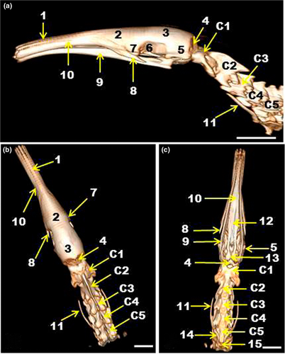
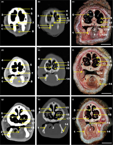
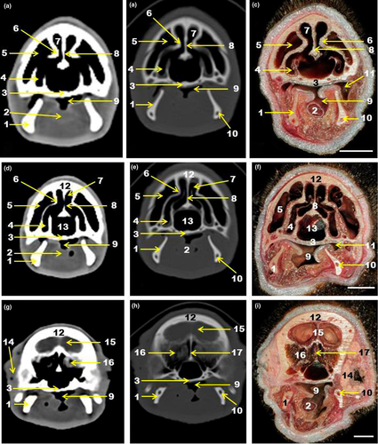
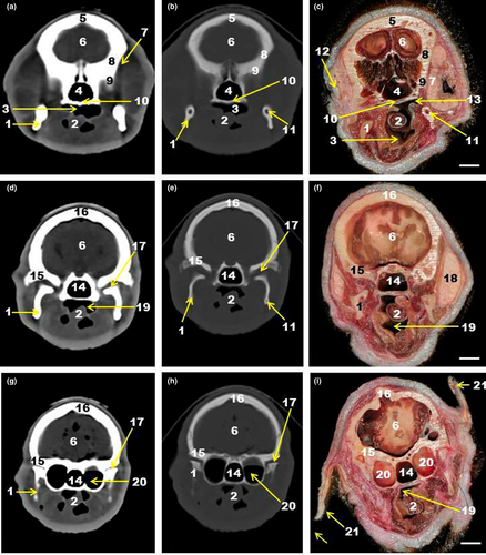
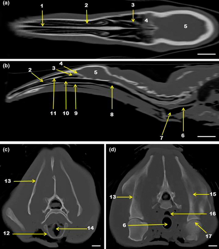
Rostrally, the bony limits of the nasal cavity were formed by the nasal bones dorsally and maxillae laterally and ventrally (Figure 1). The nasal cavity was easily visualized by CT and anatomical sections (Figures 2-4, 5). The frontal bone formed the dorsolateral part of the most caudal region of the nasal cavity (Figure 1). The nasal cavity in the giant anteater was divided by the nasal septum into two symmetrical halves (Figures 2 and 3). The nasal septum extended from the nostrils rostrally to the choanas caudally (Figure 5). The dorsal and ventral nasal conchae appeared on the lateral wall of each half of the nasal cavity (Figure 2). The dorsal nasal concha was long and unique and was continued rostrally by the straight fold, while the ventral nasal concha was short and branched in the alar and basal folds (Figure 2). Nasal meatuses were also observed under the conchae, with the dorsal nasal meatus smaller than the ventral nasal meatus (Figures 2g-i, 3a-f). The common nasal meatus was the communication space between the meatuses and was observed in the nasal cavity (Figure 2g-i). The ethmoidal labyrinth appeared in the caudal part of the nasal cavity next to the cribriform plate of the ethmoid bone (Figure 3g-i). When the bony limit of the nasal cavity was formed by the frontal bone, a relatively large frontal sinus, located caudal and lateral to the nasal cavity, was seen in tomographic and anatomical images (Figure 3a-f).
3.2 The oral cavity
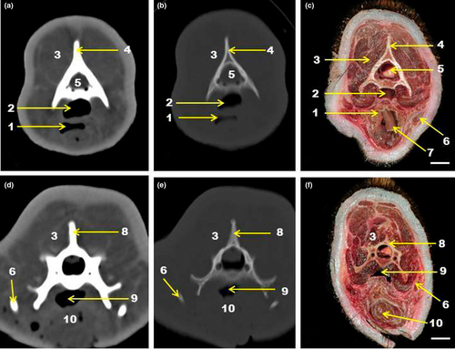
The oral cavity with the tongue, the palate and the oral vestibule was visible on tomographic images (Figures 2-4). The giant anteater oral cavity was relatively long and narrow and it was occupied mainly by the free part of the tongue, also long and narrow (Figures 2 and 3). The root of the tongue was observed in the neck at the level of the fourth cervical vertebra (C4), where the sternoglossus muscle appeared, uniting with the contralateral muscle and forming the tongue (Figure 6d-f). The well-developed hard palate was formed by the maxilla rostrally and by the palatine bone caudally (Figures 2a-f, 3a-g). The lateral walls of the oral cavity were represented mainly by the buccinator muscles, which were formed by two parts: superficial and deep (Figure 2c,f). The buccinator and masseter muscles were visible on tomographic images, although it was not possible to identify the parts of the buccinator muscle on CT (Figure 2d,g). The shape of the mandible followed the elongated and narrow shape of the oral cavity, and this was seen in all tomographic images (Figures 2-4). The temporomandibular joint also was visible on CT (Figure 4c,d,f,g). The mandibular canal was seen only in anatomical sections and in CT scans using bone window (Figures 2c,e,f,h,i,3b,c,e,f,h,i), and it was not possible to view it on tomographic images using soft tissue window (Figures 2d,g, 3a,d,g). The absence of teeth was also noticed in the tomographic images (Figures 1-4).
3.3 The pharyngeal cavity
The pharynx was divided into three portions: nasal, oral and laryngeal parts of the pharynx (Figures 4-7). The nasal portion of the pharynx or nasopharynx was continuous with the nasal cavity and the oral portion of the pharynx or oropharynx was continuous with the oral cavity. The nasal and oral portions of the Myrmecophaga tridactyla pharynx extended up to the neck, separated by a long soft palate (Figures 7 and 6). The stylohyoid bone was visible lateral to these cavities. This bone was one of the constituents of the hyoid apparatus (Figures 1, 6d-f). The sternoglossal space appeared on the oropharynx floor, which contained the sternoglossal muscle (Figure 6d-f). The laryngeal portion of the pharynx or laryngopharynx was found at the level of the fourth cervical vertebra (C4) (Figure 6d-f).
3.4 The cranial cavity
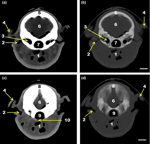
The small cranial cavity of the giant anteater was formed mainly by the parietal bones, in addition to the more caudal part of the frontal (dorsorostrally), occipital (dorsocaudally) and temporal bones (laterocaudally). All bones mentioned and the brain were visible on tomographic images (Figures 1a-c, 3g-i, 4a-h, 5a-d).
3.5 The orbital cavity
The orbital cavity or orbit was a cavity that contained the eyeball and the structures attached to the eyeball. The giant anteater had an incomplete orbit, and the bony limits were found only on the medial wall, which was formed by the orbital part of the frontal bone, lacrimal bone and orbital process of the palatine bone (Figure 4a-c). No zygomatic arch developed in the animals studied was found (Figures 1-4,7).
3.6 The ear
The ear was divided into external, middle and inner parts. The external ear consisted of the auricle and the external auditory canal. The external ear was seen in the CT. The middle ear with the tympanic cavity was also easily visible on tomographic images (Figures 4f-h, 5a-d). The inner ear was not identified in this study.
4 DISCUSSION
This study described the anatomical structures in the head region of the giant anteater (Myrmecophaga tridactyla) by CT and correlates these scans with anatomical cross-sectional images. Computed tomography was chosen as it has revealed itself as a non-invasive method used for the diagnosis in which there is no overlap of structures (Thrall, 2018; Mahdy & Zayed, 2020).
The head of the giant anteater was elongated with greatest development of the facial part in relation to the cranial part of the head. The oral and nasal cavities, edentulous maxilla and mandible, and tongue were elongated. Naples (1999) described the facial elongation in this mammal species. To this author, the nasal and frontal bones, maxillae and rostral parts of the mandible show the greatest elongation, what makes it possible to compare the giant anteater head to an elongated cylinder with a caudal base and rostral apex near the opening of the oral cavity (Naples, 1999).
Air space in the ear cavity provided good contrast in CT scans. Thus, it was possible to identify the auricle and external acoustic meatus in the external ear and the tympanic cavity in the middle ear in the giant anteater. Mahdy and Zayed (2020) reported that the air-filled tympanic cavity and tympanic bulla of the middle ear were easily identified in the CT scans of the red fox (Vulpes vulpes) head.
The nasal cavity of the giant anteater with its bony limits, nasal conchae, nasal septum, nasal meatuses and frontal sinus was visible on tomographic images, which shows the efficiency of CT in daily use when it is necessary to evaluate the nasal region. In this study, other paranasal sinuses, such as maxillary and sphenopalatine, were not observed. These paranasal sinuses were described for equines (El-Gendy et al., 2014), camels (Alsafy et al., 2014; Blanco et al., 2014) and sheep (Awaad et al., 2019). The presence of air inside the nasal cavity generates good contrast in CT images (Probst et al., 2005; Mahdy & Zayed, 2020). Tomographic findings also favourable were reported by Mahdy and Zayed (2020) in the nasal cavity of the red fox, who cite that these findings can be used as a clinical reference.
Tomographic images also made it possible to visualize the structures of the oral cavity, such as the hard and soft palate, oral vestibule, tongue and mandible. The oral cavity of the giant anteater is an elongated and narrow cavity, occupied mainly by the tongue, with a small rostral opening, which allows the tongue to pass through during feeding. It has been shown that the giant anteater has an elongated oral cavity with a mouth opening of approximately 2.75 cm in diameter (Brainard et al., 2008). For these authors, this anatomical feature limits the ability to secure endotracheal intubation in this animal.
In this study, one of the most notable anatomical features found in the head region of the giant anteater was the presence of a thin and elongated tongue that occupied a large part of the oral cavity. The presence of the tongue was observed in tomographic and anatomical images from the rostral end of the head region to the neck region. The giant anteater uses the tongue to capture prey and transfer it to the oesophagus, and the oral cavity provides the space where this tongue is left when not being used (Endo et al., 2007). Casali et al., (2017) claimed that the morphology of the tongue may reflect functional features related to myrmecophagous feeding habits. At the level of the last cervical vertebrae, it was possible to identify the sternoglossus muscle. At the root of the tongue, the sternoglossus muscle joins with the genioglossus muscle to form the free part of the tongue, although the sternoglossus muscle is the main muscle that forms the giant anteater tongue (Reiss, 1997). The sternoglossus muscle resulting from the fusion of sternohyoideus and hyoglossus muscles and is the main extrinsic muscle that contributes to the tongue in suborder Vermilingua (Casali et al., 2017).
The ability to retract the elongated tongue is supported by the also elongated hyoid apparatus, in particular by the stylohyoid bones (Naples, 1999). The cranial portion of the stylohyoid bone presented no correlation to the bony structures of the skull (Borges et al., 2017). The stylohyoid bones were visible in the tomographic and anatomical images of this study. The bones that comprised the hyoid apparatus in the giant anteater are formed by bony elements that have synovial joints between them (Naples, 1999) and that appear at the level of the second to sixth cervical vertebrae (Borges et al., 2017). Probably, this typical arrangement of the hyoid apparatus is an anatomical adaptation to the food intake process in the giant anteater (Borges et al., 2017).
The giant anteaters of this study presented the soft palate and the nasal and oral parts of the pharynx extended to the caudal level of the cervical region. These features were previously described in a study about cervical region and hyoid apparatus in living giant anteaters (Borges et al., 2017). The nasal and oral parts of the pharynx extended until the transition between the fifth and sixth cervical vertebrae, with the laryngeal part of the pharynx starting at this level (Borges et al., 2017). This is more of an anatomical adaptation to accommodate the elongated tongue (Reiss, 1997; Naples, 1999; Borges et al., 2017). Borges et al., (2017) reported that it is important to know these anatomical details to avoid incorrectly identifying the nasal part of the pharynx as the cervical part of the trachea.
Previous studies with the giant anteater showed similar coordination of the tongue and jaw movements, with medial rotation of the jaw occurring with each tongue protraction (Naples, 1999). For this author, mandibular rotation is a simple mechanism that accompanies the extremely rapid protrusion–retraction cycles of the tongue. Thus, the giant anteater rotates its mandible medially and laterally to control the tongue when it is elongated and protruded, and to retract it when it is relaxed (Endo et al., 2007). The protraction and retraction movements of the tongue are very fast (Reiss, 1997). There are suggestions that the masseter, superficial temporal (zygomatic part) and lateral pterygoid muscles are responsible for the medial rotation of the ramus of the mandible along its long axis, whereas the superficial temporal, medial and mylohyoid pterygoid muscles (rostral and medium) are responsible for the lateral rotation of the ramus of the mandible (Naples, 1999). This medial rotation is favoured by the presence of the mandibular symphysis, joining the two halves of the mandible, since the absence of fusion of the two mandible halves does not restrict the movement of the mandible (Endo et al., 2007).
In this study, lateral to the nasal and oral cavities, it was possible to observe the masseter and buccinator muscles. Naples (1999) reported that masticatory muscles in anteaters are simpler and more caudally confined than in other mammals. This may have led, in this study, to the non-identification of other masticatory muscles such as the medial and lateral temporal and pterygoid muscles. The buccinator muscle is divided into two parts: superficial and deep. In tomographic images, only the buccinator muscle was observed, without its divisions. Buccinator muscle formed by two parts, as found in the animals in this study, is an exclusive anatomical feature of Myrmecophaga (Naples, 1999), since in other mammals such as the dog, the buccinator muscle is not divided into parts (Evans & de Lahunta, 2013). The greater development of the buccinator muscle in the giant anteater and its division into parts can be explained due to its role in the masticatory function in these animals. This muscle allows the tongue to flow caudally without losing the adhered prey and decreases the oral opening when the tongue protrudes as the last means of removing ants contained in the tongue (Naples, 1999).
The present study provided the first comparison of 3D-reconstructions, CT scans and anatomical cross-sectional images in the head region of the giant anteater (Myrmecophaga tridactyla). The giant anteater has a head with structures morphologically adapted to its eating habits, such as an elongated skull, a thin and elongated tongue and, also stylohyoid bones and mandible, also elongated. Mandible with poor development of the ramus of the mandible, the presence of a mandibular symphysis and an incomplete zygomatic arch represent adaptations found in the giant anteaters that allow rotational movement of the mandible during the protraction and retraction of the tongue (Endo et al., 2007). Furthermore, our findings showed that CT is an important tool, since it allowed the identification of anatomical structures found in the head region of the giant anteater, and it is possible to use it in the routine of the veterinary clinic of wild animals.
ACKNOWLEDGEMENTS
The authors would like to thank Center of Medicine and Research of Wild Animals (CEMPAS), School of Veterinary Research and Animal Science, UNESP, for providing the animals and Coordination of Superior Level Staff Improvement (CAPES).
CONFLICT OF INTEREST
The authors declared no potential conflicts of interest with respect to the research, authorship and/or publication of this article.
Open Research
DATA AVAILABILITY STATEMENT
The data that support the findings of this study are available on request from the corresponding author. The data are not publicly available due to privacy or ethical restrictions.


