Morphological and scanning electron microscopic study of the lingual papillae in the Javan Pipistrelle (Pipistrellus javanicus)
Abstract
There are vast numbers of bats, in terms of both individuals and species, in Indonesia, although the precise species count is currently unknown. These bats demonstrate great variation in feeding patterns, with some being insectivorous, frugivorous, nectar-eating or carnivorous. One of the insectivorous bats found on Java Island, Indonesia, is the Javan pipistrelle (Pipistrellus javanicus). This paper presents a detailed morphological description of the tongue and lingual papillae of P. javanicus, using scanning electron microscopy (SEM) and light microscopy (LM). Macroscopically, the elongated tongue of P. javanicus can be divided into three sections: the apex, corpus and radix. On the dorsal part of the tongue (corpus), there is a prominence called the mediodorsal lobe, the lobe begin from the end of apex. On the radix, in the medial part, we describe the presence of a sulcus medianus. The SEM and LM observations show that the apex of the tongue bears three types of papillae: crown filiform, pronged filiform and fungiform. The corpus bears conical and fungiform papillae, whereas the radix bears vallate, large filiform, small filiform and fungiform papillae. Taken together, these results contribute to research on the tongue anatomy and feeding habits of the bat.
1 INTRODUCTION
The tongue of bats (Order Chiroptera) is a muscular organ capable of vigorous and elaborate movements in food manipulation, grooming and vocalization. The organ also participates in gustatory perception, due to the presence and location of taste receptor cells. The morphology of the tongue is related to the principal types of food and the feeding mechanism utilized by the organism, which in turn will determine the success of the animal's adaptation to its environment (Can, Atalgin, & Aydin, 2016; Iwasaki, 2002). Bat tongues show great variability in size and shape and in the features of the lingual papillae on the dorsal surface (Kilinc, Erdogan, Ketani, & Ketani, 2010; Shindo, Yoshimura, & Kobayashi, 2006). This variation in the distribution pattern, number and surface structure of the papillae is most marked between taxonomic ranks, such as orders and families, but sometimes also vary among species (Erdoğan & Pérez, 2014; Jackowiak, 2006; Kilinc et al., 2010).
The Javan pipistrelle (Pipistrellus javanicus), also known in Java as the Nighi Java bat, is insectivorous. As a natural predator of agricultural pests and mosquitoes, it plays an important role in maintaining the balance of the agricultural environment and ecosystem, using echolocation to find its prey. Echolocation is also used to control the movement of flying Javan pipistrelle in dark places. Commonly, Javan pipistrelle live and seek food on roofs or on leaf tips (Herman, Lina, & Hutson, 2000; Suyanto & Kartikasari, 2001).
Each species of bat has a distinctive tongue shape. The reason for this morphological diversity lies in the different ways in which they catch and process food, differences in grooming and variation in their vocal modulation. Important factors that influence the structure of the papillae are the type of food ingested by the bats, the way they chew their food, and how the food is transferred in the oral cavity (Abumandour, 2014; Abumandour & El-Bakary, 2013; Konig, 2004). Scanning electron microscope studies have been carried out on a variety of insect- and fruit-eating bats, including Pipistrellus savii (Park & Lee, 2009), Pipistrellus pipistrellus (Pastor et al., 1993), Hipposideros cervinus (Sharma et al., 1999), Myotis macrodactylus (Hwang & Lee, 2007), Rousettus aegyptiacus (Jackowiak, Lorych, & Godynicki, 2009; Abumandour & El-Bakary, 2013) and Rousettus amplexicaudatus (Gunawan et al., 2019). In this study, the structure of the papillae was examined using the tongue of a Javan pipistrelle (P. javanicus), also known as the Nighi Java bat.
Research on tongue morphology in animals, especially bats (both insectivorous and frugivorous), has been carried out by researchers around the world. In the insectivorous bats that have been described, there is a modification of the lingual organ due to their feeding habits and to assist with insect-pest repression (Ramteke, Zade, & Patil, 2013). In fruit-eating bats, in contrast, numerous and large fungiform papillae are widely distributed over the tongue. However, no information is yet available on the lingual papillae of P. javanicus, and recent data from other species of Pipistrellus are limited. Moreover, the biological nature of this species has been little studied due to difficulties in habitat analysis and animal tracking. In the present study, the morphology of the tongue and the three-dimensional characteristics of the lingual papillae in P. javanicus were examined using light and scanning electron microscopy (LM and SEM). The results are compared with previous reports to provide a detailed comparison of bat lingual anatomy as a literature study.
2 MATERIAL AND METHODS
2.1 Animal samples
Ten Javan pipistrelle (P. javanicus) individuals, five male and five female, were sampled in this study. They were captured in a farming area of Turi Village, Sleman, Special Region of Yogyakarta, Java Island, Indonesia. The capture process was approved by the Ethical Committee of the Faculty of Veterinary Medicine, Universitas Gadjah Mada, Yogyakarta, Indonesia (approval number 0041/EC-FKH/int./2019). Bats were captured at night using mist nets and transported in cloth bags to the laboratory. Captured bats were confirmed to be sexually mature and not pregnant or breastfeeding. Their morphological characteristics were identified according to the field key by Suyanto and Kartikasari (2001) in the Laboratory of Animal Systematics, Faculty of Biology, Universitas Gadjah Mada. In the laboratory, bats were directly euthanized and perfused.
2.2 Conservation status
Pipistrellus javanicus used in this study do not occur on any conservation list in Indonesia. According to the International Union for Conservation of Nature (IUCN) Red List of Threatened Species, P. javanicus is categorized as of Least Concern (LC).
2.3 Gross macroscopy analysis
The 10 sample bats were anaesthetized using a combination of ketamine (10 mg/kg body weight) and xylazine (2 mg/kg b.w.); for the euthanization procedure, a ketamine overdose was applied. Animals were placed in a dorsal position and fixed on a hard board. The oral cavity was opened, and the basal part of the tongue was separated from the mandible. The hyoid bone was cut, and the tongue was pulled, followed by cutting the caudal part of the larynx. Then, the sample tongue was rinsed using NaCl (physiological 0.9%) five times for 5 min each. Macroscopic observations were carried out on each sample with the use of a camera (Canon EOS 7000D). The tongue samples were divided into two group, for scanning electron microscopy and staining with haematoxylin and eosin.
2.4 Scanning electron microscopy
Five tongues from adult Javan pipistrelle (P. javanicus) were used in this study. The tongues were fixed with 0.5% glutaraldehyde (Chem Cruz), 1.5% formaldehyde (Nacalai Tesque) and 100 g HEPES (Chem Cruz) in 100 ml phosphate-buffered saline. The tongues were washed with 0.9% NaCl (Nacalai Tesque). Afterwards, the specimens were dehydrated using a graded series of ethanol (KgaA), vacuum dried (25°C, 4 Pa; Buehler 1000 Vacuum System) and sputter coated (JEOL JEC-3000FC) with platinum before being examined under a scanning electron microscope (JEOL-JSM6510LA) at an acceleration voltage of 15 kV.
2.5 Haematoxylin and Eosin (HE) staining
Five tongues were fixed in 10% formalin, then dehydrated using a graded series of ethanol, cleared in xylene (KgaA) and embedded on paraffin (Leica Biosystems). Sections were cut to 8 μm using a rotary microtome (Yamato RV 240), mounted in gelatin (KgaA) on coated glass slides and left overnight on a hot plate at 40°C. Before staining, the paraffin sections were dewaxed in xylene and rehydrated in graded ethanol. The tissue sections were stained with haematoxylin and eosin (HE; Bio-optica) and observed under a light microscope (Olympus BX51). Pictures were taken using Optilab software (Optilab).
3 RESULTS
3.1 Macroscopic observation
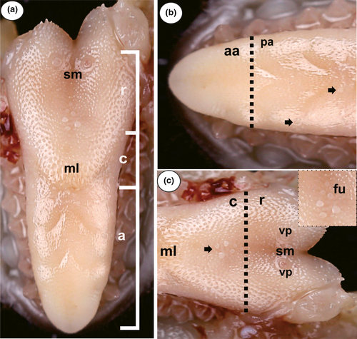
Macroscopic observation and SEM showed the tongue of P. javanicus consists of three parts: the apex, corpus and radix (Figure 1a). The boundary between the apex and corpus is marked by the frenulum, and the boundary between the corpus and radix is the caudal border of the mediodorsal lobe. The tongue has dorsal, ventral and lateral surfaces. The ventral surface of the tongue is connected to the mandible by the frenulum. The tongue of the P. javanicus specimens investigated here was 8.68 mm long, 1.44 mm wide at the apex, 2.58 mm wide at the corpus and 3.64 mm wide at the radix. A characteristic of the tongue's apex is its rounded tip that can move freely in the food collection process (Figure 1b). A characteristic of the dorsal corpus surface is a protrusion called the mediodorsal lobe (Figure 1c). On the ventral surface of the corpus the frenulum attaches directly to the mandible. The radix is characterized by the presence of vallate papillae and a median groove (Figure 1c). The radix attaches to the hyoid bone, the soft palate and the pharynx. The dorsal portion descends down and back towards the pharynx.
3.2 Scanning Electron Microscopy
3.2.1 Apex
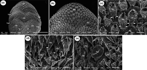
On the surface of the apex, there are more crown filiform papillae than pronged papillae (Figure 2a and Table 1). The crown filiform papillae in the caudal area lean in a posteromedial direction (Figure 2a). The pronged papillae are a small group of papillae located at the end of the apex and surrounded by crown filiform papillae. These papillae create a slightly rough texture on the anterior surface of the apex. This surface is wide and has two or three secondary processes that point towards to the caudal area (Figure 2b). Based on their morphology, which is similar in shape to a tooth, these papillae are called the pronged papillae.
| Type of papilla | Apex | Corpus | Radix | Length (µm) | Width (µm) |
|---|---|---|---|---|---|
| Crown filiform papilla | + | − | − | 100 ± 11 | 35 ± 5 |
| Pronged filiform papilla | + | − | − | 103 ± 17 | 61 ± 9 |
| Conical papilla | − | + | − | 190 ± 42 | 84 ± 15 |
| Small conical papilla | − | − | + | 62 ± 16 | 75 ± 23 |
| Large conical papilla | − | − | + | 127 ± 17 | 67 ± 13 |
| Vallate papilla | − | − | + | 205 ± 21 | 205 ± 21 |
| Fungiform papilla | + | + | + | 91 ± 16 | 91 ± 16 |
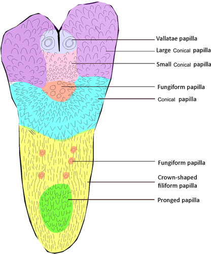
The posterior apex of the tongue is dominated by crown filiform papillae with scattered fungiform papillae (Figures 2a, 3 and Table 1). They are called crown filiform papillae because they are numerous and located very close together, almost attached to each other, in a crown-like formation.
The crown filiform and fungiform papillae found on the posterior apex have the same characteristics as those on the anterior apex. The observations revealed many fungiform papillae scattered laterally on the apex. Only a few fungiform papillae were located medially on the cranial side of the mediodorsal lobe. The fungiform papillae have a smooth and glossy surface, which contains slightly more prominent papillae relative to the other mechanical papillae (Figure 2c). These papillae were round and varied in size (Figure 2c and Table 1).
Each crown filiform papilla had a pointed end, with approximately 14 small secondary processes, that leads to the caudal lobe (Figure 2d).
3.2.2 Corpus
On the dorsal surface of the corpus, the fungiform papillae are dominated by conical papillae (Figure 3 and Table 1). The corpus of the P. javanicus tongue includes an elevated area called the mediodorsal lobe (Figures 1c and 4a).
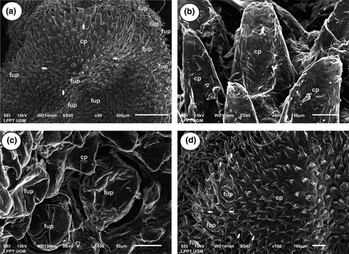
The conical papillae have a triangular or conical shape with sharp edges (Figure 4b). Conical papillae cover the semicircular mediodorsal lobe. The ends of the conical papillae in the mediodorsal lobe lean outward from the lobe. On the cranial part of the lobe, the papillae lean towards the anterior, whereas in the lateral area, the papillae lean laterally. Furthermore, on the medial part of the lobe, the papillae are directed dorsally, while on the caudal part of the lobe, the papillae bend towards the posterior (Figure 4d). As on the apex, the fungiform papillae are spread evenly over the corpus area and are mainly located lateral to the mediodorsal lobe and medial to the caudal part of the lobe (Figure 4c).
3.2.3 Radix
The radix was mostly dominated by conical papillae, although there were also fungiform and vallate papillae (Figured 5a, 3 and Table 1). The vallate papillae occur in two areas, arranged in parallel and equal in size (Figures 1c and 5a). The vallate papillae are located in the caudomedial region of the radix on either side of the median groove. The middle part of each vallate papilla is indented, which makes it look slightly concave. The surface of these papillae is generally smooth, without compartments or secondary papillae. The vallate papillae are surrounded by a canal and an outer wall that follows the papilla groove (Figure 5c). The outer wall of each vallate papilla is bounded by a large conical papilla on the lateral side and a small conical papilla on the medial side (Figure 5c). Between the two areas where vallate papillae occur, there is a median groove that increases towards the posterior area, but the papillae are not found there (Figure 5c).

The conical papillae are surrounded by the vallate papillae. The edges of the conical papillae in the radix are more rounded than those found in the corpus. Morphologically, the conical papillae are located precisely in the caudal section of vallate papillae, where they lean in a posterolateral direction. The conical papillae, which are located more laterally, were arranged towards the posteromedial area (Figure 5b). Moving towards the posterior, the conical papillae become longer (Figure 5b). The conical papillae occur in two forms; the larger ones are referred to as large conical papillae, and the others, which are smaller and blunter, are called the small conical papillae. The large conical papillae are located laterally to the vallate papillae, while the small conical papillae are located medially to the vallate papillae.
We also confirmed the presence in the radix of clustered fungiform papillae, although some were scattered between the conical papillae. The clustered papillae were located cranially from the median groove. There were approximately six fungiform papillae clusters made up of papillae of different sizes (Figure 5a), while the other papillae were located laterally to the radix (Figure 5a).
3.3 Histological observation
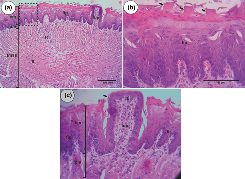
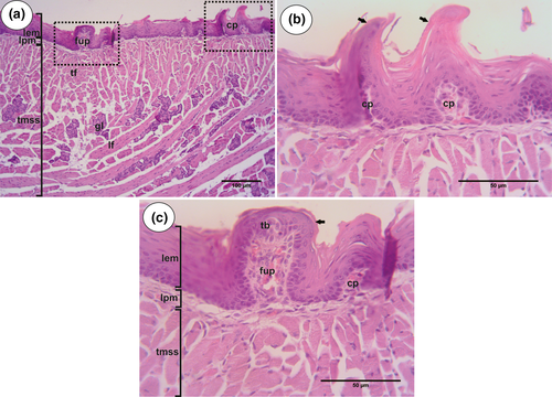
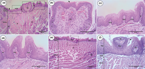
Staining with haematoxylin and eosin revealed three tongue layers in P. javanicus: lamina epithelialis mucosae, lamina propria mucosae and textus muscularis striatus syncytialis. In the lamina epithelialis mucosae, there is a squamous epithelium complex with keratinization. Confirming the SEM results, the histological findings also showed several types of papillae, namely filiform, fungiform and vallate. In the lingual mucosa, there is lamina propria, composed of connective tissue rich in blood vasa. In the textus muscularis, we found transversal fibre and longitudinal fibre (Figures 6a, 7a and 8a).
3.3.1 Apex
The observations revealed that the epithelium on the anterior part of the tongue of P. javanicus is extensively keratinized (Figure 6a). This region contains both filiform papillae and fungiform papillae (Figure 6a).
The filiform papillae appear to be the most numerous; they are spread over the anterior dorsal area of the tongue, and their connective tissue is covered by complex squamous epithelium. The tips of the filiform papillae are thin, sharp and tapered (Figure 6b).
Relatively fewer fungiform papillae were observed, and they were spread around between the filiform papillae. The surface of these papillae is round, in the shape of a fungus, and the epithelium was found to be keratinized. The connective tissue of the fungiform papillae enters the middle part of the papillae (Figure 6c).
3.3.2 Corpus
The histological findings from the corpus region are similar to those from the apex. The epithelium was covered with a thick keratinized layer. In this region, conical papillae and fungiform papillae were found. The fungiform papillae are located between conical papillae and are characterized by having one taste bud on the wall of the lamina epithelialis mucosae (Figure 7c). Conical papillae have a triangular or conical shape, with sharp edges (Figure 7b). In lingual muscle, there is a lingual gland (Figure 7a).
3.3.3 Radix
Our study shows the epithelial keratinization is thinner in the radix region than in the apex and corpus areas. Large conical papillae, small conical papillae and vallate papillae were found in the radix (Figure 8a). The large conical papillae were located laterally on the dorsal surface of the tongue. These papillae are triangular or conical, with a sharp tip on top of the surface (Figure 8d). The small conical papillae are located to the left and right of the median groove. The small conical papillae were smaller, with duller tips, than the large conical papillae (Figure 8c). Lingual glands were visible in the lingual muscle.
The vallate papillae protrude farther upward from the surface of the tongue in compared to the others papillae and are surrounded by a deep invagination in the surface of the epithelium that forms a trench and an outer wall around these papillae (Figure 8b). These papillae are located on the lateral side of the median groove and are larger than the other papillae. Vallate papillae bear 11–12 taste buds on the epithelial layer of the lateral wall (Figure 8b).
4 DISCUSSION
From the macroscopic observations, the tongue of P. javanicus was seen to consist of three parts, namely the apex, corpus and radix. This division is the same as that in the tongue of pigs, cows and horses (Konig, 2004) and in other insectivorous bats (Gregorin, 2003). However, according to Hwang and Lee (2007), the tongue of P. savii is divided into only two areas, the anterior and posterior parts. The anterior part equates to the apex, and the posterior part includes the corpus and radix. In P. javanicus, the dorsal area of the tongue possesses a prominence known as the mediodorsal lobe, running from the apex to the corpus in line with the first molar and terminating in line with the third molar. The median groove or sulcus medianus lies in the medial part of the radix. Gregorin (2003) and Ramteke et al. (2013) found a mediodorsal lobe and median groove in several species of insect-eating bats, namely Taphozous melanopogan, Chaerephon jobensis and Tadarida brasiliensis. Sharma et al. (1999) and Pastor et al. (1993) demonstrated that the tongue of insectivorous bats is smaller relative to that of frugivorous and nectar-drinking bats.
The SEM and histological staining demonstrated that the papillae of P. javanicus can be divided into mechanical papillae and gustatory papillae, based on their function. The mechanical papillae can be further divided into the filiform papillae and the conical papillae. The filiform papillae can be divided further based on their morphology, into the crown filiform papillae and the pronged papillae. The gustatory papillae can be divided into the fungiform papillae and the vallate papillae. The range of papillae found in the P. javanicus bat is the same as that found in other insectivorous bats (Gregorin, 2003; Pastor et al., 1993; Ramteke et al., 2013). The orientation and distribution of papillae are more important for insectivorous bats than the size of the tongue because their food is always a solid substance (Park & Lee, 2009). The lingual papillae of insectivorous bats show greater adaptation to food manipulation than to taste perception, as the latter is not a primary need for entomophagous animals. Pastor et al. (1993), who conducted research on P. pipistrellus, explained that the distribution and orientation of papillae allow the tongue to hold a captured insect in the mouth until it can be swallowed. Although the tongue adaptation of insect-eating bats is simpler than that of the nectar- and fruit-eating bats, the tongue of Pipistrellus demonstrates its ability to retrieve and manipulate insect prey caught by the wingtips or struck on the tail membrane. Pipistrellus is considered an aerial insectivore because it eats by catching insects on the wing.
The SEM showed that the filiform papillae are on the apex area of the tongue and can be divided into two types (crown and pronged). Filiform papillae were also identified through histological observation using HE staining. Crown filiform papillae are found in both the anterior and posterior apex. The form of the crown filiform papillae was found to be similar to that in a study conducted by Sharma et al. (1999) on Hipposideros cervinus. According to Ramteke et al. (2013), these papillae function as a structure for touching, catching prey quickly, and gripping and securing insects in the mouth while flying. In addition, the presence of these papillae makes the anterior surface of the tongue rough and cracked; this rough surface helps in the manipulation of prey and directs the insects towards the molars for mastication. The filiform papillae in these insectivorous bats differ from the filamentous type found in fruit bats. The pronged papillae are found only on the anterior apex of the tongue. These papillae aid bats in catching flying insects. These papillae also function as a barrier to hold insects and prevent their release before the bat can swallow them.
Interestingly, in P. javanicus, fungiform papillae were found to be distributed on the tongue from the apex to the radix. Our results are in line with research on Myotis macrodactylus (Hwang & Lee, 2007), Taphozous melanopogan (Ramteke et al., 2013) and P. pipistrellus (Pastor et al., 1993). According to Park and Lee (2009), the number of fungiform papillae and the size of the tongue of the species are proportional to the food consumed. Sharma et al. (1999) suggest that in insectivorous bats, the fungiform papillae have a more critical mechanical function relative to their gustatory function, even though the fungiform papillae are normally gustatory papillae that act as taste sensors. According to Park and Lee (2009), the distribution of fungiform papillae on the tongue occurs in four types. In the first type, they are distributed across the dorsal surface area, as in dogs; the second type refers to their distribution at the boundaries, including the front edges and the back, as in cats, moles and humans; the third type shows the distribution of fungiform papillae only at the front end and in the edges as in rabbits; and type four is identified by their distribution in the middle and at the front, as seen in mice. The distribution of fungiform papillae in P. javanicus, along the front edges of the tongue, is similar to the second type. In general, there are numerous taste buds on the fungiform wall. According to Mutlak et al. (2015) , taste buds are rarely found in the fungiform papillae of insectivorous bats, because taste is not a crucial factor for them when selecting food. However, our histological results show that the fungiform papillae of P. javanicus do have taste buds.
In the corpus area of the tongue, conical papillae make up the elevated portion of the mediodorsal lobe, which is a specific characteristic of the Microchiroptera. According to Gregorin (2003), in the Vespertilionidae, as in many other families, the mediodorsal lobe is less prominent than in the Molossidae, whose lobes are very prominent. According to Sharma et al. (1999) and Ramteke et al. (2013), these papillae act as a protective barrier for the lingual mucosa when holding food in the mouth while flying, and they help direct the insect fragments towards the oesophagus. Studies on Microchiroptera show that the mediodorsal lobes are formed by different mechanical papillae. In the family Molossidae, the mediodorsal lobe is formed by posteromedial papillae (Gregorin, 2003), while in P. pipistrellus (Vespertilionidae), the mediodorsal lobe is composed of filiform papillae. This protrusion plays an essential mechanical role in capturing insects while flying.
In the radix, there were only two vallate papillae were found, located in parallel. According to Sharma et al. (1999), although taste perception is a secondary requirement in food selection for insectivorous bats, the fungiform and vallate papillae still have gustatory potential. Based on our SEM and histological observations, the vallate papillae are surrounded by a groove. According to Park and Lee (2009), this groove serves to improve the accessibility of food to the taste buds in the papillae. The conformation of the vallate papillae found in this study is the same as that identified in a study by Sharma et al. (1999). Interestingly, an area between the two vallate papillae was revealed to lack papillae, and this area was also identified in P. pipistrellus (Pastor et al., 1993). According to Gregorin (2003) and Ramteke et al. (2013), the number of vallate papillae is relative to the types of food consumed by an animal species. In the Megachiroptera, there are three vallate papillae, while in the Microchiroptera, there are generally two. However, as already reported, in the family Molossidae of the Microchiroptera, there is a decreasing number of vallate papillae, and Rhinopoma hardwickii has only one (Mohebinia & Ghassemi, 2013), while no vallate papillae are found in Pipistrellus kuhli (Mutlak et al., 2015). In addition to the vallate papillae, in the radix of P. javanicus, there are large conical and small conical papillae, which are located laterally and medially to the vallate papillae. The large conical papillae with smooth and relatively wide surface may (would) two functions: as a protective structure against the lingual mucosa and to help pass insect fragments to the oesophagus for swallowing despite their smooth and relatively wide surfaces. In a study of the Molossidae conducted by Gregorin (2003), these conical papillae were referred to as basal papillae.
5 SUMMARY
In conclusion, this research provides detailed information on the lingual papillae of Pipistrellus javanicus from Yogyakarta, Java Island, Indonesia, based on scanning electron and light microscopic observation. The shape, structure and distribution of the papillae on the tongue are related to the type of food and its function.
ACKNOWLEDGEMENT
The authors would like to thank the Faculty of Veterinary Medicine, Universitas Gadjah Mada, for the Department Developing Grant in the year of 2019. The authors also thank the Integrated Laboratory for Research and Testing (LPPT), Universitas Gadjah Mada, for the use of the scanning electron microscope.
CONFLICT OF INTEREST
The authors declared no potential conflicts of interest concerning the research, authorship and publication of this article.


