Three-dimensional modelling of computed tomography images of limb bones in gazelles (Gazella subgutturosa)
Abstract
The objective of the present study was to model in 3D the detailed morphometric features of the thoracic and pelvic limb bones of the gazelle (Gazella subgutturosa), whose natural habitat is only in the Sanliurfa province, by using multidetector computerized tomography (MDCT) images. Materials from 14 (7 male and 7 female) dead gazelles were collected from the Harran University Veterinary Medicine Faculty clinics during or after treatment. MDCT images of the gazelles were taken and saved as DICOM (Digital Imaging and Communications in Medicine) files. MIMICS 20.1 software was used to generate a 3D reconstruction of the data, and morphological and morphometrical evaluation was performed. In respect of morphological features, the combined metacarpus (Mc) III and metacarpus Mc IV were the main weight-bearing metacarpal bone, while Mc II and Mc V were rudimentary. On the femur, the lateral condyle was bigger than the medial condyle at the caudodistal end and the medial wing of the trochlea femoris was larger than the lateral. As with the thoracic limb, the combined metatarsus (Mt) III and metatarsus Mt IV were the main weight-bearing metacarpal bone and they displayed a synostosis structure. There was no secondary metatarsus in the gazelles’ pelvic limb. Based on digital measurements, the tibia was the longest bone of the gazelles’ skeletal system, and there was a statistically significant sexual dimorphism throughout the skeleton at the levels of p < .05, p < .01 and p < .001. As a result, the present study's findings are useful as reference data for further studies on anatomy, surgery and archaezoology, while also being useful for classifying species and for forensic sciences.
1 INTRODUCTION
Gazella subgutturosa is a strain of Gazella which belongs to the Antilopinae subfamily of the family Bovidae (Albayrak, Pamukoğlu, & Kaya, 2007). Due to global impacts such as an increase in population across the globe, climate change, expansion of arable lands, poaching, use of excessive chemicals and chemical products, and soil erosion, the number of Gazella spp. is gradually decreasing. Similarly, its numbers have decreased and verged on extinction in Turkey (Gürler, Bozkaya, Özüt, & Durmuş, 2015; Mallon & Kindgswood, 2001). Gazella spp. was put in the red list in the reports published by International Union for Conservation of Nature and Natural Resources (IUCN) in the years 2004, 2008 and 2017, and were also reported to be under the risk of extinction (IUCN, 2004, 2008, 2017).
The three-dimensional modelling technique, being increasingly used in industry in the late 20th century, has recently been one of the most favoured methods in the medical field (D'Ursoet al., 1999). Viewing tissue and organ sections in a 3D way is called “reconstruction.” These reconstructive images help us determine treatment protocols in clinical cases, complicated pathological cases and forensic science cases that carry out re-identification procedures. They also contribute to some anthropological studies. The reconstruction technique is also crucial in the field of anatomy since it makes disordered structures easier to understand and provides educational materials (Sarıtaş, 2015; Verhoff et al., 2008).
Formed by two different words such as tomos and graphics, tomography is a radiological imaging method that examines the cross-sectional image of an object through collimated X-rays (Baykal & ve Oyar, 2003; Capello & ve Cauduro, 2008; Tuncel, 2007). Similarly, in computerized tomography, cross-sectional images are obtained by rotating the detector and X-ray tube around the object (Adapınar, 2016).
In these contexts, this study aimed to determine anatomical, osteometrical and volumetrical features of the bones that constitute ossa membri thoracici and ossa membri pelvini of the Gazella spp. (Gazella subgutturosa) which are under the risk of extinction. To do so, the present study proposes to carry out a 3D modelling via an MDCT imaging device. Moreover, a detailed anatomical description of the structure of the thoracic and pelvic limb skeletons of Gazella spp. is also presented in this study. On the other hand, it is aimed to compare osteometrical and volumetrical values of long bones between sexes. Consequently, it is hoped that obtained 3D models can be used in some scientific fields such as anatomy, surgery and archaezoology, and that they can also help classify certain species and present data for forensic science studies.
2 MATERIALS AND METHODS
In this study, 14 (7 females and 7 males) dead gazelles (2–3 years old, average weight of 15–20 kg) were used. These gazelles were brought to the hospital in the Faculty of Veterinary Sciences at Harran University for treatment but could not be saved. They had no risk of contagious diseases or other anomalies. Prior to the study, a written consent form was obtained from the General Directorate of Nature Conservation and National Parks (06.10.2017/209842) and Local Ethics Committee for Animal Experiments at Harran University (HRÜ-HADYEK) (2017/005-01-01).
2.1 Obtaining computerized tomography images and modelling
The MDCT images of the front and back extremities of the gazelles, which are the primary materials of this study, were obtained via a 64-detector, multislice, spiral computed tomography device (General Electronic Revolution) at 80 kV, 200 mA, 639 mGY and 0.625 mm slice thickness. For modelling, the MDCT images were converted into the DICOM format. Collected data were transferred to the MIMICS 20.1 program (The Materialise Group) that can carry out 3D modelling. In order to highlight osteological structures, the threshold value was first determined. By forming different masks for the selected bone tissue via “region growing” command, segmentation of all bones was completed, and they were converted into 3D forms via the “calculate 3D” command. On these 3D forms, osteometrical measurements were conducted according to the reference points suggested by Von Den Driesch (1976).
2.2 Statistical analysis
For the statistical analysis of morphometric measure values, the SPSS 13.0 program was used. The data were analysed via the Student t test. For comparisons between sexes, independent samples test was employed.
While writing the paper, the Nomina Anatomica Veterinaria (2017) was used for naming structures.
3 RESULTS
3.1 Morphological and morphometric results about ossa membri thoracici
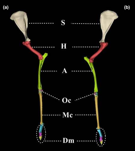
The order of the bones that constitute ossa membri thoracici from proximal to distal is presented in Figure 1.
3.1.1 Scapula
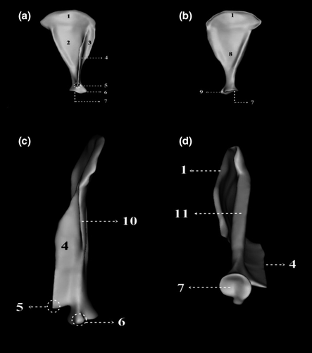
The scapula was determined to be a bone with an acute-angled triangle shape. In the facies costalis, some ambiguous facies serrata were seen. The form of the spina scapulae is low at margo dorsalis but rises from thence. The bone inclines towards the fossa infraspinata at the one-third of the middle third of the bone. The cavitas glenoidalis was found to have a round shape (Figure 2).
3.1.2 Humerus
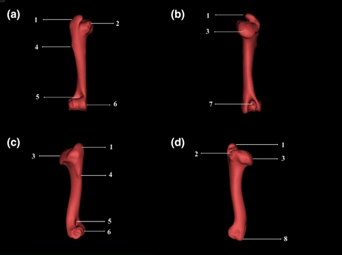
The tuberositas deltoidea of the humerus was slight in nature. It was also seen that, through a relatively clear notch that exceeded the caput humeri level, the tuberculum majus was separated into a pars cranialis and pars caudalis and had an inclination towards the caput humeri. It had an inclination towards caput humeri. Epicondylus lateralis and epicondylus medialis were found to be at the same level, and an oval but shallow fossa olecrani was seen between these two epicondylus (Figure 3). Osteometrical measurements of the humeri and their statistical analyses are presented in Table 1.
| Gender | N | Mean | Standard deviation | t-value | |
|---|---|---|---|---|---|
| Length (mm) | Female | 7 | 112.12 | 6.33 | .057NS |
| Male | 7 | 118.09 | 4.03 | ||
| DD Cr-C (mm) | Female | 7 | 12.68 | 0.77 | .074NS |
| Male | 7 | 13.51 | 0.82 | ||
| DD L-M (mm) | Female | 7 | 10.40 | 0.82 | .004 ** |
| Male | 7 | 12.01 | 0.86 | ||
| PW (mm) | Female | 7 | 34.63 | 1.53 | .436NS |
| Male | 7 | 35.37 | 1.91 | ||
| DW (mm) | Female | 7 | 22.62 | 2.04 | .023 * |
| Male | 7 | 24.97 | 1.26 | ||
| CMW (mm) | Female | 7 | 13.87 | 0.97 | .978NS |
| Male | 7 | 13.85 | 1.03 | ||
| CLW (mm) | Female | 7 | 7.89 | 0.88 | .814NS |
| Male | 7 | 8.00 | 0.75 | ||
| Surface area (mm2) | Female | 7 | 11,314.22 | 1,492.91 | .011 * |
| Male | 7 | 13,646.75 | 1,410.81 | ||
| Volume (mm3) | Female | 7 | 20,920.27 | 2,136.07 | .060NS |
| Male | 7 | 24,080.99 | 3,420.79 |
- Abbreviations: CLW, condylus lateralis width; CMW, condylus medialis width; DD Cr-C, diaphysis diameter (craniocaudal); DD L-M, diaphysis diameter (lateromedial); DW, distal width; PW, proximal width.
- ** p < .01;
- * p < .05;
- NS p > .05.
3.1.3 Skeleton antebrachii
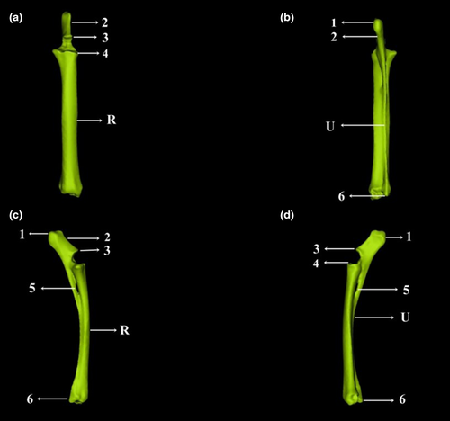
Between the radius and ulna, a spatium interosseum antebrachii proximalis was clearly seen. However, a spatium interosseum antebrachii distalis was found to be too small to distinguish. It was also observed that in radius’ proximality, the fovea capitis radii, which are an area over caput radii and on which trochlea humeri sits, were separated into two asymmetrical articular surfaces by sagittal canal, one being big in inner sider and the other being small in outer part. On the proximal ulna, the olecranon's medial face was found to be concave, while its lateral face was convex. The tuber olecrani, which is situated in the free edge of the olecranon, was divided into a tuberculum anconea mediale and tuberculum anconea laterale via a notch. It was also found to end with epiphyses, having a processus anconeus (Figure 4). Mean values for the data obtained from the radius and ulna were compared between sexes, and the results are presented in Tables 2 and 3.
| Gender | N | Mean | Standard deviation | t-value | |
|---|---|---|---|---|---|
| Length (mm) | Female | 7 | 133.09 | 4.92 | .211NS |
| Male | 7 | 135.81 | 2.38 | ||
| PW (mm) | Female | 7 | 22.80 | 1.02 | .042 * |
| Male | 7 | 24.08 | 1.09 | ||
| DW (mm) | Female | 7 | 18.61 | 0.51 | .000 *** |
| Male | 7 | 20.55 | 0.85 | ||
| DD L-M (mm) | Female | 7 | 13.22 | 1.17 | .102 NS |
| Male | 7 | 14.37 | 1.26 | ||
| DD Cr-C (mm) | Female | 7 | 7.67 | 0.38 | .018 * |
| Male | 7 | 8.30 | 0.49 | ||
| Surface area (mm2) | Female | 7 | 10,767.51 | 979.72 | .020 * |
| Male | 7 | 12,003.71 | 717.42 | ||
| Volume (mm3) | Female | 7 | 18,082.18 | 1,560.66 | .006 ** |
| Male | 7 | 21,070.47 | 1,808.33 |
- Abbreviations: DD Cr-C, diaphysis diameter (craniocaudal); DD L-M, diaphysis diameter (lateromedial); DW, distal width; PW, proximal width; surface area, total area of radius and ulna; volume, total volume of radius and ulna.
- * p < .05;
- ** p < .01;
- *** p < .001;
- NS p > .05.
| Gender | N | Mean | Standard deviation | t-value | |
|---|---|---|---|---|---|
| Length (mm) | Female | 7 | 158.15 | 8.74 | .201NS |
| Male | 7 | 163.18 | 4.48 | ||
| DD L-M (mm) | Female | 7 | 2.47 | 0.19 | .041 * |
| Male | 7 | 2.72 | 0.23 | ||
| DD Cr-C (mm) | Female | 7 | 2.98 | 0.30 | .143NS |
| Male | 7 | 3.33 | 0.51 | ||
| OL (mm) | Female | 7 | 29.06 | 2.70 | .585NS |
| Male | 7 | 29.94 | 3.17 | ||
| OW (mm) | Female | 7 | 15.79 | 0.80 | .225NS |
| Male | 7 | 16.43 | 1.07 |
- Abbreviations: DD Cr-C, diaphysis diameter (craniocaudal); DD L-M, diaphysis diameter (lateromedial); OL, olecranon length; OW, olecranon width.
- * p < .05;
- NS p > .05.
3.1.4 Ossa carpi
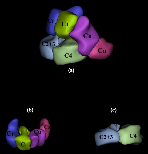
The ossa carpi consisted of six bones organized in two rows: four in the proximal row (from medial to lateral: os carpi radiale, os carpi intermedium, os carpi ulnare and os carpi accessorium) and two in the distal (from medial to lateral: os carpale II + III and os carpale IV). Os carpale I was not observed (Figure 5).
3.1.5 Ossa metacarpale
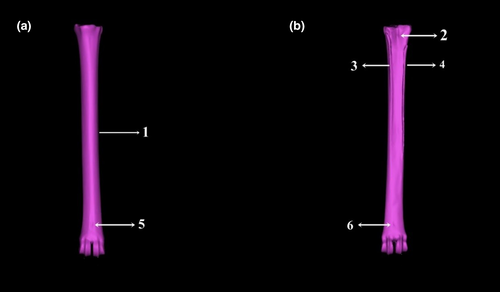
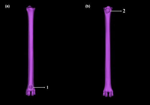
Os metacarpale III and os metacarpale IV were a fused, weight-bearing metacarpus, while os metacarpale II and os metacarpale V were rudimentary. On the dorsal face of the metacarpus, a sulcus longitudinalis dorsalis was observed. Although it was not clear proximally, it was more obvious distally. This groove had a foramen foramina were observed in the proximal part. On the palmar face, a sulcus longitudinalis palmaris was not clear. But, foramina in the proximal and distal regions were found to be clearer than those on the dorsal face. The distal end of the metacarpus was divided into two condylus via a dorsopalmar fissura (Figure 6). Mean values for the data obtained from the ossa metacarpale were compared between the sexes, and the results are presented in Table 4.
| Gender | N | Mean | Standard deviation | t-value | |
|---|---|---|---|---|---|
| Length (mm) | Female | 7 | 146.01 | 4.77 | .057NS |
| Male | 7 | 150.71 | 3.46 | ||
| PW (mm) | Female | 7 | 18.35 | 0.65 | .009 ** |
| Male | 7 | 19.58 | 0.83 | ||
| DD L-M (mm) | Female | 7 | 10.53 | 0.55 | .053NS |
| Male | 7 | 11.43 | 0.97 | ||
| DD Cr-C (mm) | Female | 7 | 9.67 | 0.38 | .421NS |
| Male | 7 | 9.88 | 0.54 | ||
| DW (mm) | Female | 7 | 18.02 | 1.01 | .077NS |
| Male | 7 | 18.91 | 0.69 | ||
| Surface area (mm2) | Female | 7 | 8,984.43 | 726.96 | .064NS |
| Male | 7 | 9,620.98 | 391.68 | ||
| Volume (mm3) | Female | 7 | 14,003.31 | 1,535.64 | .346NS |
| Male | 7 | 15,199.31 | 2,838.08 |
- Abbreviations: DD Cr-C, diaphysis diameter (craniocaudal); DD L-M, diaphysis diameter (lateromedial); DW, distal width; PW, proximal width.
- ** p < .01;
- NS p > .05.
3.1.6 Ossa digitorum manus
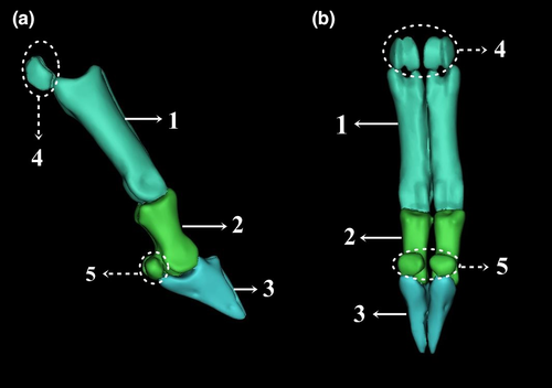
Gazella subgutturosa was observed to have a full-grown digit on its third and fourth fingers of both front legs. However, the second and third dactyli were rudimentary, and the first was not existent. Each digit consisted of three bones: phalanx proximalis, phalanx media and phalanx distalis (Figure 7).
3.2 Morphological and morphometric results about ossa membri pelvini
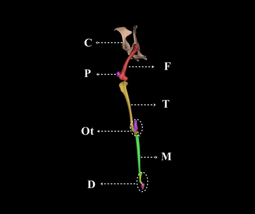
The order of the bones that constitute ossa membri pelvini from proximal to distal is presented in Figure 8.
3.2.1 Ossa coxae
Incisura (inc.) acetabuli in the acetabulum was observed to be large when compared to small ruminantia. Similarly, the fossa acetabuli was seen to be deep. In the ventrolateral acetabulum, small and shallow areas of attachment for the lateralis m. rectus femoris and medialis m. rectus femoris were present (Figure 9). The tuberculum m. psoas minoris was not clear, and the tuberculum pubicum dorsale was not present (in both sexes). A lateral sulcus was seen over the tuberculum pubicum ventrale and between the two eminentia iliopubica (Figure 9).
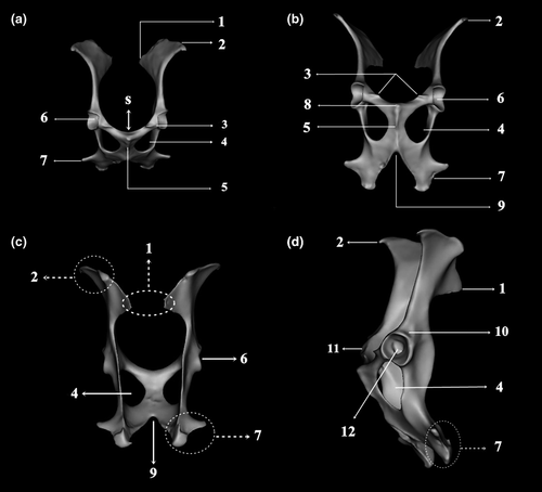
3.2.2 Femur
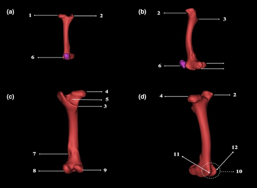
A shallow fovea capitis femoris, to which the ligamentum capitis ossis femoris attaches, was present on the caput ossis femoris. The trochanter major was observed to be a single unit and at the same level as the caput femoris. The trochanter minor was seen in the proximal part of the bone in a caudomedial camber shape. In the distal femur, the condylus lateralis was bigger than condylus medialis, and between them exists a fossa intercondylaris. The medial edge of trochlea femoris was larger than the lateral edge. The patella, which articulated with the femur's trochlea ossis femoris, was observed to be a big, reverse pyramid (Figure 10). Mean values for the data obtained from the femora were compared between sexes, and the results are presented in Table 5.
| Gender | N | Mean | Standard deviation | t-value | |
|---|---|---|---|---|---|
| Length (mm) | Female | 7 | 145.01 | 5.75 | .035 * |
| Male | 7 | 151.46 | 4.31 | ||
| PW (mm) | Female | 7 | 34.54 | 1.75 | .019 * |
| Male | 7 | 36.94 | 1.58 | ||
| DD L-M (mm) | Female | 7 | 14.12 | 0.91 | .091NS |
| Male | 7 | 15.15 | 1.17 | ||
| DD Cr-C (mm) | Female | 7 | 12.13 | 0.90 | .956NS |
| Male | 7 | 12.16 | 1.32 | ||
| DW (mm) | Female | 7 | 28.57 | 1.55 | .009 ** |
| Male | 7 | 30.45 | 0.42 | ||
| Surface area (mm2) | Female | 7 | 15,542.05 | 1,168.74 | .145NS |
| Male | 7 | 16,453.04 | 1,012.47 | ||
| Volume (mm3) | Female | 7 | 33,146.06 | 3,391.01 | .603NS |
| Male | 7 | 34,837.53 | 7,654.20 |
- Abbreviations: DD Cr-C, diaphysis diameter (craniocaudal); DD L-M, diaphysis diameter (lateromedial); DW, distal width; PW, proximal width.
- * p < .05;
- ** p < .01;
- NS p > .05.
3.2.3 Skeleton cruris
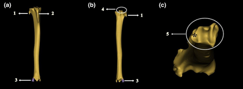
It was observed that, between the tibia's condylus medialis and lateralis, the tuberculum intercondylare lateralis and tuberculum intercondylare medialis had the same height. In the outer part of the condylus lateralis, the fibula remnant was found to be a sharp ridge. On the lateral side of the cochlea of the tibia, an articular surface for articulation with the os malleolare was present (Figure 11). Mean values for the data obtained from the tibiae were compared between sexes, and the results are presented in Table 6.
| Gender | N | Mean | Standard deviation | t-value | |
|---|---|---|---|---|---|
| Length (mm) | Female | 7 | 179.70 | 5.86 | .106NS |
| Male | 7 | 185.02 | 5.50 | ||
| PW (mm) | Female | 7 | 31.08 | 0.95 | .445NS |
| Male | 7 | 31.69 | 1.83 | ||
| DD L-M (mm) | Female | 7 | 13.71 | 0.85 | .216NS |
| Male | 7 | 14.46 | 1.25 | ||
| DD Cr-C (mm) | Female | 7 | 11.23 | 0.66 | .329NS |
| Male | 7 | 10.83 | 0.83 | ||
| DW (mm) | Female | 7 | 20.77 | 1.03 | .782NS |
| Male | 7 | 20.95 | 1.25 | ||
| Surface area (mm2) | Female | 7 | 14,667.07 | 1,871.32 | .050 * |
| Male | 7 | 16,427.41 | 1,024.45 | ||
| Volume (mm3) | Female | 7 | 28,276.14 | 3,336.44 | .594NS |
| Male | 7 | 29,663.96 | 5,806.10 |
- Abbreviations: DD Cr-C, diaphysis diameter (craniocaudal); DD L-M, diaphysis diameter (lateromedial); DW, distal width; PW, proximal width.
- * p < .05;
- NS p > .05.
3.2.4 Ossa tarsi
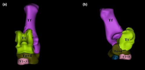
Ossa tarsi were present in three rows: proximal, middle and distal. In the proximal row, two bones named os tarsi tibiale/talus (Tt) and os tarsi fibulare/calcaneus (Tf) were present. In the middle row, the single bone called os centro quartale (Tc + IV), which was formed by the os tarsi centrale (Tc) and os tarsale quartum (TIV), was observed. In the distal row, the os tarsale I was a separate bone, while the os tarsale II and os tarsale III (TII + III) were fused and formed the os cuneiforme intermedio laterale. Os centro quartale (Tc + IV) was also found in the distal row (Figure 12).
3.2.5 Ossa metatarsalia
Metatarsus III and metatarsus IV were fused and weight-bearing (os metatarsale III and os metatarsale IV). As in the thoracic limb, these bones had a synostosis structure. There were no rudimentary metatarsals. No sulcus was seen on the dorsal and plantar faces. On the distal dorsal face, a foramen was observed. However, no foramen was seen on the distal and proximal plantar faces. On the proximal plantar face, an articular surface for articular with the sesamoid bone, os sesamoideum metatarsale, was present (Figure 13). Mean values for the data obtained from ossa metatarsalia were compared between male and female gazelles, and the results are presented in Table 7.
| Gender | N | Mean | Standard deviation | t-value | |
|---|---|---|---|---|---|
| Length (mm) | Female | 7 | 154.54 | 3.32 | .003 ** |
| Male | 7 | 161.96 | 4.27 | ||
| PW (mm) | Female | 7 | 17.66 | 1.58 | .466NS |
| Male | 7 | 18.15 | 0.78 | ||
| DD L-M (mm) | Female | 7 | 9.41 | 0.73 | .652NS |
| Male | 7 | 9.56. | 0.39 | ||
| DD Cr-C (mm) | Female | 7 | 12.01 | 0.58 | .376NS |
| Male | 7 | 11.59 | 1.07 | ||
| DW (mm) | Female | 7 | 19.05 | 0.51 | .090NS |
| Male | 7 | 20.15 | 1.49 | ||
| Surface area (mm2) | Female | 7 | 77,355.21 | 177,893.62 | .341NS |
| Male | 7 | 10,716.67 | 643.65 | ||
| Volume (mm3) | Female | 7 | 17,519.49 | 1,398.52 | .481NS |
| Male | 7 | 18,543.24 | 3,450.72 |
- Abbreviations: DD Cr-C, diaphysis diameter (craniocaudal); DD L-M, diaphysis diameter (lateromedial); DW, distal width; PW, proximal width.
- ** p < .01;
- NS p > .05.
3.2.6 Ossa digitorum pedis
The ossa digitorum pedis was similar to the ossa digitorum manus in both the number of bones and their anatomical features.
4 DISCUSSION
In this study, 3D models were reconstructed from computed tomography images of the thoracic and pelvic limb of the gazelles (Gazella subgutturosa). Using these models, the gazelle's ossa membri thoracici and pelvini bones were morphologically examined. Some morphometric measurements were also carried out.
Depending on their movement capacity, there are differences in the scapula's anatomical structure. While runner animals have a long, narrow and vertical scapula for a longer stride, fossorial and swimming mammals have a big, triangular scapula (Polly, 2007). In this study, the scapula of Gazella subgutturosa, which is a member of the antelope family, was seen to have a narrow, triangular shape.
Moreover, it was found that the cavitas glenoidalis had a round shape in the gazelles. Similarly, in former studies carried out with East African goats (Makungu & ve Merere, 2017), roe deer (Gültekin, 1962) and feral pigs (Sus scrofa) (Karan, 2012b), it was seen that the cavitas glenoidalis had a round shape. Likewise, in wild goats and hair goats, it had a roundish shape (Taşbaş, 1978). However, it was found to be oval in angora goats (Taşbaş, 1978), circular in black Bengal goats (Capra hircus) (Siddiqui, Khan, Moonmoon, Islam, & Jahan, 2008), acetabulum in Indian muntjacs (Muntiacus muntjak) (Sasan, Sarma, & Suri, 2018), and shallow and oval in Indian gazelles (Boselaphus tragocamelus) (Rohlan, Mathur, & Shringi, 2017).
In the present study, two spatium interosseum antebrachii were observed proximally and distally between the radius and ulna. The distal one was very small. However, in a study by Taşbaş (1978) carried out with wild, hair and angora goats, it was shown that there was only a proximal spatium interosseum antebrachii. Similarly, Peters (1989) argued that, in some gazelle species (Gazella blainville) and tragelaphus (Tragelaphus scriptus), there was no distal spatium interosseum antebrachii, but it sometimes was present in Bohor reedbuck (Redunca redunca). On the other hand, in some other studies, it was found that there was both a proximal and distal spatium interosseum antebrachii in spotted deer (Axis axis) (Choudhary, Mathur, Joshi, & Dangi, 2012), ruminants (Dursun, 2006) (Bahadır & ve Yıldız, 2016), black Bengal goats (Capra hircus) (Siddiqui et al., 2008) and small East African goats (Makungu & ve Merere, 2017).
In our study, a sulcus longitudinalis dorsalis was present on the dorsal surface of the metacarpus. It was further observed that this groove had a distal foramen and, in a few animals, an unclear and tiny proximal foramen. Likewise, in similar studies, it was found that Gazella gazelle bennettii (Jangir, Mathur, Dangi, & Joshi, 2012) and Antilope cervicapra (Choudhary & ve Singh, 2016) had a narrow sulcus on the dorsal surface of the metacarpus and that this sulcus had proximal and distal foramina. However, although it was claimed that metacarpus’ palmar face had a sulcus longitudinalis palmaris (Bahadır & ve Yıldız, 2016; Choudhary et al., 2016; Gültekin, 1962; Jangir et al., 2012), this structure was not clear in our research animals, but there were some clear proximal and distal foramina holes in the palmar surface. In the present study, two fully developed digits III and IV were observed in each thoracic and pelvic limb of the gazelle. This confirmed claims of Gündemir, Özkan, and Mutuş (2020) in domestic cattle, Siddiqui et al. (2008) in black Bengal goats (Capra hircus) and Taşbaş (1978) in wild, domestic, angora and hair goats.
Depending on the body weight and movement capacity, the morphology of the os coxae varies in mammals. These differences are clearly identified in ala ossis ilii's direction and the acetabulum's shape and depth (Polly, 2007). In this study, ala ossis ilii was observed to have a dorsolateral inclination in Gazelle subgutturosa, as it does in small ruminants (König & ve Liebich, 2015; Rajani, Chandrasekhar, Chandy, & Chungath, 2013). The gazelles, which are very active, were found to have a wider acetabulum depth. Taşbaş (1978) suggested that there was a horizontal sulcus that extends from the pecten osis pubis ventralae to the eminentia iliopubica in wild, domestic, angora and hair goats. Gültekin (1962) found a similar structure in roe deer and goats. Gültekin (1962) also claimed that, although rare, this sulcus might be seen in sheep, as well. In the present study, it was determined that gazelles had a similar sulcus.
Although the femur is often thought to be the longest bone of skeletal system (Bahadır & ve Yıldız, 2016; Dursun, 2006; Islam, Singh, & Guatam, 2018), it was found in the present study that the tibia was longer than the femur. Moreover, that trochanter major, on the proximal femur, was in a single piece and at the same level as the caput femoris. Similarly, in former studies, the trochanter major was also found to be at the same level as the caput femoris in feral pigs (Sus scrofa) (Karan, 2012a), sambar deer (Cervus unicolor) (Rajani et al., 2012), spotted deer (Axis axis) (Islam et al., 2018) and small ruminants (Dyce, Sack, & Wensing, 2018). However, it was revealed that it was higher than the caput femoris in Indian muntjacs (Muntiacus muntjac) (Rajani et al., 2013), cows and mares (Crişan, Damian, Dezdrobitu, & Mateaş, 2009), and large ruminants (Bahadır & ve Yıldız, 2016). But, in camels (Crişan et al., 2009), otters (Lutra lutra) (Dinç, Aydın, & Atalar, 1999) and dogs (Gültekin & ve Uçar, 1980), the trochanter major was found to be lower than the caput femoris. On the other hand, Chimming et al. (2015) claimed that in swamp deer (Blastocerus dichotomus), trochanter major was separated into cranial and caudal parts.
In the study, it was seen that the tuberculum intercondylare lateralis and tuberculum intercondylare medialis, which were situated between the tibia's condylus, were at the same level. However, Rajani et al. (2013) revealed that, in Indian muntjacs (Muntiacus muntjac), the tuberculum intercondylare mediale was longer than the tuberculum intercondylare laterale. Likewise, Crişan et al. (2009) had a similar finding in camels and cows. On the other hand, Karan (2012a) suggested that the tuberculum intercondylare laterale was higher than the tuberculum intercondylare mediale in feral pigs.
In this study, Gazella subgutturosa was found to have III and IV metatarsi, but no rudimentary metatarsus. However, Bahadır and ve Yıldız (2016) and Dursun (2006) argued that, in ruminants, a rudimentary metatarsus II was present. Similarly, Rajani et al. (2013) and Çakır, Öğüt, and Kabak (1998) showed a secondary metatarsus in Indian muntjacs (Muntiacus muntjac) and in roe deer, respectively.
There are no findings in the literature with respect to using the CT technique for morphometric and morphological analyses of the thoracic and pelvic limb of gazelles (Gazella subgutturosa). Therefore, we consider that the findings are useful as basic data set for classification of the population and for diagnosing of appendicular skeleton disorders using CT images.
CONFLICT OF INTEREST
There is no conflict of interest in the present form of the manuscript.


