Surgical Anatomical Landmarks of the Thoracolumbar Vertebral Column on Magnetic Resonance Imaging in Dogs
Summary
Magnetic resonance imaging (MRI) in veterinary medicine profoundly improved spinal cord disease investigation in canine patients. We aimed to further describe the anatomical landmarks of the thoracolumbar junction in sagittal MRI sequences. MRI studies from 90 dogs were reviewed retrospectively, representing a broad cross section of breeds and body weights. The ratio of the distance from the dorsal aspect of the vertebral canal to the dorsal aspect of the transverse process or rib articulation relative to the length of L2 vertebra was determined for T12, T13, L1 and L2 vertebrae. A statistically significant difference was noted with the transverse processes being more ventrally located than the cranial fovea costalis. The lumbar transverse processes and rib articulations dramatically varied in shape, being oval or round, respectively. The sagittal image at the level of the lateral margin of the articular facet joint proved to be the most consistent for review of these structures.
Introduction
Magnetic resonance imaging has profoundly changed how we evaluate and treat canine spinal cord disease. Since the early 1980s, MRI has routinely been used for the investigation of thoracolumbar inter-vertebral disc disease (IVDD) in dogs (Lamb and David, 2012). The ability to more accurately localize compressive lesions, along with better delineation of the margins of compression, has made MRI an invaluable tool for surgical planning and evaluation of the soft tissue structures of the central nervous system. The images created allow one to not only evaluate the location and size of a lesion but also better identify the composition of the compressive material (Tidwell et al., 2002; da Costa and Samii, 2010). This is particularly important when evaluating inter-vertebral disc disease as there are times when the degree of compression secondary to haemorrhage, haematoma or associated intramedullary changes would alter the treatment plan and the patient's prognosis (da Costa and Samii, 2010; Dennis, 2011).
When considering surgical planning for decompressive laminectomies within the thoracolumbar region, understanding of anatomical landmarks at the thoracolumbar junction is critical to aid in the identification of a specific inter-vertebral disc space. Intra-operatively, local anatomical structures are used for localization of the surgical site. This is accomplished by identifying the thoracolumbar junction and differentiation of the lumbar transverse processes and costovertebral articulations to allow accurate counting of vertebrae. In addition to using local anatomy, descriptions of using local radiographic markers such as a hypodermic needle, with or without the administration of a visible dye such as methylene blue, have been previously described (Fingeroth and Thomas, 2014). Alternative imaging modalities such as a ventrodorsal radiograph or a dorsal planar MRI series allowed the best identification of these landmarks. These imaging modalities, in combination with the sagittal and transverse images, allow quick evaluation of the features of the vertebrae that will be easily identified in surgery, including the spinous processes, articular facets and either transverse processes or the costovertebral articulations on the lateral aspect of the vertebral body (Dennis, 2011). Anatomy is not always consistent, and a number of confounding but normal variations in the vertebrae can be noted. These can include anomalous vertebrae such as hemivertebrae, block vertebrae or transitional vertebrae that may have abnormally shaped ribs, transverse processes or in some cases a transverse process on one side and a rib on the other (da Costa and Samii, 2010). With previous reports indicating that 16–30% of dogs have one of these variations, intra-operative localization can be difficult without thorough knowledge of these anomalies (Bisson, 2010).
In this pilot study, we aimed to describe the normal anatomy of the thoracolumbar junction in sagittal slices in dogs. We focused our attention on evaluation of the distinguishing characteristics between rib and transverse processes. Furthermore, our hypothesis was that we could easily and reliably determine the vertebral body location via objective measure of the height of the process on transverse T2-weighted images. We repeated evaluation of the origin of the coeliac and cranial mesenteric arteries relative to the thoracolumbar junction to confirm previously reported results.
Materials and Methods
Patient selection
A search of medical records was completed, and dogs were identified that had undergone thoracolumbar MRI at the University of Glasgow Small Animal Teaching Hospital. MRI studies were eliminated if they did not have sagittal images through the thoracolumbar spine and transverse images through at least some of the T12-L2 vertebral bodies. Cases were excluded if vertebral anomalies other than transitional vertebrae were noted. 90 patients were identified as meeting the criteria.
MR image acquisition
MRI of the thoracolumbar spine was performed using a 1.5-tesla MR imaging system (Magnetom, Siemens, Camberley, UK). T1-weighted (360-870/10-15; range TR/TE) and T2-weighted (2160-5890/86-130; range TR/TE) images were acquired in both the sagittal and transverse planes. Dorsal plane T2-weighted and transverse T1-weighted post-intravenous gadolinium injection (0.1 mmol/kg of gadopentetate dimeglumine, Magnevist; Bayer HealthCare Pharmaceuticals, UK) MR images were available in selected cases. These were not evaluated at the time of measurement but were used later as the gold standard to identify the vertebral number and location of all images.
MRI evaluation and measurements
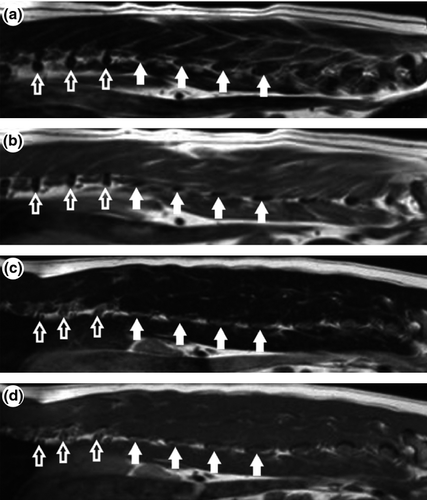
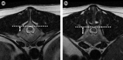
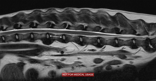
MR images were evaluated using open-source PACS Workstation DICOM viewer (ClearCanvas Workstation, ClearCanvas Inc., Toronto, ON, Canada). The T2-weighted sagittal images used at the level of lateral aspect of the articular facet were used to evaluate the shape and subjective height of the process extending from each vertebral body as illustrated in Fig. 1. The accompanying transverse images were evaluated for height of the process as compared to the dorsal aspect of the vertebral canal. Each measurement was made using a perpendicular line from the dorsal aspect of the vertebral canal ventrally until it intersected with the dorsal most aspect of either the cranial fovea costalis or transverse process. In the case of the transverse process, as it is a continuous section of bone, the line was drawn at the level of the angle change along the outer cortex of the vertebral body as demonstrated in Fig. 2b. In cases where the left and right processes did not appear to be exiting the vertebral body at approximately the same level, separate measurements were taken for each process. These measurements were completed for the T12, T13, L1 and L2 rib articulations or transverse processes, when possible. As our hospital imaging protocol only includes completing transverse images through the area of interest, transverse images through all four vertebral bodies were only identified in 15 of the included cases. The remainder had transverse images through some but not all sites. Based on the large variation in patient size, we used the previously described length of L2 vertebral body as an internal comparison and utilized the ratio of the measured height on transverse images (Fig. 2) to the length of L2 vertebral body as measured in Fig. 3 on the mid-sagittal image.
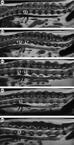
The location of the coeliac and cranial mesenteric artery was grouped into five possible locations: (1) lying within the length of the T13 vertebral body, (2) bridging the T13/L1 inter-vertebral disc space, (3) within the length of L1, (4) bridging L1/L2 disc space and (5) within the body of L2. These were evaluated on the sagittal image that showed both vessels exiting the caudal aorta as shown in Fig. 4.
Statistical method
Data were analysed using commercially available software (Minitab 16 Statistical Software, Minitab Inc. State College, PA, USA). Descriptive statistics were reported as mean, median and standard deviation (SD). The height of the rib heads and the height of the transverse processes, relative to the vertebral height, were compared using the Mann–Whitney U-test with significance defined as P < 0.05.
Results
A total of 90 dogs fit the study criteria and were used for evaluation and review of the thoracolumbar anatomy. All but one patient had image sequences that allowed evaluation of the location of the coeliac and cranial mesenteric arteries and at least one pair of ribs or transverse processes. In this patient, we were unable to visualize these structures based on the location of the saturation band. The dogs represented 31 different breeds, with the most common breeds being Labrador retriever (11), cocker spaniel (9) and dachshund (7). The ages of the dogs ranged from 6 month to 13 years with the average 6.8 year (median 7 years).
Sagittal image review (Fig. 1) – The rib heads and transverse process have distinctly different shapes on evaluation of the sagittal images. Rib articulations appeared as small round, well-demarcated hypointense areas at the level of the base of the vertebral canal. In contrast, transverse processes appeared as oval, well-demarcated hypointense areas located at the level of the middle or ventral third of the vertebral body height.
Transverse image review (Fig. 2) – The transverse processes were significantly more ventrally situated as compared to the rib articulation, when expressed as a ratio to the length of L2 (median 0.26 mm, range 0.13–0.55) (P < 0.0001). This difference became more statistically significant when comparing T12 to L2 processes.
Coeliac and cranial mesenteric arteries (Fig. 3) – The level of the arteries was consistent with previous reported data with Group 1: 5.6% of dogs, Group 2: 11.2%, Group 3: 59.6%, Group 4: 20.2% and Group 5: 3.4% (6).
Discussion
To the authors' knowledge, no previous reports of the vertebral anatomy of the thoracolumbar junction have been evaluated on MRI in the dog. Utilizing the T2w sagittal and transverse images of the thoracolumbar spine, the thoracolumbar junction can be evaluated for pre-surgical planning. A complete review of these images provides additional characterization of the thoracolumbar vertebrae and the potential for further evaluation of anatomical variation at this location. Utilization of the sagittal images provides an additional way to quickly differentiate between transverse processes and rib articulations. The subjective evaluation of the shape of the lumbar transverse processes appears to be consistent and easily utilized to count lumbar vertebrae. Furthermore, utilization of the height ratio provides a consistent objective measure that can be used to aid in the differentiation of a transverse process versus rib articulation. The distance from the dorsal vertebral column to the process itself can be used intra-operatively to guide decision-making when normal anatomical variation is encountered. Limitations of this study include lack of evaluation of a variety of anomalous vertebrae that can often be found at the thoracolumbar junction. We likely encountered a lower rate of transitional vertebrae in our study by eliminating all cases with other vertebral anomalies, therefore selecting for truly normal vertebrae at this location. Another limitation was that we were unable to measure all four vertebrae in every sequence. This is due to the retrospective nature of this study and our imaging protocols, which include only completing transverse sequences through areas of interest identified on sagittal sequences. This study would have also benefitted from the evaluation of inter- and intra-observer variation to give a better evaluation of the reliability of this method.
In review, the shape and the height of the rib articulation or transverse process on sagittal T2-weighted images can be readily and easily evaluated using sagittal images. This sequence and plane allows us to easily visualize other inconsistent, but often helpful, anatomical landmarks to aid in localization, such as the anticlinal vertebra which has previously been published to be T11 in 85% of dogs (Baines et al., 2009); the coeliac artery which has been described as arising from the ventral aspect of L1 in 71% of dogs; and the cranial mesenteric artery which has been described as arising from ventral to either the L1 or L2 vertebral body in 97% of dogs (Dennis, 2005). In our study, we verified the location of these arteries, and although helpful for general localization, this anatomy is not consistent enough to aid in vertebral localization on advance imaging studies. Similar to a previous report, we identified that 83.2% of the cranial mesenteric arteries arose from below L1 or L2 vertebral bodies, and in general, it is most common (58.9%) for both arteries to arise from below the L1 vertebral body.
In conclusion, the use of the T2-weighted sagittal images can aid in identifying surgical anatomy at the thoracolumbar junction. Rib articulations and lumbar transverse processes have easily identifiable different shapes and heights relative to the vertebral canal, with the rib articulation being round and located more dorsally in relationship to the vertebral canal and transverse processes being oval in shape and located more ventrally in relationship to the vertebral canal. The height of each process can be quantitatively verified using the height from the dorsal border of the vertebral canal as compared to the level of the process in relation to the length of the L2 vertebral bodies. As previously reported, the locations of the coeliac and cranial mesenteric artery are not reliable enough to provide localization on MRI alone. Utilizing the T2 transverse sagittal images for better localization and identification of variations in anatomy can provide added information when identifying areas of interest within the thoracolumbar vertebral column.
Further prospective studies would be warranted to evaluate the inter- and intra-observer variation in this method, particularly when applied to a population with anomalous vertebrae, to evaluate whether this method can consistently provide differentiation between thoracic and lumbar vertebral bodies at the thoracolumbar junction.
Conflict of Interest Statement
None of the authors has any financial or personal relationships that could inappropriately influence or bias the content of the paper.


