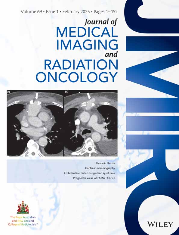
Thoracic hernias: What the radiologist should know
Felipe Aluja-Jaramillo
Radiology Department, Hospital Universitario San Ignacio – Pontificia Universidad Javeriana, Bogotá, Colombia
Search for more papers by this authorCorresponding Author
Omar Andrés Pantoja Burbano
Radiology Department, Hospital Universitario San Ignacio – Pontificia Universidad Javeriana, Bogotá, Colombia
Correspondence
Omar Andrés Pantoja Burbano, Radiology Department, Hospital Universitario San Ignacio – Pontificia Universidad Javeriana, Carrera 7 No 40 – 62, Zip code: 110231, Bogotá, Colombia.
Email: [email protected]
Search for more papers by this authorFernando R. Gutiérrez
Cardiothoracic Imaging Section, Radiology Department, Mallinckrodt Institute of Radiology, Washington University in St. Louis, St. Louis, Missouri, USA
Search for more papers by this authorCarlos Previgliano
Louisiana State University Health – Shreveport, Shreveport, Louisiana, USA
Search for more papers by this authorSanjeev Bhalla
Cardiothoracic Imaging Section, Radiology Department, Mallinckrodt Institute of Radiology, Washington University in St. Louis, St. Louis, Missouri, USA
Search for more papers by this authorFelipe Aluja-Jaramillo
Radiology Department, Hospital Universitario San Ignacio – Pontificia Universidad Javeriana, Bogotá, Colombia
Search for more papers by this authorCorresponding Author
Omar Andrés Pantoja Burbano
Radiology Department, Hospital Universitario San Ignacio – Pontificia Universidad Javeriana, Bogotá, Colombia
Correspondence
Omar Andrés Pantoja Burbano, Radiology Department, Hospital Universitario San Ignacio – Pontificia Universidad Javeriana, Carrera 7 No 40 – 62, Zip code: 110231, Bogotá, Colombia.
Email: [email protected]
Search for more papers by this authorFernando R. Gutiérrez
Cardiothoracic Imaging Section, Radiology Department, Mallinckrodt Institute of Radiology, Washington University in St. Louis, St. Louis, Missouri, USA
Search for more papers by this authorCarlos Previgliano
Louisiana State University Health – Shreveport, Shreveport, Louisiana, USA
Search for more papers by this authorSanjeev Bhalla
Cardiothoracic Imaging Section, Radiology Department, Mallinckrodt Institute of Radiology, Washington University in St. Louis, St. Louis, Missouri, USA
Search for more papers by this authorF Aluja-Jaramillo: MD, MHPE; OA Pantoja Burbano MD; FR Gutiérrez MD; C Previgliano MD; S Bhalla MD.
Conflict of interest: The authors have no relevant financial or non-financial interests to disclose.
Abstract
Thoracic hernias encompass the protrusion of thoracic contents through the thorax or intra-abdominal tissue into the thorax. They can be classified as diaphragmatic hernias – either congenital or acquired; pulmonary hernias – involving tissue protrusion through cervical fascia or intercostal spaces; and mediastinal hernias – including cardiac, intrapericardial and hiatal hernias. Prompt identification and classification of thoracic hernias rely on diagnostic imaging, primarily through computed tomography and magnetic resonance, to identify associated complications. This article comprehensively reviews thoracic hernias and their key imaging features.
Open Research
Data availability statement
Data sharing not applicable to this article as no datasets were generated or analysed during the current study.
References
- 1 Hernia - MeSH - NCBI. [Cited 7 Jan 2024.] Available from URL: https://www.ncbi.nlm.nih.gov/mesh/68006547.
- 2Chaturvedi A, Rajiah P, Croake A, Saboo S, Chaturvedi A. Imaging of thoracic hernias: types and complications. Insights Imaging 2018; 9: 989.
- 3Verhey PT, Gosselin MV, Primack SL, Kraemer AC. Differentiating diaphragmatic paralysis and eventration. Acad Radiol 2007; 14: 420–425.
- 4Gardner F. Imaging diseases of the chest (4th Edn). By D M Hansell, P Armstrong, D A Lynch, H P McAdams. pp. ix+1220, 2005 (Elsevier Mosby, London, UK) £175.00 ISBN 0-323-03660-0. Br J Radiol 2006; 79: 179.
10.1259/bjr.79.938.179a Google Scholar
- 5Eren S, Çiriş F. Diaphragmatic hernia: diagnostic approaches with review of the literature. Eur J Radiol 2005; 54: 448–459.
- 6Roberts HC. Imaging the diaphragm. Thorac Surg Clin 2009; 19: 431–450.
- 7Herling A, Makhdom F, Al-Shehri A, Mulder DS. Bochdalek hernia in a symptomatic adult. Ann Thorac Surg 2014; 98: 701–704.
- 8Vassileva CM, Shabosky J, Boley T, Hazelrigg S. Morgagni hernia presenting as a right middle lobe compression. Ann Thorac Cardiovasc Surg 2012; 18: 79–81.
- 9Hou C, Wang J, Song S, Wu Q. The grading of stomach position for postnatal outcomes in isolated left-sided congenital diaphragmatic hernia: a systematic review and meta-analysis. Prenat Diagn 2023; 43: 1008–1017.
- 10Picard E, Ben NA, Fisher D, Schwartz S, Goldberg M, Goldberg S. Morgagni hernia mimicking pneumonia in down syndrome. J Pediatr Surg 2007; 42: 1608–1611.
- 11Chick JFB, Chauhan NR, Lai JH, Khurana B. Incarcerated Morgagni hernia mimicking acute cholecystitis. Intern Emerg Med 2012; 7 (Suppl. 2): 169–171.
10.1007/s11739-012-0818-5 Google Scholar
- 12Chavhan GB, Babyn PS, Cohen RA, Langer JC. Multimodality imaging of the pediatric diaphragm: anatomy and pathologic conditions. Radiographics 2010; 30: 1797–1817.
- 13Taylor GA, Atalabi OM, Estroff JA. Imaging of congenital diaphragmatic hernias. Pediatr Radiol 2009; 39: 1–16.
- 14Tarver RD, Conces DJ, Cory DA, Vix VA. Imaging the diaphragm and its disorders. J Thorac Imaging 1989; 4: 1–18.
- 15Gaerte SC, Meyer CA, Winer-Muram HT, Tarver RD, Dewey J, Conces J. Fat-containing lesions of the chest. RadioGraphics 2002; 22 (Suppl 1): S61–S78.
- 16Sandstrom CK, Stern EJ. Diaphragmatic hernias: a spectrum of radiographic appearances. Curr Probl Diagn Radiol 2011; 40: 95–115.
- 17Wardi G, Lasoff D, Cobb A, Hayden S. Traumatic diaphragmatic hernia. J Emerg Med 2014; 46: 80–82.
- 18Mirvis SE, Shanmuganagthan K. Imaging hemidiaphragmatic injury. Eur Radiol 2007; 17: 1411–1421.
- 19Eren S, Kantarci M, Okur A. Imaging of diaphragmatic rupture after trauma. Clin Radiol 2006; 61: 467–477.
- 20Thillois JM, Tremblay B, Cerceau E et al. Traumatic rupture of the right diaphragm. Hernia 1998; 2: 119–121.
10.1007/BF01250026 Google Scholar
- 21DeBarros M, Martin MJ. Penetrating traumatic diaphragm injuries. Curr Trauma Rep 2015; 1: 92–101.
- 22Keyes S, Spouge RJ, Kennedy P et al. Approach to acute traumatic and nontraumatic diaphragmatic abnormalities. Radiographics 2024; 44: e230110.
- 23Nason LK, Walker CM, Mcneeley MF, Burivong W, Fligner CL, David Godwin J. Imaging of the diaphragm: anatomy and function. Radiographics 2012; 32: E51–E70.
- 24Groth SS, Andrade RS. Diaphragmatic eventration. Thorac Surg Clin 2009; 19: 511–519.
- 25Zia Z, Bashir O, Ramjas GE, Kumaran M, Pollock JG, Pointon K. Intercostal lung hernia: radiographic and MDCT findings. Clin Radiol 2013; 68: e412-e417.
- 26Weissberg D, Refaely Y. Hernia of the lung. Ann Thorac Surg. 2002; 74: 1963–1966.
- 27McAdams HP, Gordon DS, White CS. Apical lung hernia: radiologic findings in six cases. AJR Am J Roentgenol. 1996; 167: 927–930.
- 28Moncada R, Vade A, Gimenez C et al. Congenital and acquired lung hernias. J Thorac Imaging. 1996; 11: 75–82.
- 29Detorakis EE, Androulidakis E. Intercostal lung herniation – the role of imaging. J Radiol Case Rep 2014; 8: 16.
- 30Hauser M, Weder W, Largiadèr F, Glazer GM. Lung herniation through a postthoracoscopy chest wall defect: demonstration with spiral CT. Chest 1997; 112: 558–560.
- 31Temes RT, Talbot WA, Green DP, Wernly JA. Herniation of the lung after video-assisted thoracic surgery. Ann Thorac Surg 2001; 72: 606–607.
- 32Clark AJ, Hughes N, Chisti F. Traumatic extrathoracic lung herniation. Br J Radiol 2009; 82: e82-4.
- 33Ross RT, Burnett CM. Atraumatic lung hernia. Ann Thorac Surg 1999; 67: 1496–1497.
- 34Bhalla M, Leitman BS, Forcade C, Stern E, Naidich DP, McCauley DI. Lung hernia: radiographic features. AJR Am J Roentgenol 1990; 154: 51–53.
- 35Kahrilas PJ, Kim HC, Pandolfino JE. Approaches to the diagnosis and grading of hiatal hernia. Best Pract Res Clin Gastroenterol 2008; 22: 601.
- 36Peterson CM, Anderson JS, Hara AK, Carenza JW, Menias CO. Volvulus of the gastrointestinal tract: appearances at multimodality imaging. Radiographics 2009; 29: 1281–1293.
- 37Wright MP, Nelson C, Johnson AM, McMillan IK. Herniation of the heart. Thorax 1970; 25: 656–664.
- 38Kim HJ, Cho YS, Cho GY, Choi S II. Congenital absence of the pericardium. J Cardiovasc Ultrasound 2014; 22: 36.
- 39Chughtai T, Chiavaras MM, Sharkey P, Shulman H, Miller HA. Pericardial rupture with cardiac herniation. Can J Surg 2008; 51: E101.
- 40Kawamukai K, Antonacci F, di Saverio S, Boaron M. Acute postoperative cardiac herniation. Interact Cardiovasc Thorac Surg 2011; 12: 73–74.
- 41Schollaert E, Devos D. Traumatic intrapericardial diaphragmatic herniation. JBR-BTR 2015; 98: 141–142.
- 42Austin A, Itani W, Adams C, Beegle SH. Abnormal chest radiograph in asymptomatic patient: intrapericardial herniation of colon. Am J Med Sci 2020; 359: 135–136.


