Kinematics of whip spider pedipalps: a 3D comparative morpho-functional approach
Abstract
Amblypygi are tropical and subtropical ambush predators that use elongated, raptorial pedipalps for different activities. Although pedipalp use in predation and courtship has been explored in videography in vivo analyses, kinematic ex vivo examination of these appendages has not been conducted. Here, we rectify this lack of data by using micro-CT scans to 3D-kinematically model the appendage morphology and the range of motion (ROM) of the joints for Damon medius and Heterophrynus elaphus. We illustrate the successful application of this technique to terrestrial euarthropods in determining the maximum ROM values for each pedipalp joint. We also note that, in life, these values would be lower due to motion restricting structures like tendons, arthrodial membranes, and muscles. We further compare our maximum values obtained here with data from video-based motion analyses. The ROM of each joint shows the greatest flexibility in the femur-tibia joint (140–150°), the lowest in the basitarsus-claw joint (35–40°). ROM in the tibia-basitarsus joint is markedly distinct (D. medius: 44°; H. elaphus: 105°). This disparity reflects how H. elaphus uses the joint in the capture basket, while D. medius uses the femur-tibia joint to form the capture basket. We further illustrate notable vertical motion of the H. elaphus pedipalp compared to D. medius. This difference reflects the retro-ventral trochanter apophysis of H. elaphus. Our study opens the possibility to further whip spider kinematic understanding. Examination of other taxa using this approach will result in a more comprehensive understanding of the ecological significance and ethological implications of this unique arachnid group.
INTRODUCTION
Whip spiders (Amblypygi) are terrestrial euchelicerates consisting of ca. 220 species (Weygoldt 2000a, 2002a; Harvey 2003; Miranda et al. 2016, 2018a; Haug & Haug 2021). Amblypygids are tropical and subtropical organisms (Harvey 2003) that have dorso-ventrally flattened exoskeletons and inhabit gaps between rocks and gaps in trees (Weygoldt 2000a, 2002a). The elongate sensory antenniform appendages and elongate and spinose raptorial pedipalps make the group unique within Arachnida (Weygoldt 1996, 2002b; Fowler-Finn & Hebets 2006). As Amblypygi lack venom glands, they form a capture basket with their pedipalps to immobilize prey (Prendini et al. 2005; Garwood et al. 2017). As such, depending on the life mode, pedipalp length and spinosity varies between species and even within the sex of the same species (Weygoldt 1996, 2000a; MacArthur et al. 2018). Due to the pronounced nature of these structures, the pedipalps have been subject to different lines of research, including examination of appendage velocity and closing angles (Santer & Hebets 2009; Seiter et al. 2019) and pedipalp morphology (McLean et al. 2021). However, ex vivo kinematic analyses digitally measuring the maximum range of motion (ROM) in each joint are currently unknown. As such, we here present the first three dimensional (3D) morpho-functional analysis of amblypygid pedipalp kinematics, following Schmidt et al. (2020a,b). This novel technique has, thus far, been applied to extant swimming crabs (Schmidt et al. 2020a) and horseshoe crabs, as well as extinct sea scorpions (Bicknell et al. 2021). We therefore present the first application to terrestrial euchelicerates. We considered species within Damon C. L. Koch, 1850 and Heterophrynus Pocock, 1894. Damon is restricted to Subsaharan Africa and consists of at least 10 species (Weygoldt 1999, 2000b; Harvey 2003), while Heterophrynus occurs in South America and contains less than 20 valid species (Giupponi & Kury 2013). The genera have markedly disparate pedipalp morphologies, making them model groups to explore morpho-functional traits.
MATERIALS AND METHODS
Specimen selection, surface reconstruction, measurements
Specimens of the selected species—Damon medius (Herbst, 1797) and Heterophrynus elaphus Pocock, 1903—stored at the Bavarian State Collection of Zoology (ZSMA) were considered (Table 1). These 2 specimens were scanned with a Phoenix Nanotom micro computed tomography scanner housed at the Bavarian State Collection of Zoology using these scanning parameters: 90 kv, 110 mA, 2359 TIFFs, voxel size 15. The TIFF stacks were imported to Mimics version 23.0 (Materialise, Leuven, Belgium) to generate pedipalp surface models. Pedipalp articles were separated using the ‘Segmenting’ tool. Surface models were exported as .OBJ files for import into Autodesk Maya 2020 (Autodesk, San Rafael, USA) for kinematic analyses. Appendage measurements were taken in Mimics and used to calculate percentage length of articles relative to the entire pedipalp.
| Species | Sex, length | Preservation | Collector | Collection date | Origin | Collection number |
|---|---|---|---|---|---|---|
| Damon medius | Male, 28 mm | 75% EtOH | Roewer | 1973 | Kamerun | ZSMA19730285 |
| Heterophrynus elaphus | Female, 33 mm | 75% EtOH | Diller | 2012 | Peru | ZSMA20120286 |
Kinematic deflections and examination of maximum range of motion
The surface models were processed in Maya to examine deflection of pedipalp articles (Schmidt et al. 2020a,b). Cylindrical hypothetical joint axes were fitted to the articulation points. These were treated as bicondylar joints (typical arthropod leg joints, Boxshall 2004, 2013). As all joints were modeled as bicondylar, articles were only moved in one plane, permitting the accurate measurement of joint angles (discussed below). Artificial joints (Gatesy et al. 2010) were created using the ‘X_ROOM’ tool (Brainerd et al. 2010). After a subordination process, the distal articles followed the rotation of the proximal ones (Fig. 1).
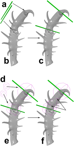
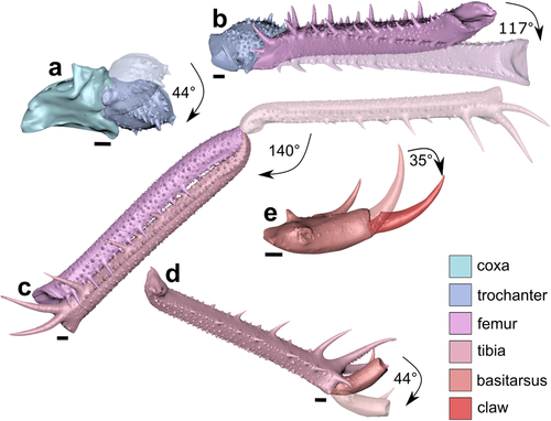
The maximum ROM (maximum excursion angle of each joint) was examined by rotating about joint axes till articles collided. This was qualified by examining pedipalp deflection in the specimens. Joint angles were calculated for all joints. The ROM of each joint represents a change of rotation about the z-axis (Schmidt et al. 2020a, Fig. 4).
Our maximum ROM data is independent of the sexes, ontogeny, and allometric growth. These parameters would have little impact on joint and podomere morphology as joint axes would not shift direction during ontogeny. Furthermore, joint and podomere morphology would not differ markedly between males and females or even show intrasexual differences among several individuals. As such, there was no need to analyze more than just one specimen per species.
The restrictive limitations of arthrodial membranes, muscles, or tendons were not modelled. The documented ROM values therefore record the maximum excursion possibilities of the appendage morphology. In reality, those values would be lower than our documented values due to membranes, muscles, and tendons, but they could not be higher. We therefore present a framework for documenting the maximum ROM based on the entire podomere morphology to encourage future research on whip spider pedipalp kinematics.
Terminology
Arachnid pedipalps often comprise of 6 articles: coxa, trochanter, femur, patella, tibia, and tarsus. However, amblypygid pedipalp terminology varies between taxa (Weygoldt 1996, 2000a, 2002a; Shultz 1999). Here, as we are examining kinematic interactions of pedipalp articles, we follow Weygoldt (2000a, 2002a) – coxa, trochanter, femur, tibia, basitarsus, and claw. Tibia is used instead of patella, as the functional morphology of a non-amblypygid arachnid patella is distinct from this article in amblypygid pedipalps. Appendage spines are referred to as dorsal or ventral spines.
RESULTS
Article length relations
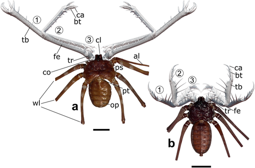
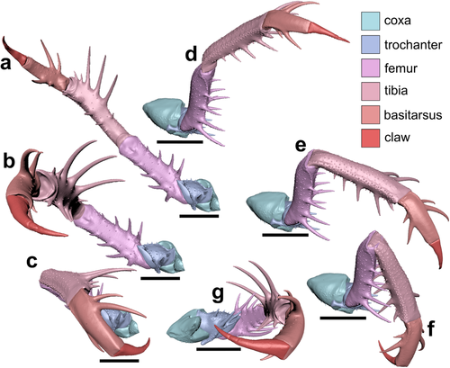
The raptorial appendages of both species have distinct spinosity and length (Fig. 2, Table 2). Damon medius pedipalps are more than twice the body length (Fig. 2a) while H. elaphus pedipalps do not exceed the post-pedipalp walking leg femur when expanded (Fig. 2b). The percentage of each article relative to the entire pedipalp differs slightly between the species (Table 2). Coxa and trochanter for both species contribute to 8–12% of the appendage length, femur and tibia ∼25% in H. elaphus, but ∼34% in D. medius. Basitarsus and claw contribute ∼11–15% in H. elaphus and ∼8% in D. medius. Except for the femur and tibia in D. medius, the size difference reflects the appendage size, as opposed to a difference in articles size.
| Species | bl | pl | Coxa | Trochanter | Femur | Tibia | Basitarsus | Claw | pl/bl ratio |
|---|---|---|---|---|---|---|---|---|---|
| Damon medius | 28 mm | 70.3 mm | 5.5 mm (7.8%) |
6.4 mm (9.1%) |
24.1 mm (34.3%) |
23.5 mm (33.4%) |
5.6 mm (8%) |
5.2 mm (7.4%) |
2.51 |
| Heterophrynus elaphus | 33 mm | 44.5 mm | 5.3 mm (11.9%) |
5 mm (11.2%) |
11.2 mm (25.2%) |
11.3 mm (25.4%) |
6.7 mm (15.1%) |
5 mm (11.2%) |
1.38 |
Pedipalp orientations
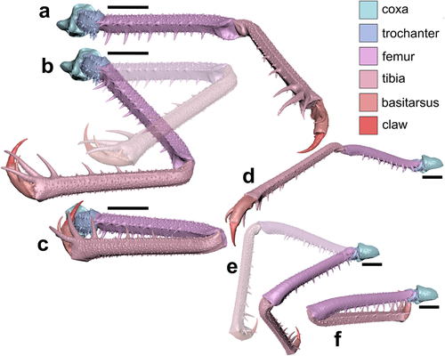
Three distinct orientations were modeled (Figs 2–4; Seiter et al. 2019; McLean et al. 2019). Orientation 1 (Figs 3a,d and 4a,d; Figs S1 and S4, Supporting Information) shows all joints maximally extended and results in completely open pedipalps. This emulates the behavior in fighting or in threatening predators (Seiter et al. 2019; McLean et al. 2019). The D. medius trochanter enables more distal pedipalp sections to exhibit a pronounced lateral extension (Fig. 3a,d). Conversely, the H. elaphus coxa-trochanter joint moves more distal elements dorsally (Fig. 4a,d). Orientation 2 (Figs 3b,e and 4b,e; Figs S2 and S5, Supporting Information) shows the femur-tibia joint bent to 90°. This position emulates the awaiting prey position (Figs 3b,e and 4b,e). Orientation 3 (Figs 3c,f and 4c,f; Figs S3 and S6, Supporting Information) is the maximally closed position present while resting. While forming the capture basket to keep prey, whip spider's pedipalps are not fully flexed.
Maximum ROM
Examined maximum ROM (Figs 5 and 6; Table 3) differs between the two species. The trochanter rotates ∼40° inside the coxa-trochanter joint in both species (Figs 5a and 6a). In both species, the trochanter-femur joint moves the appendage dorsally. The coxa-trochanter joint moves the trochanter proximal to the chelicerae. The D. medius femur can rotate ∼117° (Fig. 5b) along the trochanter-femur joint, while this rotation in H. elaphus is ∼80° (Fig. 6b). In H. elaphus, the ventral-located spines are longer than the dorsal spines, while the dorsal and ventral femoral spines of D. medius do not differ substantially. The femur-tibia joint allows the raptorial action and has a maximum ROM of 140° (Fig. 5c) and 150° (Fig. 6c). The two distal sections (Figs 5d,e and 6d,e) do not show any torsion and movement within the same plane as the femur-tibia joint. Contrary, in other taxa [such as Charinus kakum Harms, 2018 (fig. 3 in Harms, 2018)], torsion of the distalmost articles is prominent. In D. medius, the basitarsus movement towards the tibia is restricted by the dorsal tibial spines and extends only ∼44° (Fig. 5d), while in H. elaphus, this joint attains a maximum ROM of ∼105° (Fig. 6d). The difference here reflects the appendage section length, use in prey catching, and prey item retention in the capture basket. Damon medius uses the femur and tibia to construct the capture basket, while H. elaphus also employs the basitarsus in this construction. Finally, in the distal-most joint (basitarsus-claw joint), the maximum ROM is 35° (Fig. 5e) and 40° (Fig. 6e).

| Joint | Damon medius | Heterophrynus elaphus |
|---|---|---|
| Coxa-trochanter joint | 44° | 37° |
| Trochanter-femur joint | 117° | 80° |
| Femur-tibia joint | 140° | 150° |
| Tibia-basitarsus joint | 44° | 105° |
| Basitarsus-claw joint | 35° | 40° |
Retro-ventral apophysis
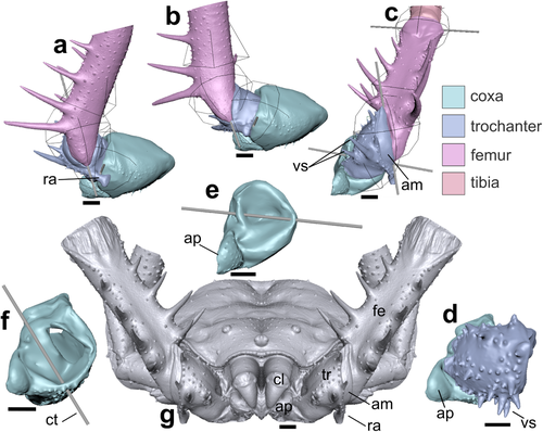
An intriguing feature is the anvil-shaped retro-ventral apophysis on the H. elaphus trochanter (Fig. 7). The structure (∼2 mm long) is an autapomorphy of the genus Heterophrynus. It precludes the ventral and some dorsal movement of the appendage and limits the lateral trochanteral rotation (Fig. 7a). When the pedipalp is lowered, the retro-ventral trochanter apophysis is moved away from the coxa (Fig. 7b,c). To move the posterior pedipalp ventrally, the femur must move towards the trochanter (Fig. 7c).
Coxa-trochanter joint axis orientation
A key distinction between H. elaphus (Fig. 7d) and D. medius (Fig. 7f) is coxa-trochanter joint axis orientation. This difference aids in prey-holding motions, especially as the H. elaphus ventro-medial spines are longer than those of D. medius. Figure 7e shows a H. elaphus specimen with uplifted pedipalps, illustrating the location of chelicerae relative to the anterior coxal process and presents an overview of the general distance between the left and right proximal-most pedipalp articles.
DISCUSSION
Kinematic approaches of arthropod joints
Several euarthropod groups have been subject to kinematic examination using various methods (deVries et al. 2012; Hwang & Choi 2015; Vagts et al. 2017). Earlier studies considered arachnid appendage joints with in vivo measurements of dissected animals or live specimen observations (Petrunkevitch 1909; Ellis 1944; Manton 1977; Shultz 1989; Wolff et al. 2016), while more recent studies have explored strike kinematics and run video-based ROM analyses to obtain angle values (Santer & Hebets 2009; Seiter et al. 2019; McLean et al. 2020). As such, exploring kinematics of arachnid appendages with 3D surface models is novel, building on applications to crustaceans (Schmidt et al. 2020a) and extinct euarthropods (Schmidt et al. 2020b; Bicknell et al. 2021). Our approach represents a simple but informative tool for deriving precise kinematic data and allows examination of hypothetical joint axes in 3D. These data are fundamental to thoroughly understanding species-specific morphologies and may present insight into other aspects of whip spider ecology.
Pedipalp length patterns among whip spiders
Amblypygid pedipalp length relative to body length varies markedly in the group. Several taxa (e. g., Sarax; Rahmadi et al. 2010) have pedipalps that are half the body length, while other taxa [e.g., Euphrynichus bacillifer (Gerstaecker, 1873)] have tibial sections that are double the body length (Weygoldt 2000a, 2002a). Species of Trichodamon Mello-Leitão, 1935 additionally bear elongate, thin, and needle-like pedipalps (Miranda et al. 2018b). Within the examined genera, morphological variation is notable (Weygoldt 2000a, 2002a). Damon diadema (Simon, 1876) has pedipalps twice the body length and D. medius has pedipalps more than three times the body length (McLean et al. 2020; this study). In Heterophrynus species, H. armiger Pocock, 1902 and H. alces Pocock, 1902 have small and robust pedipalps, while H. batesii Butler, 1873 exhibits a slender, elongate morphology (Víquez et al. 2014; Chapin & Hebets 2016; Seiter & Gredler 2020), and H. elaphus has pedipalps comparable to the body length (Seiter & Gredler 2020; this study).
Examining pedipalp spine length was beyond the scope of our study, as we focused on gross morphology. However, spinosity may play roles in the motion of certain taxa. Phrynus Lamarck, 1801 for example, has small pedipalps with dorsal tibial spines that prevent pedipalp closure and restrict femur-tibia flexion. As spine length can vary between sexes and individuals, these features must be considered when developing kinematic parameters for larger scale projects.
Maximum ROM values
Values of joint ROM are sparse in literature. This reflects the inaccuracies in gathering such data—direct in vivo studies that manually deflect appendage elements likely underestimate values and video-based in vivo analyses are commonly hampered by camera angles. The maximum ROM of the femur-tibia joint for D. medius was reported to be 55.5°; much smaller than our calculated 140° (fig. 5 in McLean et al. 2020). This disparity reflects the measuring of angles during predator-prey interactions (our Orientation 2), compared to calculation of the ROM range for article morphology (Orientation 1). Furthermore, as muscles and membranes were not modelled, the angle values must be lower than those derived here.
The 3D kinematic data on orientations and article deflection gathered here further the knowledge on amblypygid autecology and ethology. Such data could be combined with video-based in vivo studies of predation and courtship. While entire pedipalp movement is primarily driven by the femur-tibia joint, we illustrate that the maximum ROM and orientation of movement in distinct species differs. Further examination of comparable models may yield a comprehensive understanding of pedipalp range motion and its significance in the ecology of these euchelicerates.
Trochanter apophyses among Amblypygi
The anvil-shaped retro-ventral apophysis is autapomorphic of Heterophrynus (Weygoldt 1996, 2000a, 2002a). However, the function of this structure varies between species. While H. elaphus can lift the pedipalps dorsally, taxa with longer pedipalps (e.g., H. guacharo de Armas, 2015) can only hold pedipalps horizontally (Weygoldt 1999, 2005, 2008). As such, one might question whether this lifting motion is not possible (or unnecessary) for species with longer pedipalps. Article flexibility in pedipalps of Heterophrynus could therefore be explored. Such examination may also present insight into the relationship between entire pedipalp length and apophysis size. These data may explain whether Heterophrynus species with long pedipalps have lower coxa-trochanter joint flexibility. Furthermore, coxa-trochanter flexion of other amblypygid taxa with other trochanter apophysis morphologies could be considered (figs. 7 and 8 in Weygoldt 1996). For example, Charinus kakum Harms, 2018—Charinidae (fig. 7 in Garwood et al. 2017)—has a spinous, anterior-facing ventral trochanter apophysis with 14 strong spines (fig. 4a–c in Harms 2018) that restricts pedipalp motion towards the walking legs (see fig. 3b in Harms 2018). This morphology resulted in pedipalp coxae being located slightly dorsally to the first walking leg coxa, with a slightly inclined pedipalp orientation, and limited basitarsus torsion.
In Paracharon caecus Hansen, 1921 (Palaeoamblypygi), a similarly sharp trochanter apophysis is expressed (fig. 2e in Garwood et al. 2017). Further, coxa of antenniform legs are not aligned with coxae of adjacent appendages. This species may therefore be the only extant example of a whip spider with nearly complete horizontally articulated pedipalps (Garwood et al. 2017; Haug & Haug 2021). Such a morphology was more common in some extinct groups (fig. 3 in Garwood et al. 2017; Haug & Haug 2021). Thus, kinematic analyses of the joints of extant taxa exhibiting this trochanteral organization could enhance our understanding of joint morpho-functionality across amblypygid groups.
Sexual selection as a driving evolutionary factor
Amblypygid pedipalp length and morphology are interesting to consider in the context of intra- and intersexual selection (Darwin 1871; Fairbairn et al. 2007; Hunt et al. 2009). Intrasexual selection results in stronger males for male-male-competition or male mate-guarding behavior (Alexander 1962) and intersexual selection results in morphological display structures (Chapin & Reed-Guy 2017; McArthur et al. 2018; McLean et al. 2019). Indeed, hypertrophied male amblypygid pedipalps are not necessarily correlated with more effective predation success (McLean et al. 2019). Therefore, a trade-off between morpho-functional pedipalp characters used in foraging and mating exists (comparable to fiddler crabs, Latruffe et al. 1999; Rosenberg 2002). For Amblypygi, McLean et al. (2019) showed that Damon variegatus C. L. Koch, 1850 have statistically significantly longer pedipalp tibiae than females (McLean et al. 2019). However, the male pedipalps are more gracile and less spinose than females. As such, these raptorial structures may function as display structures, as opposed to male-male competition or mate guarding.
CONCLUSIONS
Examination of euarthropod appendages with 3D kinematics is a cardinal direction for furthering the understanding of euarthropod appendage movement and motion. Our study establishes the application of 3D kinematics for terrestrial euchelicerates and, in doing so, provides a standardized framework for further studies. The data derived from our analyses represent maximum ROM values and are therefore hypothetical values that in vivo, video-based tests can be used as a null hypothesis. Further 3D kinematic studies should consider variation in sexes and larger sample sizes with individuals of different ontogenetic stages to uncover any major differences in the two main aspects not explored here. Furthermore, our method highlights a new direction for more completely understanding the motion of extinct amblypygid through the lens of extant forms.
ACKNOWLEDGMENTS
This research was supported by funding from the Deutsche Forschungsgemeinschaft, Award Number: Me-2683/10-1 (to MS), an Australian Research Council Discovery grant (DP200102005), and a UNE Postdoctoral Research Fellowship (to RDCB).
Open access funding enabled and organized by Projekt DEAL.
CONFLICT OF INTEREST
No potential conflict of interest was reported by the authors.
Open Research
DATA AVAILABILITY STATEMENT
Three-dimensional data supporting the findings of this study (e.g. Autodesk Maya files) are available from the corresponding author upon request.


