First record of Tanaostigmatidae (Hymenoptera: Chalcidoidea) in South Korea
Abstract
Protanaostigma kyushuana Masi 1940 (Chalcidoidea: Tanaostigmatidae) is newly recognized from South Korea, and its finding represents the first record of the phytophagous chalcid family in the country. A detailed redescription and photographs of the species are given. Images of the holotype specimen are also provided for comparison. In addition, the relationship and taxonomic status between Protanaostigma and Tanaostigmodes are briefly discussed.
Introduction
Tanaostigmatidae, a small to moderately sized parasitic wasp family within Chalcidoidea (Hymenoptera) is composed of 99 species in nine genera worldwide and two species in two genera from the Eastern Palaearctic (Gupta & Joshi 2016; Masi 1940; Noyes 2016; Yang et al. 2004). The family can be easily distinguished from other chalcidid families by the enlarged and distinctly swollen prepectus. Most of the members in the family are known as gall inducers, on plants in Fabaceae and a few inquilines in the galls of other insects as well (La Salle 2005).
During our taxonomic study on Korean Chalcidoidea, a species of Tanaostigmatidae has been newly recognized. We provide a detailed redescription and photographs of Protanaostigma kyushuana along with images of a type specimen for reference.
Materials and methods
The terminology used in the present study follows that of La Salle (1987). The images were captured with a Leica DFC 495 camera attached to a Leica M205A Stereozoom stereomicroscopy (Leica, Microsystems, Solms, Germany) and produced with LAS software (version 4.1.0., Leica Microsystems, Heerbrugg, Switzerland). Figure plates were prepared in Adobe Photoshop CS6 (Adobe Systems Incorporated, San Jose, CA, USA). The following abbreviations are used: ELKU, Entomological Laboratory, Kyushu University, Fukuoka, Japan; KNA, Korea National Arboretum, Pocheon, South Korea; POL, distance between the inner edges of the two lateral ocelli; LOL, distance between the inner edges of a lateral ocellus and median ocellus; OOL, distance from the outer edge of a lateral ocellus to the compound eye; F1 − F4, funicular segments 1 through 4; CC, costal cell; MV, marginal vein; PMV, postmarginal vein; SV, stigma vein.
Taxonomy
Order Hymenoptera
Family Tanaostigmatidae Ashmead, 1904.
Genus Protanaostigma Ferrière 1929
Type species: Protanaostigma milletiae Ferrière 1929.
Diagnosis
Members of the genus can be distinguished from all other genera in the family by having the ventral edge of the hind femur expanded into a tooth-like lobe.
Distribution
Oriental (Indonesia), Palearctic (Japan) and Australian (Australia).
Remark
Bouček (1988) mentioned the existence of additional undescribed species from Africa (Madagascar), South and East Asia, and North Australia.
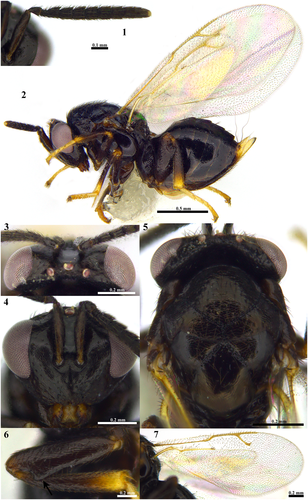
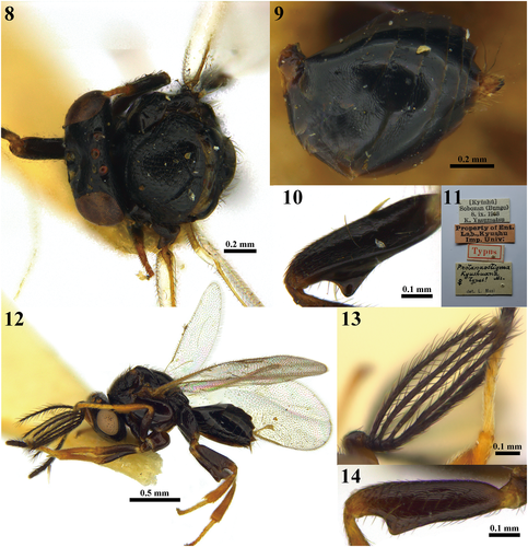
Protanaostigma kyushuana Masi, 1941 (Figs 1–7, 8–14).


Protanaostigma kyushuana Masi, 1941: 40.
Type material (1♀). Holotype, 1♀, JAPAN: Kyushu, Sobosan (Bungo), 8.IX.1938, K. Yasumatsu (ELKU) [examined].
Material examined (3♀♀4♂♂). SOUTH KOREA: 1♀, National DMZ Native Botanic Garden, Yanggu-gun, Gangwon-do, 11–30.VI. 2015, I.K. Kim leg. (KNA); JAPAN: 1♀, Kyushu, Mt. Hiko, Fukuoka, Japan, 11.VII.1969, K. Kanmiya leg. (ELKU); 1♀, Honshu, Mt. Hakusan, Kaga, Japan, 29–31.VIII.1960, T. Hidaka leg. (ELKU); 3♂♂, Kamiozoegawa, Fuji, Saga Pref. Kyushu, 4.VII.1973, K. Yamagishi leg. (ELKU); 1♂, ditto, 25. IX. 1973, K. Yamagishi leg. (ELKU).
Redescription
Female
Body length 1.80–2.42 mm.
Head
Head imbricate to coriaceous with scattered setae, 1.44× as broad as long in dorsal view, slightly narrower than mesosoma, 1.35× as wide as high in frontal view; POL: LOL: OOL = 5: 2: 3; eyes bare; vertex behind ocelli nearly flat in lateral view; scrobal impression glabrate; interantennal projection well defined with weak longitudinal median carina ventrally; subocular sulcus weakly visible.
Antenna
Scape 4.71–5.12× as long as wide, with more or less flattened ventral expansion; pedicel 1.33–1.50× as long as wide; first anellus almost equal in width and 0.73× as long as second anellus; proportions (length: width) of funicle segments as follows: F1 (42: 33); F2 (42: 37); F3 (38: 39); F4 (38: 39); F5 (42: 39); F6 (38: 38); club 2.00–2.35× as long as wide.
Mesosoma
Mesosoma 1.08× as long as metasoma in lateral view; mesoscutum reticulate to imbricate with scattered setae; scutellum coriaceous to smooth, slightly elongate hexagonal, 1.50–1.52× as long as wide; propodeum almost glabrate with one median carina and strong plicae; mesopleuron smooth and bare; hind coxa reticulate to imbricate on dorsally; hind femur on apical edge with a subtriangular tooth ventrally.
Wing
Fore wing with marginal fringe reaching to postmarginal vein; stigmal vein almost straight, not perpendicular to postmarginal vein; basal cell with 20–25 setae; propotions of wing veins as follows: CC/MV 0.43–0.46; MV/PMV 0.64–0.75; MV/SV 0.56–0.57; PMV/SV 0.76–0.88.
Metasoma
Metasoma imbricate to coriaceous, elliptical in dorsal view, 0.72× as broad as width of mesosoma with distinct median line dorsally.
Colour
Body black except mandible yellowish brown, with teeth reddish brown, prepectus and tegula dark brown, last tergite and ovipositor sheath whitish yellow; antennae uniformly black except scape brown to dark brown; legs dark brown except basal and apical part of all tibiae, tarsi yellow; wings hyaline with veins yellow and covered with brown setae.
Male
Body length 1.88–2.04 mm. Similar to female, but dark brown to blackish body, antenna dark brown except scape yellow, legs brown to dark brown except basal and apical part of femora, tibiae and tarsi yellow; scape 2.14–2.85× as long as wide, with flattened ventral expansion; F1–F4 with long rami; length of rami of each funicular segment as follows: 48: 47: 43: 39; basal cell with 22 setae; CC/MV 0.35–0.39; MV/PMV 0.93–0.96; MV/SV 0.71–0.79; PMV/SV 0.77–0.81.
Biology
Unknown.
Distribution
South Korea (Gangwon-do, new record), Japan (Honshu, Kyushu).
Remarks
The original description of Protanaostigma kyushuana by Masi was based on a single female specimen. Later, Bouček (1988) mentioned briefly that the male of the species has four branches in an antenna. Therefore, we provide the detailed diagnostic characters of the male, which was not included in the original description. In addition, collection date of the holotype reported in the original publication is not the same as the label data; it should be 8.IX.1938, not 3.IX.1938.
Discussion
The generic status of Protanaostigma is questionable as a few authors have previously discussed (Bouček 1988; Yang et al. 2004). In regards to the traditional generic classification concept, Protanaostigma is separated from Tanaostigmodes, the most common genus in the family, only by having a tooth-like lobe on the ventral edge of the hind femur (Bouček 1988; Ferrière 1929). However, we conceded that this character may not be an apomorphic character to define this genus as Bouček (1988) and Yang et al. (2004) discussed that Protanaostigma may only be an eccentric species group in Tanaostigmodes. This is because some species of Tanaostigmodes (e.g. T. puerariae) and members of Protanaostigma have a protruding lobe on the ventral edge of hind femur. It requires an extensive morphological and molecular phylogenetic study to clarify the generic status of the two genera.
Acknowledgments
The authors thank Toshiharu Mita (Kyushu University, Fukuoka, Japan) for providing the type and non-type specimens. This work was supported by a grant from the Korea National Arboretum (KNA) of the Republic of Korea (as Project no. KNA1-1-15, 14-3).


