Removal of Infected Canine Cemented Total Hip Prostheses Using a Femoral Window Technique
Presented at the American College of Veterinary Surgeons Symposium, September 21 to 24, 2000, Washington, DC.
Address reprint requests to Jonathan Dyce, MA, VetMB, College of Veterinary Medicine, Veterinary Teaching Hospital, The Ohio State University, 601 Vernon L. Tharp St, Columbus, OH 43210–1089.
Abstract
Objective— To evaluate a femoral window technique for retrieval of cemented total hip prostheses.
Study Design— Retrospective clinical study.
Animals— Twelve dogs with infection of a cemented modular total hip prosthesis.
Methods— Implant removal was performed by an extended craniolateral approach to the hip and proximal femur without trochanteric osteotomy. The femoral cement mantle was fragmented and removed with simple orthopedic instrumentation by a lateral femoral window that was repaired using cerclage wires. Surgical technique, intraoperative and postoperative complications, bacterial culture results, histopathologic findings, and completeness of cement removal were recorded. Follow-up radiographs were taken 5 to 9 weeks postoperatively. Long-term follow-up information was obtained by client questionnaire. Functional outcome was assessed by scoring ability to stand, sit, walk, run, play, climb stairs, and get into a car.
Results— Prosthesis retrieval was performed 2 to 41 months after implantation (median, 14 months). Complete removal of femoral cement was achieved in 10 dogs. A nondisplaced femoral fissure, extending proximally from the window, was an intraoperative complication in 2 dogs. Staphylococcus spp was most commonly isolated (6 dogs) from interfacial membrane samples. Systemic antibiotic therapy, dependent on susceptibility testing, was administered for 3 to 10 weeks postoperatively. There was radiographic evidence of osteotomy healing at 5- to 9-week reassessment. Recurrence of osteomyelitis was not observed. Long-term functional outcome was considered mildly abnormal.
Conclusions— The lateral window was an effective technique for retrieval of retained femoral cement.
Clinical Relevance— Removal of an infected prosthesis using this technique generally resulted in a clinical outcome comparable to that with ab initio femoral head and neck excision.
Infection is a devastating complication of human and canine total hip replacement (THR). The pathogenesis of biomaterial-centered infection after THR is complex and has been described in detail by Gristina.1 Bacterial contaminants adhere to the implant surface and can produce a polysaccharide glycocalyx that affords protection from opsonizing antibodies and antibiotics. Chronic exposure to a bulk or particulate foreign body such as polymethylmethacrylate (PMMA) can exhaust local macrophage oxidative responses and negate local microbicidal competence. The development of a so-called immuno-incompetent fibro-inflammatory zone adjacent to the implant allows bacterial colonization to occur. Excision of all infected or necrotic tissue and removal of the prosthesis and cement mantle is generally required to eradicate such infection.2–5
The removal of retained femoral PMMA cement, with preservation of surrounding bone stock, is a particularly challenging task. The purpose of our report was to evaluate a femoral window technique for cement retrieval when THR infection occurs.
Materials and methods
Medical records of dogs that had retrieval of a total hip prosthesis to manage bacterial infection were reviewed. All dogs had a commercially available cemented modular canine hip prosthesis (Biomedtrix Inc, Allendale, NJ) implanted at The Ohio State University Veterinary Teaching Hospital between 1991 to 2001, using a previously reported technique.6
Dogs were included in this study if femoral cement retrieval was performed using a window technique, and infection was confirmed by a positive bacterial culture of the interfacial membrane. Animal patient data retrieved included signalment, body weight, diagnosis, implant size, intraoperative complications and bacteriologic culture and susceptibility results during initial THR, postoperative complications, and interval to retrieval. The following details of implant removal were reviewed: surgical technique, intraoperative and postoperative complications, aerobic and anaerobic bacteriologic culture and susceptibility results, histopathologic findings, length of femoral window, and completeness of cement removal. Follow-up radiographs were obtained 5 to 9 weeks postoperatively. Long-term follow-up information was obtained by client questionnaire. Owners were asked to rate, using a 5-point scoring system, the ability of their dog to perform 7 common activities: standing, sitting, walking, running, playing, climbing stairs, and getting into a car.6 Outcome was expressed as the mean of the grades for each activity.
Surgical technique
The surgical approach used was a modification of a standard exposure for canine THR.6 With the dog in lateral recumbency, an extended craniolateral surgical approach was made to the hip and proximal femur, with excision of the previous skin wound proximally. Identification of normal tissue planes was complicated by postsurgical fibrosis. Deep gluteal muscle tenotomy was performed adjacent to the greater trochanter. Trochanteric osteotomy was not necessary. The grossly thickened joint capsule was incised radially from the acetabular border to the vastus ridge on the cranial aspect of the femur. The capsulotomy can be developed into an inverted T- or L-shaped incision. The hip was then luxated craniodorsally using a combination of external rotation, distraction, and leverage from a Lewin bone clamp placed around the femoral neck. The femur was externally rotated 90° and elevated proximally with a broad-tipped Hohmann retractor. The aperture of the femoral canal at the level of the femoral neck osteotomy could be enlarged laterally with an osteotome at this stage to facilitate proximal extraction of the stem with an intact cement mantle. The entire mantle may be recovered from this modified aperture, particularly when there is massive bone-cement interface failure, a stable stem-cement interface, and no cement extension distal to the femoral isthmus.
The femoral implant was expelled using a dull 1-inch (25.4-mm) osteotome driven proximally against the collar of the stem where it overhung the calcar. If the entire femoral cement mantle was recovered by this maneuver (confirmed by comparison to the radiographic silhouette), there was no indication to proceed to the femoral window. The femur was returned to a neutral position and retracted caudally to allow retrieval of the acetabular cup and associated cement. To remove the cup, a circumferential cleavage plane was developed, with a mallet and sharp ¼- inch osteotome, along the acetabular polyethylene-cement interface. The underlying cement mantle was then broken up and extracted. The interfacial membrane was removed and submitted for bacteriologic culture and susceptibility testing and histopathologic examination.
Cefazolin sodium (20 mg/kg intravenously) was not administered at induction but immediately after collection of material for bacteriologic culture. A reamer (generally 1 size larger than that required to seat the cup) was an efficient means of debriding the cement-depleted acetabulum. In the case of cranial intramedullary extension of ilial cement, fenestration of the lateral ilial cortex may be required to recover the PMMA. Cement that had extruded ventrally through an open acetabular fossa can generally be recovered after dissection of the joint capsule from the ventral acetabulum. In the case of cement that has extruded through inadvertent medial cortical penetration, the perforation can be enlarged with a small acetabular reamer to allow cement retrieval. Synovectomy was performed routinely, whereas capsulectomy was discretionary.
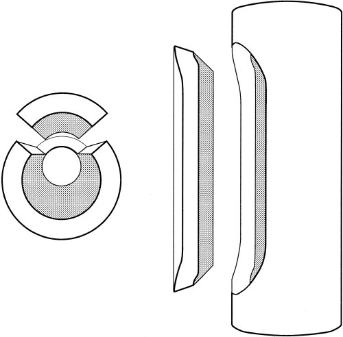
A standard lateral approach to the femoral diaphysis was then developed, with cranial retraction of the vastus lateralis.7 A rectangular subtrochanteric lateral femoral window, with convex rounded proximal and distal margins, was fashioned using an oscillating saw with a thin 3/8-inch blade (Fig 1). The caudal border of the window lies immediately cranial to the linea aspera. The window was extended to the level of the distal cement mantle, as determined from a preoperative lateral radiograph, and should occupy less than one third of the femoral circumference. All edges are beveled to prevent medial collapse of the repaired osteotomy (Fig 2). The cortical window was tucked between the adductor and biceps femoris muscles until reduction. Great care was taken when manipulating the windowed femur to avoid fracture.

The lateral femoral window extends from the subtrochanteric region to the distal aspect of the cement mantle.

The window should not occupy more than one third of the circumference of the cortex. The beveled cuts prevent axial displacement of the repaired osteotomy.
The femoral cement was carefully fragmented, using a mallet and sharp ¼- inch osteotome, and was then removed piecemeal through the window (along with the cement restrictor if present), and proximal femoral opening. The endosteal surfaces of the femur and window were debrided of interfacial membrane, and the field lavaged copiously with saline solution (0.9% NaCl). If a pedestal of endosteal bone had formed distal to the implant, it was foraged. When complete cement recovery was confirmed, the osteotomy was repaired using multiple cerclage wires. In general, calcar osteolysis dictated no further modification of the femoral neck osteotomy. If the remaining joint capsule was adequate, it was closed over the acetabulum, and no interpositional sling was used. Otherwise closure was routine, without use of drains.
Perioperative analgesia routinely involved administration of intravenous opioids and nonsteroidal anti-inflammatory drugs, with options of supplementary epidural or transdermal opioids. Pen rest with light leash-walking exercise was recommended for 6 weeks after surgery, with a progressive return to normal exercise thereafter.
Results
Prosthesis retrieval and femoral cement extraction by a window was performed in 12 dogs with infection (Table 1). Breeds were German shepherd dog (5), rottweiler (3), Newfoundland (2), Labrador retriever (1), and mixed breed (1). Sex distribution was 7 males and 5 females. The right limb was involved in 10 dogs. Mean body weight at the time of initial THR was 42 kg (range, 27 to 62 kg), and mean age was 4 years (range, 10 months to 11 years).
| Dog No. | Age at THR (mos) | Sex | Breed | Weight (kg) | Time to Retrieval (mos) | Revision Culture | Complications of Revision | Follow-Up Time (mos) | Outcome* |
|---|---|---|---|---|---|---|---|---|---|
| 1 | 12 | FS | German shepherd | 27 | 20 | Pseudomonas spp | Femoral fissure | NA | NA |
| 2 | 57 | MC | German shepherd | 39 | 19 | Coagulase negative Staphylococcus spp | Femoral fissure | 3 | 5 |
| 3 | 49 | MC | German shepherd | 44 | 5 | Serratia spp | NR | 39 | 3 |
| 4 | 96 | M | German shepherd | 48 | 24 | Pseudomonas spp Coagulase negative Staphylococcus spp | NR | 19 | 3 |
| 5 | 54 | FS | German shepherd | 30 | 5 | Enterococcus spp | NR | 11 | 2 |
| 6 | 30 | F | Rottweiler | 53 | 38 | Staphylococcus aureus | NR | 20 | 2 |
| 7 | 36 | MC | Rottweiler | 38 | 41 | S aureus | NR | NA | NA |
| 8 | 75 | FS | Rottweiler | 48 | 8 | Enterococcus spp | NR | NA | NA |
| 9 | 10 | M | Newfoundland | 62 | 2 | Pseudomonas spp Enterobacter spp | NR | 3 | 4 |
| 10 | 15 | FS | Newfoundland | 48 | 4 | S aureus | NR | 6 | 3 |
| 11 | 132 | MC | Labrador retriever | 43 | 7 | Coagulase negative Staphylococcus spp | NR | 8 | 2 |
| 12 | 18 | M | Cross-bred | 28 | 21 | Coagulase negative Staphylococcus spp | NR | 5 | 2 |
- Abbreviations: THR, Total hip replacement; F, female; FS, female spayed; M, male; MC, male castrate; NR, none recorded; NA, not available.
- *Outcome was defined by the mean rating of the graded ability to stand, sit, walk, run, play, climb stairs, and get into a car,6 where Grade 1 = normal, Grade 2 = near normal, Grade 3 = mildly abnormal, Grade 4 = moderately abnormal, and Grade 5 = severely abnormal.
In 11 dogs, the indication for THR was hip dysplasia and osteoarthritis. In dog 12, THR was performed to address acetabular malunion and articular impingement of an acetabular C plate. The material of the femoral stem was titanium alloy in 4 dogs and cobalt chromium in 8 dogs. Inadvertent penetration of the medial acetabular cortex was the only intraoperative complication documented during initial THR (dog 8).
Four dogs had postoperative complications that required revision surgery. Luxations of the prosthesis were managed by open reduction (dogs 3 and 5) or acetabular revision and 3 further open reductions (dog 9). Acetabular bone-cement interface loosening was addressed by acetabular revision in dog 1.
Pseudomonas spp was isolated from the deep surgical wound around the prosthesis in dogs 1 and 4, and the same organism was isolated at the time of implant removal in those dogs. In dogs 5 and 9, positive bacterial cultures for Enterococcus spp, Pseudomonas spp, and Enterobacter spp, respectively, were obtained during previous revision surgery to manage luxation and were predictive of the bacteria grown from the interfacial membrane on retrieval (Table 1).
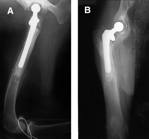
The presumptive diagnosis of THR infection was based primarily on clinical and radiographic signs (Fig 3).8,9 In each case, definitive management involved prosthesis retrieval and cement extraction by the femoral window, performed 2 to 41 months after implantation (median, 14 months). In dog 5, the degree of femoral osteolysis about the stem tip at the time of initial diagnosis was so severe that there was a substantial risk of pathologic fracture after implant retrieval. After a 4-month period of antibiotic therapy, the magnitude of periosteal new bone formation and cortical remodeling was such that the implant could be safely removed.

Total hip replacement infection (dog 10). Lateral (A) and ventrodorsal (B) radiographic projections. Note circumferential bone-cement interface deterioration, femoral cortical osteolysis, and periosteal new bone formation. (Lateral wire stabilization had been performed previously for stabilization of a cranial cruciate ligament rupture.).
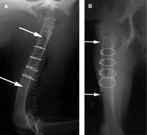
The mean length of the femoral window was 10 cm (range, 7 to 12 cm). The window was secured using 2 to 6 full cerclage wires (diameter 0.9 mm [18 g] to 1.25 mm [14 g]) of either hand twist or ASIF loop configuration (Synthes, Paoli, PA) (Fig 4). Autogenous cancellous bone grafts were not used but could have been applied when there was a large defect or cortical fissure.

Immediate postoperative implant retrieval (dog 10). Lateral (A) and ventrodorsal (B) radiographic projections. There is no radiographic evidence of residual polymethylmethacrylate. Arrows indicate the proximal and distal extent of the window.
A nondisplaced femoral fissure, extending proximally from the window, was an intraoperative complication in dogs 1 and 2. In dog 1, the fissure was stabilized immediately using cerclage and an intramedullary pin, and in dog 2, routine femoral window repair was performed and the fissure was not specifically addressed.
Postoperative radiography documented complete retrieval of cement in 9 dogs. There was a small amount of residual PMMA medial to the acetabulum in dog 6, and in the distal femur and proximal lateral metaphysis in dogs 1 and 7.
At the time of diagnosis of THR infection, remote bacterial infections were identified in 5 dogs: pyotraumatic dermatitis (dogs 1 and 11), pyoderma (dog 10), discospondylitis and pyotraumatic dermatitis (dog 6), and urinary tract infection (dog 2). These infections were managed concurrently with treatment of femoral and pelvic osteomyelitis.
Aerobic and anaerobic bacterial culture of intraoperative swabs showed Staphylococcus spp (6 dogs), Enterococcus spp (2), Serratia spp (1), Pseudomonas spp (1), and mixed cultures of Pseudomonas spp and Staphylococcus spp (1), and Pseudomonas spp and Enterobacter spp (1). In 8 dogs (67%), bacterial isolates were resistant to the initial perioperative antibiotic (first-generation cephalosporin). In each dog, systemic antibiotic therapy, dependent on susceptibility, was administered for 3 to 10 weeks (median, 4 weeks) postoperatively. Chronic sinus discharge in dog 12 resolved within 48 hours of implant removal, without excision of the sinus tract. A sinus tract was excised at the time of implant removal in dog 7.
Histopathologic examination of the synovial-like interfacial membrane was performed in 6 dogs and uniformly showed neutrophilic, lymphoplasmacytic, and histiocytic inflammation with hemorrhage, edema, and granulation tissue formation. There was variable tissue necrosis and dystrophic mineralization. This chronic suppurative inflammation was consistent with infection; however, microorganisms were rarely found.
There was radiographic evidence of osteotomy healing at 5- to 9-week reassessment with bridging periosteal new bone formation and remodeling of the osteotomy margins. The periosteal response apparent at the time of diagnosis had reduced in volume and taken on a more regular character, and there was more uniform medullary density compared with immediate post-retrieval radiographs. The previously sclerotic bone margin of the acetabular bed also remodeled, and any acetabular defects filled rapidly with bone. Cerclage wires appeared stable. The overall appearance was of resolving osteomyelitis.
Long-term follow-up (3 to 39 months postoperatively) was available in 9 dogs. The mean outcome for an individual dog was grade 3 (mildly abnormal function). Dog 2 was diagnosed with degenerative myelopathy 3 months after implant retrieval. Dog 5 returned to mildly abnormal function after implant retrieval but was euthanized 19 months postoperatively because of congestive heart failure. Recurrence of osteomyelitis or sinus discharge was not observed. In 3 dogs with bilateral THR at the time of revision, the contralateral prostheses remained unaffected.
Discussion
Infection after THR can occur as a result of intraoperative contamination, local extension of wound infection, or hematogenous infection.9 A 3-stage classification system is commonly used in humans to describe the mode and timing of infection.10 Early postoperative infections (type I) commonly involve complications of wound healing, including purulent discharge, and the patient can have signs of systemic infection. Late chronic infections (type II) also originate at the time of surgery but have a delayed presentation (6 to 24 months) because of a small inoculum or low bacterial virulence. Progressively increasing pain and deteriorating function are typical, but systemic signs are rare. Acute hematogenous infections (type III) are the least common in humans and are associated with antecedent bacteremia, generally more than 2 years after surgery. Hematogeous Brucella canis osteomyelitis complicating THR has been reported in 2 dogs.11 A fourth clinical setting is the patient with positive intraoperative cultures at the time of revision arthroplasty.12
In the current series, the majority of dogs had features that were most suggestive of type II infection. There was convincing circumstantial evidence for type III infection in dog 6, which had a normally functioning prosthesis for 3 years until discospondylitis occurred. Positive intraoperative cultures at the time of revision arthroplasty were obtained in 2 dogs.
Dog 3 has been reported previously.13 A nosocomial Serratia spp infection was introduced in a contaminated disinfectant during management of pyotraumatic dermatitis overlying the prosthetic hip and open reduction of luxation.
Staphylococcal organisms were the sole organisms cultured in 6 dogs (50%), and this strongly resembles the bacterial flora cultured from THR infection in humans.2,3,12 The finding of patterns of resistance to the perioperative antibiotic in 8 dogs (67%) is also similar.14 The numbers of individual infectious agents were too small to suggest that the bacterial population had any determining effect on prognosis. The hypothesis that infections caused by more virulent organisms are more difficult to eradicate after exchange arthroplasty in humans is controversial.2,12
The goal of management is to eradicate infection and restore function.3 In humans, treatment options for the infected prosthesis include 1-stage or 2-stage reimplantation or resection arthroplasty. Single-stage revision involves excision of infected tissue, removal of the entire prosthesis, copious lavage, then reimplantation of prosthesis with antibiotic impregnated PMMA to achieve high local concentrations of antibiotic and administration of systemic antibiotics. Using single-stage revision, elimination of infection has been reported in 77% to 91% cases.2,15,16
Two-stage revision additionally incorporates a period of temporary antibiotic impregnated cement and intravenous antibiotic therapy to eliminate infection before definitive reconstruction. Two-stage revision permits uncemented reconstruction and allograft use when there is massive proximal bone loss.4 Intraoperative bacterial culture results direct the correct choice of antibiotic for cement impregnation at reimplantation.17 Eradication of infection is achieved in over 90% cases.3,4 A more limited debridement with cup exchange, preservation of the femoral implant in situ, and aggressive antibiotic treatment can be successful in selected cases with a positive intraoperative culture and acute postoperative infection.12 Resection arthroplasty after implant retrieval in humans can offer pain relief and allow elimination of infection; however, this results in leg length inequality, and the functional recovery is inferior to that offered by successful revision.18,19
The complete removal of PMMA cement is a prerequisite of implant retrieval or reimplantation to eliminate possible persistence of a nidus of infection associated with the residual PMMA.1,17,18,20 It is a challenge to accomplish this without inadvertent perforation or fracture of the potentially weakened femoral cortex. A number of devices and techniques for removal of PMMA proximally by the neck osteotomy or trochanteric osteotomy are described in humans, eg, hand tools, power tools, segmental cement extraction, laser, lithotripter, and ultrasound.21–23
Femoral diaphyseal osteotomy provides better direct observation of the bone-cement interface, facilitating PMMA removal using simple orthopedic instrumentation and therefore reduces the risk of femoral shaft perforation or fracture.24 Diaphyseal osteotomy techniques include the extended trochanteric osteotomy and the femoral window.20,24–28 Our report describes a modification of the lateral femoral window technique.25,27 The rounded corners of the osteotomy avoid potential stress risers generated by converging saw cuts. In an investigation of femoral biopsy hole shape and size, Clark et al suggested that a rectangular hole with round ends afforded greatest residual bone strength, and that whereas increasing the width of the hole caused a significant reduction in strength, increasing length did not.29 The beveled edges prevented medial collapse of the window when secured with cerclage.27
The window allowed exposure of the diaphyseal medullary canal for cement recovery that was superior to that offered by the femoral splitting technique, described in dogs for exchange arthroplasty of an aseptically loose prosthesis.30,31 Surgical morbidity was comparable.30,31 Cement recovery from the trochanteric region was consistently the difficult part of the procedure because of restricted direct observation of the endosteal surface. The technique of extended trochanteric osteotomy could be considered to improve access to this region.20,26 Unfortunately there was no long-term follow-up information on the 2 dogs with retained femoral cement. Bourne and others documented retained femoral cement in 11 human animal patients undergoing resection arthroplasty to manage infected THR. In only 1 case was surgical removal of that cement required to control infection.19 It is possible that incomplete cement retrieval is more detrimental to exchange arthroplasty; however, complete cement removal should remain the goal for resection arthroplasty.12 Removal of the acetabular component is relatively straightforward; however, preoperative radiographs should be scrutinized for intrapelvic cement. In dog 6 where there was a small quantity of residual intrapelvic cement, no complications were identified 20 months postoperatively.
It is occasionally possible to remove the loose femoral stem and intact cement mantle proximally without creating a lateral femoral window. For this reason, we would recommend first attempting to expel the implant proximally through an expanded neck osteotomy before fashioning a window. The femoral window technique is particularly indicated in cases with a curved proximal femur, cement filling the canal distal to the femoral medullary isthmus, and with a well-fixed prosthesis.
Complications of the canine femoral window technique included nondisplaced fissure fractures that extended proximally from the window. These fissures were caused during cement recovery in 2 dogs. Fracture of the femur after THR in humans is most likely to occur during revision arthroplasty, and care during femoral manipulation and cement recovery is advised.32 Such fractures should be assessed individually with regard to the requirement for repair. Creation of the femoral window did not lead to postoperative femoral fracture in any of our dogs.
The majority of femoral windows used to facilitate revision THR in humans were healing 3 months postoperatively.24,33 Rapid healing was also observed in our dogs, and sequestration of the window did not occur.
In our experience, retrieval of the infected prosthesis by a femoral window generally resulted in a clinical outcome comparable to that of ab initio femoral head and neck excision (FHNE).34,35 There was a clinical impression that dorsal displacement of the greater trochanter, and therefore effective limb shortening, was not as evident, possibly because of the stabilizing effect of periarticular postsurgical fibrosis. Function after implant retrieval was obviously inferior to that of uncomplicated THR.6
Successful revision arthroplasty of an infected cementless canine THR has been described in a research model.36 However, there are a number of concerns regarding exchange arthroplasty for cemented THR infection in canine patients. Femoral and acetabular osteomyelitis is associated with periprosthetic bone loss and will dictate selection of larger femoral stem. Loss of dorsal acetabular bone can complicate acetabular component selection. Osteopenic bone stock, and calcar osteolysis, associated with subsidence, compromise successful femoral reimplantation. As endosteal polishing is likely to have occurred with the chronically loose prosthesis, a secure interlock between PMMA and bone is relatively difficult to achieve.
If exchange arthroplasty is performed with a femoral window in humans, the recommendation is that the femoral stem bypass the end of the window by at least 2 times the femoral diameter to minimize the effect of cumulative stress risers.24 Such long-stemmed prostheses are not available for use in dogs. However, we have performed exchange arthroplasty using the femoral window technique and standard length femoral stems in a small number of dogs with aseptic loosening (unpublished data).
A period of immobilization as enforced in humans after exchange arthroplasty would not be as practical in dogs.2 Two-stage revisions are unlikely to be feasible for veterinary patients, and therefore determination of antibiotic susceptibility before single-stage revision would require preoperative synoviocentesis or biopsy.
Failure to eliminate the original infectious agent when exchange arthroplasty is performed can necessitate further revision, and there is a heightened risk of development of new infection after contamination of the surgical wound at revision.37 Overall, there is an increased likelihood of complication after THR revision compared with the initial arthroplasty.2,12
It is our current recommendation that implant retrieval and revision to FHNE should be the procedure of first choice for the infected canine THR. The femoral window technique can be used when direct proximal extraction of the cement mantle is not possible. The probable outcome of the procedure is a comfortable arthrosis and fair clinical function.
Acknowledgment
The authors thank Tim Vojt and Marc Hardman for preparation of the figures.


