Laparoscopic cholecystectomy for cholecystolithiasis in a case with situs inversus totalis
Abstract
We report a case of cholecystolithiasis associated with situs inversus totalis (SIT) that had been successfully treated with a laparoscopic cholecystectomy. Although cholecystolithiasis associated with SIT is rare, trained surgeons who perform unusual surgical procedures, even if they have not experienced such a case, would perform laparoscopic cholecystectomy for cholecystolithiasis associated with SIT easily and safely.
Introduction
Situs inversus totalis (SIT) is a form of heterotaxia that is usually found incidentally while performing a medical examination for other causes, including preoperative examinations.1 As a complete transposition of the viscera including SIT has been known as an autosomal recessive disease,2,3 the incidence of SIT has been reported to range between 1/20 000 and 1/5000.2,4–6
Among the surgical treatment for diseases in gastroenterological fields, the anatomical significance of SIT has been occasionally considered gravely in treating appendicitis,7 gastrointestinal carcinoma,8,9 extrahepatic bile duct carcinoma10 and liver transplantation.11
Surgeons might find more difficulty performing a surgical procedure for patients with cholecystolithiasis associated with SIT. While laparoscopic cholecystectomy is indispensable for all surgeons in this era of less-invasive surgery, we herein show a case of cholecystolithiasis associated with SIT that had been successfully treated with a laparoscopic cholecystectomy.
Case report
A 49-year-old man who had been suffering from repeated epigastralgia for 2 years was considered to be indicated for surgical treatment. However, preoperative chest X-ray revealed the evidence of dextrocardia (Fig. 1), suggesting the coexistence of situs inversus totalis. Abdominal ultrasound sonography demonstrated a gallbladder with stones located in the left side of the abdomen. A drip-infused cholangiogram showed the gallbladder on the left side without any anomalies in the cystic duct and common bile duct (Fig. 2).
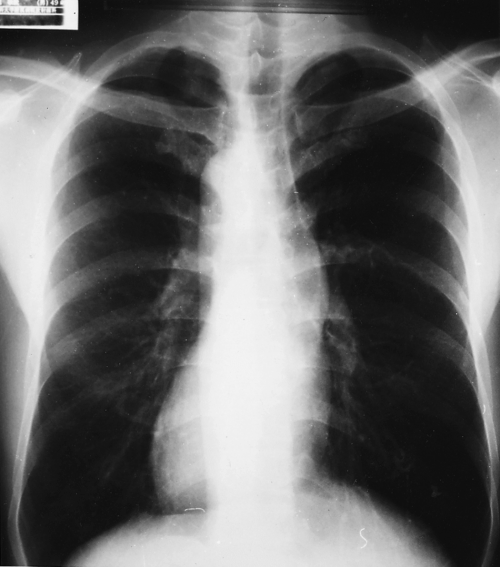
Preoperative chest X-ray demonstrating dextrocardia.
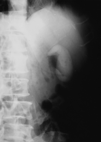
Preoperative cholangiogram showing the gallbladder on the left side of the abdomen.
A 10-mm trocar to put the laparoscope into the abdomibal cavity was inserted through an umbilical portion in a usual manner. Additional trocars were placed on the upper and left sides of the abdomen in a mirror image location of the ordinal portion of the instruments for usual laparoscopic cholecystectomy (Fig. 3). The videoscopic view also demonstrated the evidence of SIT with the liver and gallbladder located in the left upper quadrant (Fig. 4). An intraoperative cholangiogram showed no other anomalies of the bile duct. Cholecystectomy was completed easily and safely because the surgical procedures including grasping, dissecting, parting and ligating did not differ from those in usual laparoscopic cholecystectomy.
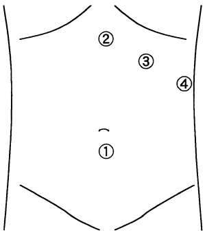
A 10-mm trocar for the insertion of the laparoscope was put through an umbilical portion. Additional trocars were placed on the upper and left sides of abdomen on the contrary to the usual procedure for laparoscopic cholecystectomy.
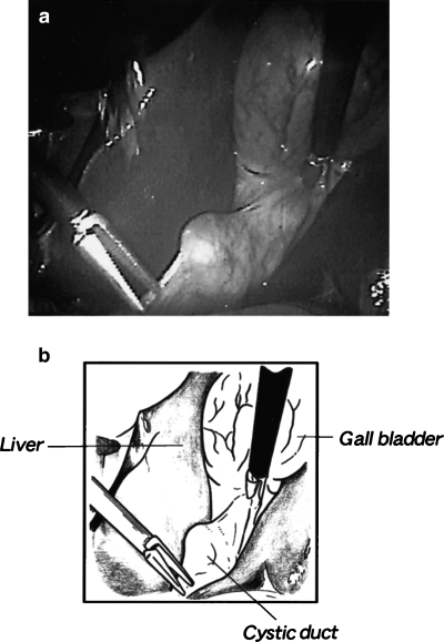
Videoscopic demonstration of situs inversus totalis with the liver and gallbladder located in the left upper quadrant portion.
Reports with regards to the application of laparosopic cholecyctectomy for cholecystolithiasis associated with situs inversus totalis (SIT)12–21 are reviewed in Table 1.
| Case | Author | Age/sex | Associated diseases | Symptoms |
|---|---|---|---|---|
| 1 | Campos and Sipes12 | 39/F | Kartagener's syndrome | LUQP |
| 2 | Lipschutz et al.13 | 80/M | None | Mid-abdominal pain |
| 3 | Drover et al.14 | 29/F | CHD | LUQP |
| 4 | Takei et al.15 | 51/F | HT | Epigastralgia, LUQP |
| 5 | Goh et al.16 | 62/M | IHD, DM, HT | LUQP |
| 6 | Huang et al.17 | 36/M | None | LUQP |
| 7 | McDermott and Caushaj18 | 66/M | HT, RA | Abdominal discomfort |
| 8 | Crosher et al.19 | 63/M | None | Colic pain |
| 9 | Demetriades et al.20 | 61/F | None | LUQP |
| 10 | Demetriades et al.20 | 37/M | None | LUQP |
| 11 | Djohan et al.21 | 20/F | None | Abdominal discomfort |
- CHD, congenital heart disease; DM, diabetes mellitus; HT, hypertension; IHD, ischemic heart disesase; LUQP, left upper quadrant abdominal pain; RA, rheumatoid arthritis.
Discussion and conclusion
Since the development and spread of the laparoscopic cholecystectomy for biliary diseases, there have been many reports with regards to application of laparosopic cholecyctectomy for cholecystolithiasis associated with SIT.12–21
These cases include some clinical problems of diagnostic points and perioperative management, except for deciding the surgical indications and procedures. Situs inversus commonly involves the transposition of the thoracic viscera, and case was initially supposed to have SIT by a chest X-ray demonstraing dextracardia. The location where the patients with SIT feel pain can be confusing,22 and discrepancy between the symptom and the diagnosis acquired by the physical examination is occasionally found.23 Moreover, as Kartagener's syndrome, congenital heart disease or ischemic heart disease occasionally coexists in the patients with SIT,12,16 systematic examination should also be performed before laparoscopic cholecystsctomy is performed in patients with SIT.
In the surgical treatment for our case, trocars were placed in the mirror image position in reversion with the ordinal laparoscopic cholecystectomy, and the operation was performed without any intraoperative trouble. Of course, the absence of other anomalies in the viscera, including the cystic duct and the common bile duct, must be ascertained, and then cholecystectomy in patients with SIT can be treated safely and easily with a usual laparoscopic procedure.
Although it has been emphasized that surgical treatment for a case with extrahepatic bile duct carcinoma combined with SIT proved to be quite difficult when preduodenal portal vein was coexisted,10 laparoscopic cholecystectomy could be performed easily for our case with cholecystolithiasis. In addition, cholecystolithiasis associated with SIT is comparatively a rare modality, trained surgeons with general experience could perform laparoscopic cholecystectomy for a patient with cholecystolithiasis associated with SIT without any intraoperative difficulty. Physicians should also realize that it might be more important and difficult to prepare for the unpredictable disorder derived by the coexistence of SIT than to laparoscopically treat cholecystolithiasis associated with SIT.
Acknowledgment
The authors thank Dr Soichirou Maekawa of the Department of Surgery, Fukuoka City Hospital, for drawing the illustrations used in this report.


