Facial pattern categories of sleep breathing-disordered children using Ricketts analysis
Abstract
Abstract The facial patterns of 29 children under the age of 15 years with tonsil and/or adenoid and sleep disorder problems was analysed. The lateral-cephalograms of these patients was digitized in a zero-based computer program using Ricketts analysis to examine facial patterns. Results of the analysis were compared with the mean of the control group, n = 41 (9-year-old Japanese children). There were significant differences (P < 0.01–0.001) between the patient group and the control group when comparing the facial axis, lower facial height, mandibular arc, total facial height, and McNamara–Pogonion. The facial pattern of children with sleep breathing disorders was discovered to be the dolico facial pattern.
INTRODUCTION
As adult patients with obstructive sleep apnea have a dolico facial pattern,1 we analysed the facial patterns of children with sleep breathing disorders to determine whether or not there was any correlation.
SUBJECTS AND METHOD
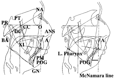
The study analysed 29 children under the age of 15 years. Each child had habitual loud snoring and/or sleep apnea that had been noticed by the parents of the subjects. After taking patients’ cephalograms, we found enlarged tonsils and adenoids. We then scanned and digitized these for computer analysis (Fig. 1). The data were summarized and compared against the control group, n = 41.2

. Cephalometric measurements. The cephalometric points and planes were: BA (basion), anterior margin of the foramen magnum; NA (nasion), junction of nasal and frontal bones; PT (pterygoid point), antero-inferior border of foramen rotundum; CC (center of cranium), intersection of cranial base plane and facial axis; O (orbitale), lower border of orbit; PR (porion), upper border of porus acusticus externus; POG (Pogonion), anterior border of symphysis of mandible; PM (protuberance menti), upper border of protuberance menti; GN (gnathion), intersection of facial plane and mandibular plane; XI, center of mandibular ramus; DC (condyle center), center of crossing part of condyle to cranial base plane; ANS, anterior nasal spine; A (point A), junction of alveolar bone of maxilla with anterior nasal spine; cranial base plane, BA–NA; Frankfort horizontal (FH), PR–O; facial axis, PT–GN; facial plane, POG–NA; mandibular plane, lower border of mandible; condylar axis, DC–XI; corpus axis, XI–PM; McNamara line, perpendicular to FH through NA.
RESULTS
Five (17.2%) children had tonsil problems, seven (24.1%) had adenoid problems, and 17 (58.6%) had both tonsil and adenoid problems. There were significant differences (P < 0.005) between the patient group and control group when comparing the facial axis (FX), lower facial height (LFH), mandibular arc, total facial height (TFH), and McNamara–Pogonion. Data analysis found that children with sleep breathing disorders had a dolico facial pattern (Table 1).
| Control | Patient | |
|---|---|---|
| n | 41 | 29 |
| Age | 9.0 | 6.6 ± 3.1 |
| Ricketts analysis | ||
| FX | 86.0 ± 3.0 | 81.8 ± 3.1*** |
| FD | 86.0 ± 3.0 | 84.8 ± 3.2 |
| MP | 30.0 ± 4.0 | 31.9 ± 5.6 |
| LFH | 49.0 ± 4.0 | 54.6 ± 5.3*** |
| MA | 25.0 ± 4.0 | 21.2 ± 4.8*** |
| TFH | 64.0 ± 3.0 | 68.4 ± 4.5*** |
| McN–A | 1.0 ± 2.0 | 0.7 ± 3.9 |
| McN–Pog | – 6.0 ± 2.0 | – 9.1 ± 5.4** |
| L. pharynx | 12.1 ± 2.6 | 13.1 ± 4.4 |
- ** P < 0.01,
- *** P < 0.001.
- The cephalometric measurements were: (i) facial axis angle (FX), BA-CC-GN; (ii) facial depth angle (FD), crossing of facial plane to FH; (iii) mandibular plane angle (MP), crossing of mandibular plane to FH; (iv) lower facial height angle (LFH), ANS-XI-PM; (v) mandibular arc angle (MA), crossing of condylar axis to corpus axis; (vi) total facial height angle (TFH), crossing of corpus axis to cranial base plane; (vii) McNamara–Point A (McN-A), the shortest distance from McNamara line to point A; (xiii) McNamara–
- Pogonion (McN-Pog), the shortest distance from
- McNamara line to Pogonion; (ix) lower pharynx (L. pharynx), the shortest distance from the pharyngeal back wall to the intersection of the posterior border of the tongue and lower border of the mandible.
DISCUSSION
According to Ricketts analysis, there are three facial pattern classifications among orthodontic patients: (i) brachyo facial pattern (short face); (ii) mesio facial pattern (medium face: Japanese standard of facial axis is 86 ± 3); and (iii) dolico facial pattern (long face).3 The facial pattern of the present population series was dolico facial pattern.
The facial pattern of adults and children with sleep breathing disorders is the dolico facial pattern. However, the data of the children demonstrated a lesser degree of dolico facial pattern compared with the adults. (The data of adult patients with obstructive sleep apnea were as follows: FX 79.8, LFH 55.7, and TFH 69.8) When the tonsil and/or adenoid obstructs the airway, the negative pressure in the chest deforms it into a ‘funnel chest’ condition. This negative pressure in the chest causes the retraction of the tongue as well as the mandible, thereby possibly developing a dolico facial pattern in children with sleep breathing disorders.
The facial pattern of children with sleep breathing disorders was determined to be the dolico facial pattern (long face).


