Fabry cardiomyopathy presenting with a high defibrillation threshold: A short case report
Abstract
Fabry disease is an X-linked recessive glycosphingolipid storage disorder caused by a deficiency of lysosomal enzyme α-galactosidase A. It is recognized that Fabry disease patients often have ventricular arrhythmias. Although the effectiveness of implantable cardioverter-defibrillator (ICD) therapy in patients with ventricular fibrillation is established, there is little evidence regarding ICD therapy for Fabry disease. Here, we report the case of patient with Fabry disease who was treated with an ICD and presented with high defibrillation thresholds.
1 Case Report
We report the case of a 45-year-old man with a familial history of Fabry disease. He had clinical manifestations from childhood and was diagnosed with Fabry disease by an enzyme assay at 19 years of age. He began enzyme replacement therapy with agalsidase alpha at age 34; however, by age 37, his kidney status progressed to end-stage-renal disease requiring hemodialysis. Technetium-99m-labelled methoxyisobutyl isonitrile myocardial perfusion scintigraphy revealed no ischemic lesions at 43 years of age.
He was transferred to our hospital due to cardiopulmonary arrest at age 45. An electrocardiogram revealed ventricular fibrillation, and sinus rhythm was restored by cardiac defibrillation. After intensive therapy, we planned an implantable cardioverter-defibrillator (ICD) implantation. He had normal vital signs (blood pressure: 110/78 mmHg; heart rate: 75 bpm; O2 saturation: 97% on room air; and body temperature: 36.4 °C). Chest X-ray revealed mild cardiomegaly and no pulmonary congestion. The plasma brain natriuretic peptide level was 300.5 pg/mL. An electrocardiogram during sinus rhythm showed signs of left ventricle hypertrophy with a normal QRS duration (QRS duration: 124 ms; QTc: 0.435; S wave amplitude in leads V1+R wave amplitude in leads V5=4.92 mV). QTc was defined as QT interval/square root of RR interval. Transthoracic echocardiography revealed left ventricular hypertrophy (interventricular septal thickness at end-diastole 14.6 mm; left ventricular posterior wall thickness at end-systole 15.0 mm; left ventricle mass 221 g) with a preserved ejection fraction (left ventricular dimension at end-diastole 39.1 mm; left ventricle dimension at end-systole 26.4 mm; ejection fraction 61.4%). Oral drug therapy included carvedilol (2.5 mg/day), but no antiarrhythmic drugs were taken.
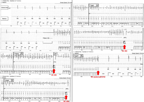
An ICD (FORTIFY ST VR; St. Jude Medical, St. Paul, MN, USA) with a single-coil shock lead (DURATA 7122Q-52; St. Jude Medical, St. Paul, MN, USA) was implanted in the right precordium because he had an arteriovenous shunt for dialysis in his left forearm. The defibrillation thresholds (DFTs) were tested. He demonstrated high DFTs (Fig. 1), which were defined as safety margins of less than 10- J between the DFTs and the maximum energy output of the implanted device [1]. The initial defibrillation setting was as follows: waveform, “Biphasic”; waveform mode, “Tilt”; shock configuration, “RV to Can”. Based on the patient's high DFTs, we modified the device settings, changing the waveform mode to Pulse Width, but the DFTs did not improve. We then extracted the generator and the lead, and, although the patient had a shunt in his left forearm, we moved the generator to the left precordium and replaced the single-coil shock lead with a dual-coil shock lead (DURATA 7120Q-58; St. Jude Medical, St. Paul, MN, USA) in order to improve the DFTs. Finally, after these changes, we achieved an adequate DFT.

Defibrillation threshold (DFT) testing; the implantable cardioverter-defibrillator (ICD) failed to terminate ventricular fibrillation (intracardiac 20J – failure; intracardiac 30J – failure; intracardiac 40J – failure; and external 270J – success).
2 Discussion
To the best of our knowledge, this is the first report of a patient with Fabry disease presenting with high DFTs.
Predicting high DFTs in patients for whom ICD implantation is planned is important; however, the association between Fabry disease and high DFTs has not been systematically studied. Rhythm and conduction abnormalities have been reported to exist in Fabry disease as a result of the glycosphingolipid storage in cardiomyocytes, conduction system cells, and endothelial cells [2]. As left ventricular hypertrophy is the most common manifestation of Fabry disease, the high DFTs observed in this patient may be attributable to myocardial hypertrophy, which is a well-known substrate of high DFTs [3].
By managing the device settings and using a dual-coil lead, it was possible to implant an ICD with an adequate defibrillation safety margin in this patient with high DFTs. Patients undergoing right-sided ICD implantations have higher DFTs compared with left-sided implants [4].
In the case presented here, a man with Fabry disease and ventricular fibrillation had high DFTs after ICD implantation. Further reports should be accumulated in order to determine whether Fabry disease is generally complicated with high DFTs.
Conflicts of interest
The authors have no conflicts of interest to disclose.


