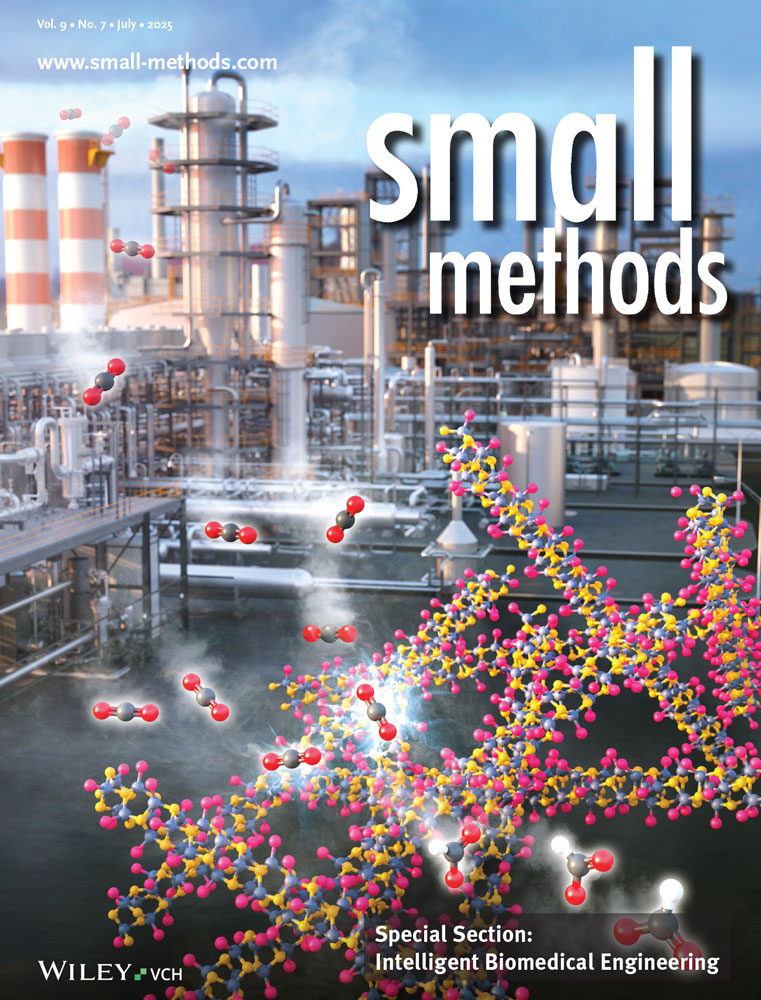
Glycan-Anchored Fluorescence Labeling of Milk-Derived Extracellular Vesicles for Investigating Their Cellular Uptake and Intracellular Fate
Xueqi Su
College of Ocean Food and Biological Engineering, Jimei University, Xiamen, Fujian, 361000 China
Search for more papers by this authorSiqin Zhang
College of Ocean Food and Biological Engineering, Jimei University, Xiamen, Fujian, 361000 China
Search for more papers by this authorTianyu Zhang
College of Ocean Food and Biological Engineering, Jimei University, Xiamen, Fujian, 361000 China
Search for more papers by this authorXueping Pan
College of Ocean Food and Biological Engineering, Jimei University, Xiamen, Fujian, 361000 China
Search for more papers by this authorYingying Ke
College of Ocean Food and Biological Engineering, Jimei University, Xiamen, Fujian, 361000 China
Search for more papers by this authorYalan Fan
College of Ocean Food and Biological Engineering, Jimei University, Xiamen, Fujian, 361000 China
Search for more papers by this authorJian Li
College of Ocean Food and Biological Engineering, Jimei University, Xiamen, Fujian, 361000 China
Search for more papers by this authorLingyu Zhang
College of Ocean Food and Biological Engineering, Jimei University, Xiamen, Fujian, 361000 China
Search for more papers by this authorCorresponding Author
Chaoxiang Chen
College of Ocean Food and Biological Engineering, Jimei University, Xiamen, Fujian, 361000 China
E-mail: [email protected]
Search for more papers by this authorXueqi Su
College of Ocean Food and Biological Engineering, Jimei University, Xiamen, Fujian, 361000 China
Search for more papers by this authorSiqin Zhang
College of Ocean Food and Biological Engineering, Jimei University, Xiamen, Fujian, 361000 China
Search for more papers by this authorTianyu Zhang
College of Ocean Food and Biological Engineering, Jimei University, Xiamen, Fujian, 361000 China
Search for more papers by this authorXueping Pan
College of Ocean Food and Biological Engineering, Jimei University, Xiamen, Fujian, 361000 China
Search for more papers by this authorYingying Ke
College of Ocean Food and Biological Engineering, Jimei University, Xiamen, Fujian, 361000 China
Search for more papers by this authorYalan Fan
College of Ocean Food and Biological Engineering, Jimei University, Xiamen, Fujian, 361000 China
Search for more papers by this authorJian Li
College of Ocean Food and Biological Engineering, Jimei University, Xiamen, Fujian, 361000 China
Search for more papers by this authorLingyu Zhang
College of Ocean Food and Biological Engineering, Jimei University, Xiamen, Fujian, 361000 China
Search for more papers by this authorCorresponding Author
Chaoxiang Chen
College of Ocean Food and Biological Engineering, Jimei University, Xiamen, Fujian, 361000 China
E-mail: [email protected]
Search for more papers by this authorAbstract
Milk-derived extracellular vesicles (mEVs) are promising therapeutic delivery platforms due to their natural bioactivity, biocompatibility, and ability to cross biological barriers. However, analyzing their cellular uptake and trafficking is limited by existing fluorescent labeling methods, which often cause dye leakage and disrupt vesicle integrity. Here, a glycan-anchored fluorescence labeling strategy for mEVs is developed, involving periodate oxidation of surface sialic acids followed by aniline-catalyzed ligation of hydrazide-functionalized fluorophores. Nano-flow cytometry characterization confirmed ≈100% labeling efficiency without compromising mEVs integrity or uptake behavior. This approach enabled quantitative analysis of mEVs internalization, identifying clathrin-mediated endocytosis and macropinocytosis as the primary pathways and confirming mEVs’ capacity for lysosomal escape. Comparative analyses showed that traditional lipophilic dyes induced vesicle aggregation, dye leakage, and transfer, potentially misrepresenting mEVs behavior. Additionally, co-labeling mEVs with glycan-anchored fluorophores and FITC-conjugated paclitaxel enabled real-time tracking of drug delivery, revealing a burst release from lysosomes that led to significant cytotoxicity. Overall, the glycan-anchored fluorescence labeling allows precise analysis of mEVs uptake and intracellular fate, paving the way for further research and application in targeted drug delivery.
Conflict of Interest
The authors declare no conflict of interest.
Open Research
Data Availability Statement
The data that support the findings of this study are available from the corresponding author upon reasonable request.
Supporting Information
| Filename | Description |
|---|---|
| smtd202401996-sup-0001-SuppMat.pdf9.3 MB | Supporting Information |
Please note: The publisher is not responsible for the content or functionality of any supporting information supplied by the authors. Any queries (other than missing content) should be directed to the corresponding author for the article.
References
- 1Y. Couch, E. I. Buzàs, D. Di Vizio, Y. S. Gho, P. Harrison, A. F. Hill, J. Lötvall, G. Raposo, P. D. Stahl, C. Théry, K. W. Witwer, D. R. F. Carter, J. Extracell. Vesicles. 2021, 10, e12144.
- 2G. van Niel, G. D'Angelo, G. Raposo, Nat. Rev. Mol. Cell Biol. 2018, 19, 213.
- 3D. K. Jeppesen, A. M. Fenix, J. L. Franklin, J. N. Higginbotham, Q. Zhang, L. J. Zimmerman, D. C. Liebler, J. Ping, Q. Liu, R. Evans, W. H. Fissell, J. G. Patton, L. H. Rome, D. T. Burnette, R. J. Coffey, Cell. 2019, 177, 428.
- 4C. Marar, B. Starich, D. Wirtz, Nat. Immunol. 2021, 22, 560.
- 5E. I. Buzas, Nat. Rev. Immunol. 2023, 23, 236.
- 6M. A. Kumar, S. K. Baba, H. Q. Sadida, S. A. Marzooqi, J. Jerobin, F. H. Altemani, N. Algehainy, M. A. Alanazi, A.-B. Abou-Samra, R. Kumar, A.-S. Akil, S. A., M. A. Macha, R. Mir, A. A. Bhat, Signal Transduction Targeted Ther. 2024, 9, 27.
- 7R. Tenchov, J. M. Sasso, X. Wang, W.-S. Liaw, C.-A. Chen, Q. A. Zhou, ACS Nano. 2022, 16, 17802.
- 8R. Kalluri, K. M. McAndrews, Cell. 2023, 186, 1610.
- 9M. Kou, L. Huang, J. Yang, Z. Chiang, S. Chen, J. Liu, L. Guo, X. Zhang, X. Zhou, X. Xu, X. Yan, Y. Wang, J. Zhang, A. Xu, H. F. Tse, Q. Lian, Cell Death Dis. 2022, 13, 580.
- 10M. Prasadani, S. Kodithuwakku, G. Pennarossa, A. Fazeli, T. A. L. Brevini, Int. J. Mol. Sci. 2024, 25, 5543.
- 11S. R. Marsh, R. G. Gourdie, Nat. Rev. Bioeng. 2024, 2, 806.
- 12Y. Zhong, X. Wang, X. Zhao, J. Shen, X. Wu, P. Gao, P. Yang, J. Chen, W. An, Pharmaceutics. 2023, 15, 1418.
- 13J. Munir, A. Ngu, H. Wang, D. M. O. Ramirez, J. Zempleni, Pharm. Res. 2023, 40, 909.
- 14L. Tong, S. Zhang, Q. Liu, C. Huang, H. Hao, M. S. Tan, X. Yu, C. K. L. Lou, R. Huang, Z. Zhang, T. Liu, P. Gong, C. H. Ng, M. Muthiah, G. Pastorin, M. G. Wacker, X. Chen, G. Storm, C. N. Lee, L. Zhang, H. Yi, J. W. Wang, Sci. Adv. 2023, 9, eade5041.
- 15L. Tong, H. Hao, Z. Zhang, Y. Lv, X. Liang, Q. Liu, T. Liu, P. Gong, L. Zhang, F. Cao, G. Pastorin, C. N. Lee, X. Chen, J. W. Wang, H. Yi, Theranostics. 2021, 11, 8570.
- 16S. L. Ong, C. Blenkiron, S. Haines, A. Acevedo-Fani, J. A. S. Leite, J. Zempleni, R. C. Anderson, M. J. McCann, Nutrients. 2021, 13, 2505.
- 17J. Zhong, B. Xia, S. Shan, A. Zheng, S. Zhang, J. Chen, X. J. Liang, Biomaterials. 2021, 277, 121126.
- 18F. Aqil, R. Munagala, J. Jeyabalan, A. K. Agrawal, A. H. Kyakulaga, S. A. Wilcher, R. C. Gupta, Cancer Lett. 2019, 449, 186.
- 19Q. Jiang, Y. Liu, X. Si, L. Wang, H. Gui, J. Tian, H. Cui, H. Jiang, W. Dong, B. Li, Annu. Rev. Food Sci. Technol. 2024, 15, 431.
- 20M. Cieślik, K. Nazimek, K. Bryniarski, Int. J. Mol. Sci. 2022, 23, 7554.
- 21M. Boudna, A. D. Campos, P. Vychytilova-Faltejskova, T. Machackova, O. Slaby, K. Souckova, Cell Commun. Signal. 2024, 22, 171.
- 22M. Dehghani, S. M. Gulvin, J. Flax, T. R. Gaborski, Sci. Rep. 2020, 10, 9533.
- 23C. Bao, H. Xiang, Q. Chen, Y. Zhao, Q. Gao, F. Huang, L. Mao, Int. J. Nanomed. 2023, 18, 4567.
- 24J. B. Simonsen, J. Extracell. Vesicles. 2019, 8, 1582237.
- 25D. Fortunato, D. Mladenovic, M. Criscuoli, F. Loria, K. L. Veiman, D. Zocco, K. Koort, N. Zarovni, Int. J. Nanomed. 2021, 22, 10510.
- 26M. Cha, S. H. Jeong, S. Bae, J. H. Park, Y. Baeg, D. W. Han, S. S. Kim, J. Shin, J. E. Park, S. W. Oh, Y. S. Gho, M. J. Shon, Anal. Chem. 2023, 95, 5843.
- 27L. Loconte, D. Arguedas, R. El, A. Zhou, A. Chipont, L. Guyonnet, C. Guerin, E. Piovesana, J. L. Vazquez-Ibar, A. Joliot, C. Thery, L. Martin-Jaular, J. Extracell. Vesicles. 2023, 12, e12384.
- 28C. Chen, N. Cai, Q. Niu, Y. Tian, Y. Hu, X. Yan, J. Extracell. Vesicles. 2023, 12, 12351.
- 29O. M'Saad, J. Bewersdorf, Nat. Commun. 2020, 11, 3850.
- 30X. Zhou, J. Zhang, Z. Song, S. Lu, Y. Yu, J. Tian, X. Li, F. Guan, Chem. Commun. 2020, 56, 14869.
- 31J. Pan, X. Li, B. Shao, F. Xu, X. Huang, X. Guo, S. Zhou, Adv. Mater. 2022, 34, 2106307.
- 32Y.-J. Liu, C. Wang, Cell Commun. Signal. 2023, 21, 77.
- 33D. K. Jeppesen, Q. Zhang, J. L. Franklin, R. J. Coffey, Trends Cell Biol. 2023, 33, 667.
- 34M. T. Roefs, J. P. G. Sluijter, P. Vader, Trends Cell Biol. 2020, 30, 990.
- 35A. Giovanazzi, M. J. C. van Herwijnen, M. Kleinjan, G. N. van der Meulen, M. H. M. Wauben, Sci. Rep. 2023, 13, 8758.
- 36E. Lazaro-Ibanez, F. N. Faruqu, A. F. Saleh, A. M. Silva, T.-W. Wang, J. Rak, K. T. Al-Jamal, N. Dekker, ACS Nano. 2021, 15, 3212.
- 37B. S. Joshi, M. A. de Beer, B. N. G. Giepmans, I. S. Zuhorn, ACS Nano. 2020, 14, 4444.
- 38C. Williams, F. Royo, O. Aizpurua-Olaizola, R. Pazos, G. J. Boons, N. C. Reichardt, J. M. Falcon-Perez, J. Extracell. Vesicles. 2018, 7, 1442985.
- 39V. Vrablova, N. Kosutova, A. Blsakova, A. Bertokova, P. Kasak, T. Bertok, J. Tkac, Biotechnol. Adv. 2023, 67, 108196.
- 40A. R. Martins, C. C. Ramos, D. Freitas, C. A. Reis, Cells. 2021, 10, 109.
- 41M. Lin, X. Xu, X. Zhou, H. Feng, R. Wang, Y. Yang, J. Li, N. Fan, Y. Jiang, X. Li, F. Guan, Z. Tan, Cell. Mol. Biol. Lett. 2024, 29, 46.
- 42N. Cai, X. Zhan, Q. Zhang, H. Di, C. Chen, Y. Hu, X. Yan, Angew. Chem., Int. Ed. 2024, 1, 202413946.
- 43B. Wang, J. Brand-Miller, Eur. J. Clin. Nutr. 2003, 57, 1351.
- 44A. M. Zivkovic, D. Barile, Adv. Nutr. 2011, 2, 284.
- 45Y. Wang, X. Ze, B. Rui, X. Li, N. Zeng, J. Yuan, W. Li, J. Yan, M. Li, Front. Nutr. 2021, 8, 766606.
- 46C. Chen, X. Pan, M. Sun, J. Wang, X. Su, T. Zhang, Y. Chen, D. Wu, J. Li, S. Wu, X. Yan, Small. 2024, 20, 2310712.
- 47C. Chen, M. Sun, J. Wang, L. Su, J. Lin, X. Yan, J. Extracell. Vesicles. 2021, 10, e12163.
- 48Y. Zeng, T. N. Ramya, A. Dirksen, P. E. Dawson, J. C. Paulson, Nat. Methods. 2009, 6, 207.
- 49B. Cheng, Q. Tang, C. Zhang, X. Chen, Annu. Rev. Anal. Chem. 2021, 14, 363.
- 50M. Kaksonen, A. Roux, Nat. Rev. Mol. Cell Biol. 2018, 19, 313.
- 51R. R. Kay, Cells Dev. 2021, 168, 203713.


