Hypericin and resveratrol anti-tumor impact through E-cadherin, N-cadherin, galectin-3, and BAX/BCL-ratio; possible cancer immunotherapy succor on Y79 retinoblastoma cells
Abstract
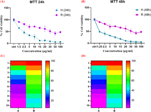
One of the detrimental features of retinoblastoma is highly invasiveness of this type of cancer; the ability to metastasize into distal organs even in the early stages. This phenomenon highlights the importance of experiments targeting this characteristic to diminish the disquieting outcomes. In this study we assessed the impact of hypericin, and resveratrol (two main herbal extracts with known anti-cancer properties on the important metastasis/cancer progression pathways in Y79 retinoblastoma cell line). MTT assay performed for 24 and 48 h, and the kind of cell death, investigated by Annexin V/PI flowcytometry. Finally, the expression of important metastatic/apoptotic genes assessed in different samples by the aid of real-time PCR. The 24 and 48 h IC50 for resveratrol, and hypericin was about 10.87 and 7.697, 2.267, and 1.732 μg/mL, respectively. Flowcytometry results revealed that the kind cell death after treatment of Y79 cell with both compounds was mostly apoptosis. Both compounds induced up-regulation of E-cad, and down-regulation of N-cad, and Gal-3; furthermore, BAX/BCL-2 ratio increased significantly in cells treated with hypericin. Hypericin seems to exert more cytotoxicity effect on Y79 cells compared with resveratrol. As both compounds enhanced the expression of the E-cadherin, along with the decreasing the galectin-3, they can be proposed as beneficial accompanying reagents in cancer immunotherapy.
1 INTRODUCTION
Retinoblastoma is a highly malignant tumor which can invade the neighboring tissues like the brain and the distal organs as well.1-4 For the latter, the tumor cells benefit from the bloodstream and before entering to it they should invade the extracellular matrix and adjoining tissues/cells impeding between the bloodstream and the tumor.5 According to the literature, retinoblastoma occurs mostly in children. As its symptoms are modest at early stages, the disease will have a chance to develop and metastasize, and this will result in an even poorer prognosis.6-8 However, we cannot address a preclinical model which plays as a touchstone to investigate the metastasis of retinoblastoma and assess the impact of treatments targeting different stages of metastasis in this cancer.
Some studies have introduced models for this aim, e.g. an orthotropic zebra fish model as a retinoblastoma metastatic model.5 Based on their study, retinoblastoma has the ability to metastasize even at the early stages, and soon after transplantation to the model zebra fishes. This outcome further underscores the importance of studying the metastasis procedure of this cancer and finding efficient treatment that can target this pathway in retinoblastoma, so that the patient's life may be secured.9, 10
Cell adhesion molecules (CAMs) are important factors in human embryo development and the regulation of different tissues homeostasis and regulation.11, 12 There are various families in the CAMs superfamily which have been well studied; e.g. Ca2+ dependent or independent families including the cadherins, selectins, and integrins, IgSF CAMs (Immunoglobulin superfamily CAMs).13-15
Galectin-3 is a member of the galectin family. This glycoprotein mainly exists in the cytoplasm; however, it may translocate to the nucleus, or even may be secreted and so can be found in body fluids including serum or urine. It has been shown that cancer cells augment the expression of galectin-3 and exploit it to evade from the immune cells.16-18
Resveratrol is a naturally occurring agent which can be found in plant sources like grape, red wine, etc. It is categorized under the polyphenolic compounds and exerts pleiotropic anti-cancer effects.3 Amounting studies have revealed the anti-cancer potential of the agent in different steps of cancer including apoptosis induction in cancer cells, inhibition of tumor growth, and angiogenesis.19 Furthermore, some researches have demonstrated the capacity of this agent in targeting cancer stem cells20; hence, it plays a crucial role in inhibiting cancer cell resistance to conventional treatments, as well as tumor recurrence. These findings together with the potential of resveratrol to affect specific cancer-related signaling pathways make it a potential candidate of interest for treating cancers including breast, colorectal, prostate, and variable other cancers.21
Hypericum perforatum L. also renowned as Tipton Amber, Hardhay weed, Klamath weed, Goat weed, and St.John's wort is a precious herbal remedy from the Clusiaceae or Hypericaceae family and is an aborigine of European and Eastern countries. Various biological compounds can be found in this plant including hyperforin and hypericin. Amounting studies unravel the medicinal benefits of hypericin including its anti-bacterial, anti-inflammatory, anti-depression, anti-cancer effects on solid tumors and hematologic malignancies, etc.22, 23 The anti-tumor effects of resveratrol and hypericin have been demonstrated with the amounting studies. Our main aim in this study was to assess the effect of these extracts on apoptosis induction, and important genes in metastasis including E-cadherin (E-Cad), N-cadherin (N-Cad), and galectin-3 (Gal-3) in Y79 cells, as a delegacy of retinoblastoma in vitro model.
2 MATERIALS AND METHODS
2.1 Cell culture
Y79 retinoblastoma cell line purchased from Pasture Institute of Iran (NCBI code: C201). These cells were cultured in Roswell Park Memorial Institute (RPMI) medium (Gibco) supplemented with 10% V/V of fetal bovine serum and 1% V/V of mixed penicillin/streptomycin antibiotics (all from Thermo Fisher Scientific) in an incubator humidified with 5% CO2 and 95% air (Memmert). The medium surrounding cells was replaced with fresh medium every 4 days. When the confluency of the cells reached the appropriate count, the cells were counted, and 10,000 cells were added to each well of a 96-well plate for MTT assay (each concentration was performed at least in three replicates).
2.2 MTT assay
To assess the cytotoxicity of resveratrol (Catalogue number: TCI-R0071) or hypericin (Catalogue number: H26425.MB) (from Thermo Fisher SCIENTIFIC) on Y79 cells, an MTT assay was performed. To this aim, as mentioned in the previous section, cells were seeded in each well of a 96-well plate overnight. After that, a concentration range of these compounds was added to the wells (from concentration 0 (the control) up to 100 μg/mL) for 24 and 48 h (for 24 h 8000 cells were seeded, and for 48 h 6000 cells were seeded to each well). Post this time, the MTT powder (Sigma Aldrich, Catalogue number: M5655) added to each well (concentration of the stock was 5 μg/mL and 20 μL added to 180 μL of the medium in each well). After 3 h, formazan crystals emerged in the wells. These crystal sediments were dissolved in 200 μL of dimethyl sulfoxide (DMSO) (from Merck, Catalogue number: 102952), and the absorbance of each well was calculated by an ELISA reader device (BioTek) at 570 nm.
2.3 Flow cytometry
The type of cellular death was assessed following treatment with resveratrol or hypericin using Annexin V/Propidium Iodide (AV/PI) (Sigma Aldrich, Catalogue number: 11 858 777 001) flow cytometry. For this aim, after the treatment period (24 h IC50 concentration of each compound), Y79 cells were centrifuged and the above medium was discarded (about 200 000 cells were used for each replicate of this experiment); solutions of the AV/PI kit were added to these cells exactly the same way as the manual's instruction of the kit and analyzed with a BD FACS Calibur (BD biosciences) device. The population of cells with low AV/low PI was reported as live cells, with high PI/ low AV reported as necrotic, with high AV/low PI as early apoptotic, and with both reporters high considered as late apoptotic (all experiments performed at least in duplicates).
2.4 Real-time PCR
To investigate the impact of resveratrol or hypericin on the expression of important genes in the metastasis/apoptosis pathway, real-time PCR was performed. To reach this goal, Y79 cells were added to each well of a 6-well plate (100 000 cells were seeded in each well). We designed the experiment in a way to have 3 different groups: the control (abbreviated as Cntr.); untreated Y79 cells under normal cell culture conditions for these cells (mentioned above), the R group; Y79 cells treated with 10.87 μg/mL of resveratrol for 24 h, and the H group; Y79 cells treated with 2.267 μg/mL of hypericin for 24 h (all experiments performed at least in duplicates).
After 24 h of treatment, RNA from these samples was extracted by using TRIzol reagent (Thermo Fisher Scientific, Catalogue number: 15596026). The quality and quantity of the extracted RNAs were assessed by NanoDrop device (Thermo Scientific). The extracted RNAs (1 μg) from each sample were utilized for cDNA synthesis (with Vivantis cDNA synthesis kit, Catalogue number: cDSK01-050). These cDNAs were the template for the real-time PCR. Primers were designed to specifically amplify cDNA from the Bax, Bcl-2, E-cadherin (abbreviated as E-Cad), N-cadherin (abbreviated as N-Cad), and galectin-3 (abbreviated as Gal-3) genes (primers sequence are shown in Table 1). The real-time PCR process program performed as follows with the Ampliqon SYBR green master mix (catalogue number: A323499): holding stage: 95°C/15 min; cycling stage: denaturing step: 95°C/15 s, followed by annealing step 60°C/30 s, amplification step 72°C/30s (Number of Cycles: 40), 3-melt curve analysis stage.
| Gene name | Forward | Reverse |
|---|---|---|
| E-cadherin | GAGGTCAGCGTGTGTGACTG | ACAGCAAGAGCAGCAGAATCA |
| N-cadherin | GGGAATCCGACGAATGGATGA | GGGAGTCATATGGTGGAGCTG |
| Gapdh (Internal control) | AAGTTCAACGGCACAGTCAAGG | CATACTCAGCACCAGCATCACC |
| Bax | CCAAGAAGCTGAGCGAGTGT | CCCAGTTGAAGTTGCCGTCT |
| Bcl-2 | TCTTTGAGTTCGGTGGGGTC | GTTCCACAAAGGCATCCCAG |
2.5 Statistical analysis
All experiments were performed at least in duplicate. The statistical analysis was done by the GraphPad prism (version 8.00). To compare results attained from samples, a t-test was performed and p-values less than 0.05 were considered as significant.
3 RESULTS
3.1 Cytotoxicity assay
MTT assay results revealed that the toxicity of resveratrol was augmented by increasing its concentration. As we can see in Figure 1, 10.87 μg/mL of resveratrol induced about 50% cellular death after 24 h (the 24 h IC50). Forty-eight-hour MTT assay results demonstrated that the 48 h IC50 of resveratrol was calculated to be 7.697 μg/mL. Hypericin IC50 was calculated to be 2.267 μg/mL and 1.732 μg/mL μg/ml after 24 and 48 h, respectively (Figure 1).

3.2 Flow cytometry
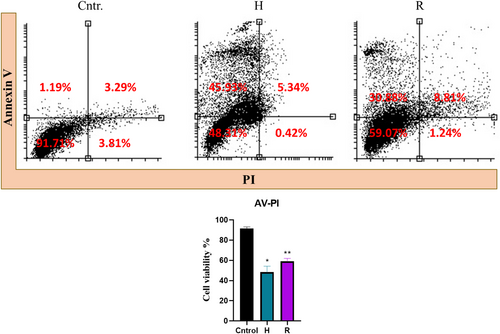
AV/PI flow cytometry results revealed that hypericin or resveratrol induced apoptosis in treated Y79 cells. Samples treated with hypericin underwent much more apoptosis compared with other samples; this group underwent about 53%, while Y79 cells treated with 10.87 μg/mL of resveratrol for 24 h showed about 40% apoptosis (Figure 2).

3.3 Real-time PCR
Investigating the expression of pro-apoptotic Bax or anti-apoptotic Bcl-2 mRNAs revealed alteration of these genes' expression in treated samples. After treatment of Y79 cells with hypericin, Bax expression level increased, and Bcl-2 expression decreased in all treated samples compared with the Y79 control/untreated cells. The Bax/Bcl-2 ratio increased significantly in cells treated with hypericin; although this ratio increased in cells treated with resveratrol compared with the control group, this increase was not statistically significant (Figure 3).

E-Cad mRNA expression in R and H samples increased significantly compared with the control group. However, the expression of this mRNA decreased in the N group (this decreasing was not significant; the p-value was more than 0.05). Real-time PCR results revealed that the N-Cad mRNA expression level decreased significantly in all groups compared with the control group (Figure 4). Investigating the mRNA expression level of Gal-3 in different samples revealed that the expression of this gene at the mRNA level decreased in R and H samples (Figure 3).

4 DISCUSSION
Retinoblastoma is considered as a highly malignant tumor; if not diagnosed and treated properly at the early stages of the disease development, it may be fatal. One of the early organs of metastasis is the brain, and by recruiting the bloodstream, retinoblastoma tumor cells can begin their lethal adventure to distant organs.5, 9, 24 It is undoubtedly important to investigate the metastasis of retinoblastoma, and developing more animal models to be a yardstick for assessing treatments impedes this procedure. In this study, we examined the effect of hypericin and resveratrol on the expression level of important genes in metastatic and apoptosis pathways.
In the current study, we tried to assess the anti-cancer impact of two renowned medicinal herbs, resveratrol, and hypericin on a retinoblastoma cell line. To this aim, this impact was traced in various levels including cytotoxicity (MTT), kind of cell death induction (AV/PI), alteration at the expression level of anti/pro-apoptotic as well as metastasis-related genes. According to the MTT result, hypericin exerts a more cytotoxic effect on Y79 cells compared with resveratrol in both 24 and 48 h of treatment. This is in accordance with the previous studies investigating the anti-tumor activity of these compounds.2, 22, 25-29 The IC50 of resveratrol was calculated to be 10.87 μg/mL after 24 h; however, hypericin induced about 52% cell death (about 48% of cells were alive) after this time in a 2.267 μg/mL dose. The cytotoxicity of both compounds revealed to be time-dependent; after 48 h, the IC50 of resveratrol decreased to 7.697 μg/mL, and after the same time, hypericin induced about 50% cell death at a 1.732 μg/mL dose. According to the AV/PI flow cytometry results, the kind of death was mostly apoptosis. On the other hand, resveratrol with the dose of 7.697 (μg/ml) induced 40% apoptosis, which may declare that the cytotoxicity of hypericin is much higher in Y79 cells compared with resveratrol. These results are concordant with the previous results, which have been reported that resveratrol in general seems to exert cancer cells cytotoxicity in higher doses compared with the documents existing for hypericin.29, 30 According to this result, hypericin revealed more cytotoxicity in Y79 compared to resveratrol. To find out the possible underlying molecular features of this phenomenon, we assessed the alteration of apoptotic-related genes which seemed to be important in apoptosis induction in Y79 cells. According to the study performed by Li et al., the Bax (a pro-apoptotic gene) Bcl-2 (an anti-apoptotic gene) axis is a very important pathway in this way.31
Real-time PCR data further confirmed the results obtained from the flow cytometry. According to our study, hypericin apoptotic induction is dependent on the Bax and Bcl-2; on the other hand, resveratrol seems not to induce apoptosis in Y79 cells independent of the Bax and Bcl-2 pathway. As we can see in Figure 2, hypericin induced more early apoptosis in Y79 cells compared with resveratrol; on the other hand, resveratrol induced more late apoptosis and necrosis compared with hypericin. This may demonstrate that the kind of cell death induction by resveratrol in Y79 cells differs from that of hypericin; furthermore, this data reveals that compounds/interventions like hypericin, which can trigger apoptosis and no other kinds (e.g. necroptosis, etc.) in Y79 cells may result in better impact in the final emission of this tumor. After treatment with hypericin, the Bax/Bcl-2 ratio increased significantly compared with the control sample; this increase can violate the one-to-one ratio of Bax to Bcl-2. This can derive the Bax-Bax dimers formation instead of Bax-Bcl-2 and infer apoptosis by the aid of mitochondria. Different studies have reported that resveratrol induces up-regulation of some pro-apoptotic BCL-2 family members like BAX and inhibits the expression of the anti-apoptotic ones like BCL-2.32, 33 This discrepancy may in some parts arise from different cell lines utilized for experiments.
In a study performed by Andrew K. Joe et al., they investigated the anti-tumor activity of resveratrol on six different human cell lines including MCF-7, SW480, etc. In their experiment, the 48-hour IC50 of resveratrol was calculated to be 70–150 μM (16–34 μg/mL).34 Our results revealed that this IC50 for resveratrol on Y79 retinoblastoma cells is about 7.697 μg/mL, which demonstrates higher toxicity of this compound in Y79 cells compared with MCF7, SW480, HCE7, Seg-1, Bic-1, and HL60.
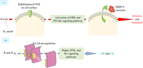
E-Cad is an epithelial marker which plays an important role in cells' adherent junction by interactions with the actin cytoskeleton filaments and different catenins.35, 36 Predominantly, when tumor cells are to metastasize, they will undergo a process named epithelial to mesenchymal transition (EMT).37-39 To this aim, they shall down-regulate the expression of the E-Cad.40-42 Treatment of the Y79 cells with hypericin and merely treatment of cells with resveratrol enhanced the expression of the E-Cad. This result proposes that resveratrol and hypericin may be important factors impeding the EMT process in Y79 cells and hence can induce different contact inhibitions (mentioned in Figure 4) by inducing the activation of Hippo, RTK, and Src signaling pathways.43-46 N-Cad is the other member of the cadherin family which is considered a metastasis progression factor in different cancer types.47-56 Our results revealed that treatment of the Y79 cells with hypericin or resveratrol can decrease the expression of the N-Cad. As it is depicted in Figure 4, N-Cad can induce the activation of the important tumor progression pathways, e.g. the ERK and PI3/Akt pathway, which can eventually result in the secretion of the matrix metalloproteinase-9 (MMP-9) which plays an important role in the degrading of the ECM.57
Among all treatments, from the statistical point of view, resveratrol could down-regulate the expression of Gal-3; similarly, hypericin treatment resulted in the decrease of this gene expression but was not statistically significant. Gal-3 is reported to be secreted by cancer cells and plays an important role in suppressing immune cells on behalf of cancer cells.16, 58, 59 This proposes that treatment with resveratrol or hypericin can be beneficial in some cancer immunotherapy approaches. Although the apoptosis pathways are interrelated, hypericin seems to induce apoptosis in a somewhat different pathway compared with resveratrol. Figure 4 depicts and proposes possible anti-metastatic mechanisms of action that may occur in the R or H groups in Y79 cells after treatment. As shown in this figure, both treatments can suppress metastasis by down-regulating N-Cad gene expression; on the other hand, R and H treatments exert more anti-metastatic impact by up-regulating E-Cad. Both reagents induce down-regulation of MMP9 (Figure 4).
5 CONCLUSION
Hypericin and resveratrol show anti-metastatic and apoptotic activity in Y79 retinoblastoma cells. From a molecular point of view, these agents may exert this effect mostly by down-regulating the N-Cad gene expression, as well as enhancing the expression level of E-Cad; it seems that hypericin induces apoptosis in Y79 cells in a BAX/BCL-2 dependent pathway. Moreover, they exert anti-tumor activity in Y79 cells by down-regulating the Gal-3 gene, which can be deemed that cancer immunotherapy can benefit from these compounds.
Along with the possible achievements, the current study suffers from some limitations, including a lack of in vivo experiments and tracing other kinds of programmed cell death induction rather than apoptosis. Although the current in vitro study is a crucial and inevitable step prior to clinical trials, monitoring the metastasis lesions in retinoblastoma mice models after receiving hypericin or resveratrol can pave the way for the utilization of such treatments at bedside.
AUTHOR CONTRIBUTIONS
FSh, MB, MD, and SSh performed the main experimental tests and drafted the first version of the manuscript; HZA was involved in data analysis. MAJ conceived the idea of the study, and MAJ and AA supervised the team. All authors reviewed the manuscript.
ACKNOWLEDGMENTS
The authors intend to thank Motamed Cancer Institute for their support and good suggestions in this project.
CONFLICT OF INTEREST STATEMENT
The authors declare that there is no conflict of interest.
ETHICS STATEMENT
Not applicable.
Open Research
DATA AVAILABILITY STATEMENT
The data that support the findings of this study are available on request from the corresponding author. The data are not publicly available due to privacy or ethical restrictions.


