Shoulder structures and strength in competitive preadolescent swimmers: A longitudinal ultrasonographic study
Abstract
Background
Repetitive shoulder movements during competitive training may cause changes in the strength of periarticular shoulder structures in preadolescent swimmers.
Objective
To prospectively determine the effects of training on shoulder periarticular structures and muscle strength in preadolescent swimmers.
Design
Prospective cohort study.
Setting
Community-based natatorium.
Participants
Twenty-four preadolescent swimmers aged 10–12 years.
Interventions
Not applicable.
Main Outcome Measures
Measurements were repeated in three periods as preseason, midseason, and postseason. Ultrasonographic measurements (supraspinatus tendon thickness, humeral head cartilage thickness, deltoid muscle thickness, and acromiohumeral distance) were performed using a portable device and a linear probe. Shoulder (flexion, extension, abduction, internal and external rotation) and back (serratus anterior, lower, and middle trapezius) isometric muscle strength were measured with a handheld dynamometer.
Results
Supraspinatus tendon thickness and acromiohumeral distance were similar in all periods (all p > .05); however, deltoid muscle and humeral head cartilage thicknesses increased throughout the season (p = .002, p = .008, respectively). Likewise, whereas shoulder muscle strength increased (all p < .05), back muscle strength was similar in all periods (all p > .05).
Conclusions
In preadolescent swimmers, acromiohumeral distance and supraspinatus tendon thickness seem to not change; but humeral head cartilage and deltoid muscle thicknesses as well as shoulder muscle strength increase throughout the season.
INTRODUCTION
Competitive swimmers often train a distance of ~8000–14,000 meters per day, 6–7 days a week, for 10–12 months a year.1, 2 During these training sessions, ~2500–9600 shoulder rotations are completed per day2, 3 making these athletes prone to injuries of the shoulder complex.4 Likewise, shoulder pain is commonplace in swimmers,5-8 with decreased acromiohumeral distance (AHD) and increased supraspinatus tendon thickness (STT) reported as predisposing factors.9-14
Biomechanical studies showed that propulsive forces in swimming are mainly shoulder adduction and internal rotation,15, 16 and swimmers are at risk of shoulder injury, especially during front crawl-style arm rotation.17 Shoulder stabilizers (mainly the rotator cuff muscles ie, supraspinatus, infraspinatus, subscapularis, teres minor) significantly contribute to the maintenance of AHD by keeping the humeral head in the glenoid cavity during dynamic arm elevation.18, 19 As such, it is necessary to maintain the balance of strength among several (ie, rotator cuff, shoulder adductor, and internal rotator) muscles to reduce the risk of injury, although swimming training might cause imbalance between the shoulder rotators in (preadolescent) swimmers20-22 and increase the risk for injury and shoulder pain.23
The effects of swimming training on shoulder periarticular structures in an adolescent population have not been studied.11, 13, 14 The objective of this study was to prospectively explore the effects of training season on various shoulder structures and strength in competitive/elite preadolescent swimmers.
METHODS
Design and participants
This was a prospective cohort study in competitive preadolescent swimmers. Its protocol was approved by the local ethics committee (December 21, 2021; approval number: GO21/1341) and it was conducted in accordance with the Declaration of Helsinki and the international principles governing research in humans and animals. The study was also registered at clinicaltrials.gov (NCT054268876).
Thirty nationally qualified swimmers aged between 10 and 12 years were recruited. Inclusion criteria were being elite-level member of the team and training at least five times a week (with at least 1–2 hours per session). Swimmers were excluded if they had less than 2 years of competitive swimming experience, limited participation in training (due to back, neck, or shoulder pain) for more than 2 weeks during the season, inability to complete full training, and history of shoulder surgery. All eligible swimmers and their legal guardians were informed of the study procedure and gave written consent.
Outcomes
Sitting/standing height, body weight, arm span/length, and shoulder width were measured and recorded. Shoulder width was defined as the distance between the lateral edges of the acromion processes.
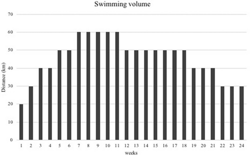
Data were collected throughout the training season at the natatorium in which training was completed. Swimmers participated in three data collection sessions: baseline (preseason, T1), 12 weeks (midseason, T2), and 24 weeks (postseason, T3) after the initial assessment–January, April, and July, 2022, respectively. During the 24-week season, the swimmers were supervised by coaches during swimming training sessions twice daily three times per week, once daily three times per week. A standard/prescribed swimming training volume that swimmers are exposed to during the season is summarized in Figure 1. In addition to 24-week swimming training, the swimmers completed land-based training that included general strengthening/stretching exercises for the trunk and upper/lower limbs. Participants were instructed to avoid vigorous physical activity during the 24 hours before testing and did not participate in the training 1 day before the measurements to avoid any/possible fatigue. Measurements were taken after rest, at the beginning of each period.

Strength measurements
Shoulder/back isometric muscle strength was measured with a hand-held dynamometer (HHD) (Model-01165, Lafayette Instrument Company, Lafayette, IN, USA), which is a valid and reliable tool for assessing isometric muscle strength in swimmers.24 Before the measurements, participants were verbally informed about the technique. To ensure correct movement, participants were asked to perform submaximal contraction against the assessor's hand before the test and the measurements were explained. The “break test” technique, which requires isometric contraction, was applied.25 Bilateral shoulder (flexion, extension, abduction, and internal/external rotation) and back (serratus anterior and middle/lower trapezius) muscle strengths were measured with three repetitions (average values were included in the analyses). A 30-second rest was given between measurements during which the tester otherwise verbally motivated the swimmers.
Strength measurements were performed according to previous protocols.26 Flexion and extension isometric strength were measured at 140° abduction of the shoulder in the scapular plane, with the elbow elongated (simulating hand entry and early pull-through phase during swimming). Internal/external rotation (IR/ER) measurements were done at 90° abduction of the arm, with the elbow at 90° flexion (simulating mid-pull-through and recovery phases of the swim). For measuring abduction, participants were in the sitting position with the arms positioned at 90° of abduction, resistance was applied superior to the elbow.
For middle trapezius, the swimmers were placed in prone position at 90° glenohumeral abduction, with the elbow at 90° flexion whereby the dynamometer was placed on the posterolateral corner of the acromion. Resistance was applied laterally. The clinician stabilized the contralateral scapula while matching the force exerted by the participant (Appendix A: Figure A1). For the lower trapezius, the arm was positioned in 140° glenohumeral abduction whereby the dynamometer was placed on the posterolateral corner of the acromion. Resistance was applied in the lateral and superior directions with the dynamometer. The clinician stabilized the contralateral hip while matching the force of the participant (Appendix B: Figure A2). Serratus anterior was tested in supine position with the elbow and shoulder in 90° flexion. Resistance was applied over the ulna along the humeral axis (Appendix C: Figure A3).27
Because the study included swimmers of different body weights, the mean maximum contraction force was standardized to body weight (N/kg × 100).26 ER/IR ratio was calculated from the values standardized for body weight in each swimmer.
Ultrasonographic measurements
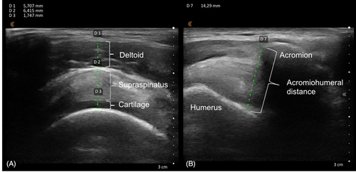
All images were obtained by a physiatrist with at least 4 years of experience in musculoskeletal ultrasound. A smartphone-based portable ultrasound device and a linear probe (Clarius Mobile Health, 205-2980 Virtual Way, Vancouver, British Columbia, Canada) were used. To avoid compression, a generous amount of gel was applied and the probe was held using the suspension technique, that is, touching only the skin. Measurements were done before training in the swimming pool setting in accordance with the European Musculoskeletal Ultrasound Study Group/Ultrasound Study Group of the International Society of Physical and Rehabilitation Medicine (EURO-MUSCULUS/USPRM) protocols.28, 29 Participants were positioned so that the dorsal aspect of the hand was placed in the low back region, while the arm was in extension, adduction, and internal rotation. Anterior deltoid muscle thickness (DMT) was measured between its inner and outer fasciae. STT was measured between the subdeltoid bursa and the underlying cartilage. Humeral head cartilage thickness (HCT) was measured as the anechoic space over the bone and AHD was measured between the humerus and acromion in neutral position (Figure 2).

Statistical analysis
Data were evaluated with SPSS 23.0 (IBM SPSS Statistics version 23.0, IBM Corp. Armonk, New York, USA). Numerical values are given as mean and SD or median and quartiles, depending on data normality. Categorical variables are given using frequency (n) and percentage (%) values. Normal distribution was tested using both graphical (eg, Q-Q plot, histogram, etc.) and analytical (eg, Shapiro–Wilk's normality test) techniques. Repeated measure analysis of variance was used to analyze differences between periods. The Greenhouse–Geisser correction was performed if the sphericity assumption was violated. Pairwise comparisons with Bonferroni correction were conducted when there was a significant time effect by period interactions. The eta-squared (η2) test was used to quantify the percentage of the variance explained by each covariate (effect size) and interpreted as follows: 0 < η2 < 0.04 trivial effect, 0.04 < η2 ≤ 0.24 small, 0.25 ≤ η2 < 0.64 moderate, and η2 > 0.64 large effect. Statistical significance was set at p < .05.
G*Power 3.1 statistical program was used for sample size calculation that is, with a two-tailed type 1 error of .05 and a statistical power of 85% using the pre- and postseason AHD mean ± SD values (1.21 ± 0.3; 1.04 ± 0.1, respectively) obtained from the study of Hibbert et al.12 Twenty-four swimmers were calculated to be included in the current study.
RESULTS
Thirty preadolescent swimmers volunteered to participate and six who did not complete the training season due to injury and/or inability to continue swimming regularly during the season were excluded. Therefore, 24 swimmers (aged 11.7 ± 0.9 years) participated in all three data collection sessions. Their anthropometric characteristics are summarized in Table 1.
| No. | % | |
|---|---|---|
| Gender | ||
| Female | 11 | 45.8 |
| Male | 13 | 54.2 |
| Swimming style | ||
| Front crawl | 7 | 29.2 |
| Butterfly | 5 | 20.8 |
| Back crawl | 5 | 20.8 |
| Breaststroke | 7 | 29.2 |
| Mean ± SD | Range | |
|---|---|---|
| Age (year) | 11.7 ± 0.9 | 10.2–12.9 |
| Swimming experience (year) | 4.8 ± 1.4 | 3–6 |
| Body height (cm) | 157.5 ± 8.5 | 140.0–176.0 |
| Body weight (kg) | 46.9 ± 8.3 | 30.7–63.1 |
| Body mass index (kg/m2) | 18.5 ± 2.8 | 14.0–22.6 |
| Arm span (cm) | 161.0 ± 10.9 | 138.2–182.2 |
| Shoulder width (cm) | 38.7 ± 2.6 | 33.2–44.3 |
| Upper arm length (cm) | 80.5 ± 5.4 | 69–91 |
| Sitting height (cm) | 82.6 ± 4.2 | 74.5–91.2 |
Changes in muscle strengths
Comparative muscle strength measurements are presented in Table 2. Although shoulder IR, ER, flexion, extension, and abduction significantly increased, serratus anterior and lower/middle trapezius strengths were similar throughout the season. In more detail, mean IR increased by 15.8% (p = .015) between T1 and T2, by 31.6% (p < .001) between T1 and T3, by 13.6% (p = .022) between T2 and T3. Mean ER increased by 15.0% (p = .003) between T1 and T2, by 21.5% (p < .001) between T1 and T3. Mean flexion increased by 16.0% (p = .006) between T1 and T2, by 31.7% (p = .001) between T1 and T3. Mean extension increased by 30.0% (p < .001) between T1 and T3, by 21.6% (p < .001) between T2 and T3. Mean abduction increased by 9.0% (p = .048) between T1 and T3.
| Preseason (T1) | Midseason (T2) | Postseason (T3) | Time effect | |||
|---|---|---|---|---|---|---|
| Mean ± SD | Mean ± SD | Mean ± SD | F | p | η2 | |
| Shoulder | ||||||
| Internal rotation | 191.8 ± 41.4 | 222.2 ± 49.5b | 252.3 ± 54.7c,d | 19.149 | <.001a | 0.50 |
| External rotation | 173.5 ± 34.5 | 199.4 ± 37.6b | 210.7 ± 28.9c | 14.910 | <.001a | 0.44 |
| ER/IR ratio | 0.9 ± 0.1 | 0.9 ± 0.1 | 0.8 ± 0.1 | 1.635 | .212 | 0.08 |
| Flexion | 129.0 ± 20.6 | 149.7 ± 27.2b | 169.9 ± 39.4c | 13.403 | <.001a | 0.41 |
| Extension | 126.8 ± 27.8 | 135.6 ± 22.0 | 164.9 ± 17.9c,d | 27.954 | <.001a | 0.60 |
| Abduction | 238.7 ± 27.8 | 256.7 ± 37.2 | 260.2 ± 40.4c | 3.440 | .042a | 0.15 |
| Back | ||||||
| Serratus anterior | 375.4 ± 55.2 | 381.2 ± 73.4 | 390.3 ± 64.2 | 0.644 | .531 | 0.03 |
| Middle trapezius | 215.0 ± 59.6 | 216.0 ± 37.0 | 219.7 ± 43.7 | 0.133 | .875 | 0.01 |
| Lower trapezius | 171.0 ± 46.2 | 180.1 ± 36.0 | 188.4 ± 29.8 | 1.716 | .193 | 0.08 |
- Abbreviations: ER, external rotation; IR, internal rotation.
- a Significant time effect (p < .05).
- b Significant change between T1 and T2 (p < .05).
- c Significant change between T1 and T3 (p < .05).
- d Significant change between T2 and T3 (p < .05).
Changes in ultrasonographic measurements
Comparative measurements are presented in Table 3. DMT significantly changed between the periods (F = 7.077; p = .002; η2 = 0.24 “small effect”), that is, increased by 9.2% (p = .014) between T1 and T2, increased by 13.9% (p = .010) between T1 and T3, similar between T2 and T3 (p = .720). HCT also changed significantly between the periods (F = 6.842, p = .008, η2 = 0.25) that is, increased by 15.5% (p = .015) between T1 and T3, similar between T1 and T2 (p = .117), T2 and T3 (p = .130). STT and AHD measurements were similar in all periods (F = 1.85, p = .182; F = 1.463, p = .245, respectively).
| Preseason (T1) | Midseason (T2) | Postseason (T3) | Time effect | |||
|---|---|---|---|---|---|---|
| Mean ± SD | Mean ± SD | Mean ± SD | F | p | η2 | |
| Deltoid muscle thickness | 4.46 ± 0.79 | 4.87 ± 0.85b | 5.09 ± 0.97c | 7.077 | .002a | 0.24 |
| Supraspinatus tendon thickness | 5.24 ± 0.69 | 5.11 ± 0.81 | 5.41 ± 0.88 | 1.850 | .182 | 0.08 |
| Cartilage thickness | 1.50 ± 0.46 | 1.58 ± 0.42 | 1.74 ± 0.53c | 6.842 | .008a | 0.25 |
| Acromiohumeral distance | 10.85 ± 1.80 | 10.47 ± 1.39 | 10.40 ± 1.37 | 1.463 | .245 | 0.06 |
- a Significant time effect (p < .05).
- b Significant change between T1 and T2 (p < .05).
- c Significant change between T1 and T3 (p < .05).
DISCUSSION
This study provides information regarding the changes of shoulder structure/strength in preadolescent swimmers throughout the competitive season. Structurewise, HCT and DMT increased; AHD and STT did not change. Strengthwise, ER/IR ratio and back strength did not change, but other muscle strengths increased during the swimming season.
Decreased AHD can cause mechanical stress on soft tissues in the subacromial space and make swimmers more vulnerable to shoulder pain/injury.30, 31 Hibberd et al.11 found that AHD was not different between swimmers and nonoverhead athletes in preseason measurements; however, another study showed that AHD significantly decreased in swimmers during the training season for 12 weeks.12 Our study revealed that AHD did not significantly decrease throughout the competitive season, because of the land-based training program that includes strengthening of the muscles that prevent excessive cranial translation of the humeral head.32, 33 Another possible reason could be the preadolescent age of our population with naturally shorter exposure to swimming training. Nonetheless, slight narrowing of the AHD could have also stemmed from scapular motion disorders, that is, the lack of sufficient increase in back muscles strength (responsible for scapular stabilization). However, the influence of muscles responsible for scapular stabilization on scapular movement is controversial. A recent randomized controlled study reported that exercises aiming to improve scapular biomechanics and stabilization did not have sufficient effect on scapular movement.34 Contrary to this study, Hotta et al. showed that intersegmental coordination exercises could modulate the scapular tilt.35
Although there are previous reports on STT in swimmers,8, 13, 14, 36 longitudinal follow up throughout the season is limited.37 Examining the acute effects of swimming, Porter et al.13, 14 showed increased STT, whereas Tate et al.37 reported that it did not significantly change during the season. Our results were in keeping with the latter study. At this point, it would be noteworthy that although the response of the tendon to acute loading is increased thickness, the absence of significant changes during the season could indicate adaptation to chronic loading.38, 39 This would also be important to avoid mechanical impingement.
When compared with the reported reference values of healthy children aged 10–12 years,40 HCT was lower in our study, though it showed significant/progressive increase throughout the season. Whether these results are favorable or due to overload/edema is not yet known.
Concerning strength assessment, Manske et al.41 reported increased shoulder flexion, extension, IR, ER, and abduction strengths after land and swimming training throughout the season. Similarly, our results yielded consistent increase in shoulder flexion, IR, ER, and abduction strengths during the competitive 24-week season. As low shoulder extension strength is known to be potentially related to shoulder pain,23 despite having not evaluated any pain scores in our patients, we can speculate that the increased extension strength may be protective against the development of pain in swimmers.
The balanced ratio of shoulder ER and IR is more important than absolute IR and ER strength, especially in overhead athletes.21, 42 The literature provides evidence that IR becomes stronger than ER from the beginning to the end of the season, with an eventual strength imbalance during the pull (push) phase in swimmers.21, 42, 43 This imbalance changes the centralization of humeral head in the glenoid space, possibly causing impingement/injury. Contrary to the aforementioned studies, although both increased, ER and IR strength balance was maintained throughout the season in our study. Although the strength ratios did not change after 24-week follow-up, increased IR strength from midseason to the end of the season was greater than that of ER. As our study provides data concerning a period of 24 weeks, these preadolescent swimmers need to be followed to more advanced levels for detecting possible muscle imbalances and reducing the risk of injury. Particularly, participants who have completed their growth/maturation with longer swimming background can be enrolled and also assessed in correlation with the pertinent clinical signs/symptoms.
Swimming is a sport in which upper limb repetitive movements are inevitable; therefore, good scapular stabilization is required to perform the stroke. In this aspect, lower trapezius and serratus anterior muscles play an important role.44 Our results failed to show any gain in back muscle strength during the swimming season, which might predispose competitive swimmers to relevant injuries. For example, the lower trapezius enables the scapula to rotate upward, helping to prevent compression during swimming (stroke).45
CONCLUSIONS
Our findings showed that the AHD and STT did not change during the competition season and that the HCT and DMT increased. Additionally, progressive increase in shoulder, but not back, muscle strength was observed during the 24-week period. These results showed that the 24-week training period caused changes in the periarticular shoulder structures (that could be detected with ultrasound) and progressive increase in the shoulder muscle strength.
ACKNOWLEDGMENTS
The authors would like to thank all the swimmers, their parents and coach Efe Orhan of the sports club.
DISCLOSURES
None.


