Metabolic Effects of Light at Night are Time- and Wavelength-Dependent in Rats
Abstract
Objective
Intrinsically photosensitive retinal ganglion cells are most sensitive to short wavelengths and reach brain regions that modulate biological rhythms and energy metabolism. The increased exposure nowadays to artificial light at night (ALAN), especially short wavelengths, perturbs our synchronization with the 24-hour solar cycle. Here, the time- and wavelength dependence of the metabolic effects of ALAN are investigated.
Methods
Male Wistar rats were exposed to white, blue, or green light at different time points during the dark phase. Locomotor activity, energy expenditure, respiratory exchange ratio (RER), and food intake were recorded. Brains, livers, and blood were collected.
Results
All wavelengths decreased locomotor activity regardless of time of exposure, but changes in energy expenditure were dependent on the time of exposure. Blue and green light reduced RER at Zeitgeber time 16-18 without changing food intake. Blue light increased period 1 (Per1) gene expression in the liver, while green and white light increased Per2. Blue light decreased plasma glucose and phosphoenolpyruvate carboxykinase (Pepck) expression in the liver. All wavelengths increased c-Fos activity in the suprachiasmatic nucleus, but blue and green light decreased c-Fos activity in the paraventricular nucleus.
Conclusions
ALAN affects locomotor activity, energy expenditure, RER, hypothalamic c-Fos expression, and expression of clock and metabolic genes in the liver depending on the time of day and wavelength.
Study Importance
What is already known?
- ► Acute exposure to bright morning light increases plasma triglyceride levels in healthy men and glucose levels in men with obesity with type 2 diabetes; blue-enriched light in the morning and in the evening impacts glucose metabolism in healthy adults..
What does this study add?
- ► Nocturnal light exposure in rats acutely induces glucose intolerance, depending on the time of day, intensity, and wavelength of the exposure.
- ► Here, we show that also the effects of light at night on energy metabolism are wavelength and time dependent.
Introduction
With our modern lifestyle, our daily pattern of light exposure is very different from that of the 24-hour solar cycle. Artificial light at night (ALAN) has been associated with multiple disturbances to the environment and to virtually every organism, being considered an endocrine disruptor ((1)). Studies in animals and in humans have shown how ALAN interferes with the normal secretion of hormones such as melatonin ((2)) and glucocorticoids ((3)); alters locomotor activity patterns, sleep, and alertness ((4, 5)); and even alters body weight, food intake, and glucose metabolism ((6-10)). Our group reported that ALAN caused the upregulation of the enzyme phosphoenolpyruvate carboxykinase (PEPCK) in the liver of rats. Because PEPCK is well known to play an important role in gluconeogenesis, this finding suggested that aberrant light exposure could affect glucose metabolism ((11)). More recently, our lab showed that 2 hours of ALAN in rats impaired glucose tolerance and that this effect was depending both on the time of the day and the wavelength ((12)). Most recently, we showed that 1 hour of ALAN induces changes in the hepatic transcriptome and that this effect was at least partially mediated via the autonomic nervous system (ANS). Additionally, it has been shown that light can acutely increase plasma triglycerides in healthy men and plasma glucose and triglycerides in men with obesity with type 2 diabetes, suggesting that exposure to artificial light can also alter lipid metabolism ((13)).
Light stimuli perceived by the retina reach the biological clock in the suprachiasmatic nucleus (SCN) via the retinohypothalamic tract. The entrained rhythmic information from the SCN subsequently is transmitted to the paraventricular nucleus of the hypothalamus (PVN), amongst others. The preautonomic neurons in the PVN are responsible for balancing the activity of the sympathetic and parasympathetic branches of the ANS ((14, 15)), which is key for the control of daily carbohydrate and lipid metabolism in the liver. Hence, we hypothesized that (nocturnal) light may reach the PVN via the SCN and subsequently cause changes in the liver and energy metabolism. Therefore, the first and main objective of this work was to provide a detailed description of the time- and wavelength-dependent effects of light at night on energy metabolism in rats. The second aim was to investigate the time and wavelength dependency of light-induced changes in neural activity in the SCN and the PVN and, ultimately, the expression of hepatic clock and metabolic genes.
Methods
Animals and housing
Sixty-four male Wistar rats (Charles River Breeding Laboratories, Sulzfeld, Germany) weighing 240 to 280 g were individually housed under a controlled ambient temperature of 21°C to 23°C and 12-hour light/dark cycle (lights on at 07:00, Zeitgeber time 0 (ZT0); lights off 19:00, ZT12) with white fluorescent light (maximum, 150 lux) during the light phase and dim red light during the dark phase (< 5 lux). All animals had ad libitum access to chow food (Teklad Global Diet; Harlan, Horst, the Netherlands) and water. Body weight was measured weekly. All the experiments were performed in accordance with the Council Directive 2010/63EU of the European Parliament and the Council of 22 September 2010 on the protection of animals used for scientific purposes. All procedures were also approved by the Animal Ethics Committee of the Royal Dutch Academy of Arts and Sciences (KNAW, Amsterdam, the Netherlands) and in accordance with the guidelines on animal experimentation of the Netherlands Institute for Neuroscience.
Experimental design
After an acclimatization week, animals were divided randomly into the following four groups: dark controls and blue, green, and white light (n = 16 each). Light-treated animals were divided further into two subgroups (n = 8 each), using blocked randomization by body weight; both subgroups received two different light pulses, one at the beginning and one toward the end of the dark phase. Subgroup 1 received a light pulse at ZT14-16 and ZT18-20, whereas subgroup 2 received light at ZT16-18 and ZT20-22 (Supporting Information Figure S1A).
Metabolic cages
To measure locomotor activity, food intake, energy expenditure, and respiratory exchange ratio (RER) before, during, and after the light exposure, animals were placed in Metabolic Pheno Cages (TSE systems, Bad Hombourg, Germany). Animals were individually housed and kept in these cages for 96 hours. The first 24 hours were taken as acclimatization and were not used for the analysis, the second day was used as a control day, the light treatment was given on the third day, and the fourth day was used as a control day to make sure the possible effects we would see were exclusively light dependent (Supporting Information Figure S1B, data of control days not shown). Data were acquired every 15 minutes for all the variables. For each parameter, the effects of light exposure were analyzed as the absolute change (Δ) between the eight measurements obtained from the metabolic cages during the light exposure and the last time point before lights went on.
Light exposure protocol
Metabolic cages were placed inside rectangular metal frames with LED strips around the perimeter. To determine which light intensity to use, all lights were measured with a spectrometer (Avaspec-3648-USB2; Avantes, Apeldoorn, the Netherlands). Using the rodent toolbox designed by Lucas et al. ((16)) we selected intensities such that roughly the same photon flux was obtained for each wavelength. Detailed characteristics of the light conditions used can be found in Supporting Information Table S1. All animals in total received three 2-hour light pulses with ~2 wash-out weeks in between each light exposure (Supporting Information Figure S1).
Blood and tissue sampling
In the last week of the experiment, each group of eight animals was further divided into two subgroups of four animals each. Each subgroup received a third light pulse at one of the two time points they had been treated before (Supporting Information Figure S1). Immediately after this third 2-hour light pulse, animals were deeply anesthetized with an overdose of pentobarbital (intraperitoneal). A thoracotomy was performed, and blood was collected with a cardiac puncture from the left ventricle. A sample of liver tissue was collected, frozen immediately in liquid nitrogen, and kept at −80℃ until RNA isolation. All animals were perfused transcardially with paraformaldehyde (PFA) 4% weight/volume in 0.1 M phosphate buffer (pH = 7.2) and brains recovered for further analysis.
Plasma measurements
Blood glucose levels were determined by using blood glucose test strips with an accuracy of 0.1 mmol/l (FreeStyle; Abbot Diabetes Care, Alameda, California). Plasma insulin and corticosterone levels were measured by using a radioimmunoassay (Millipore, St. Charles, Missouri, and MP Biomedicals, Santa Ana, California, respectively).
RNA isolation and complementary DNA synthesis
Liver tissues were homogenized with tri-reagent using an Ultra-Turrax device (IKA, Staufen, Germany) in order to prevent RNA degradation. RNA isolation and complementary DNA synthesis were performed as described elsewhere ((17, 18)) and in the Supporting Information.
Quantitative polymerase chain reaction
To measure the expression profiles of hepatic clock and metabolic genes, diluted complementary DNA (2:38) was used for all quantitative polymerase chain reaction (qPCR) in duplicate. The expression levels of the tested genes were standardized by dividing over the geometric mean of three reference genes glyceraldehyde-3-phosphate dehydrogenase (GAPDH), 40S ribosomal protein S18 (S18), hypoxanthine-guanine phosphoribosyltransferase (HPRT). Reverse transcription quantitative polymerase chain reaction (RT-qPCR) was performed by using a Light Cycler 480 (Roche). Gene expression levels were calculated with the software for linear regression of qPCR data LinRegPCR. Melting curves of the RT-qPCR were used as quality controls. Primer sequences are listed in Supporting Information Table S2.
Immunohistochemistry for c-Fos
Perfused brains were kept in PFA 4% for 24 hours and then changed to 30% sucrose with 0.05% sodium-azide until they were cut. Brains were cut at 30-µm thickness with the Cryostat NX50 (Thermo Fisher Scientific, Waltham, Massachusetts) and stored in cryoprotectant solution (30% v/v glycerol, 30% v/v ethylene glycol, 40% 0.1M Tris-buffered saline with 30% sucrose, and 0.05% sodium azide) at −20°C until the immunohistochemical analysis was performed with a protocol previously described ((17)), using a different primary antibody (c-Fos polyclonal rabbit SC-52, 1:1000; Santa Cruz Biotechnology, Dallas, Texas). A CCD camera (Sony Model 77CE; Tokyo, Japan) attached to a microscope (Zeiss Axios-kop with Plan-NEOFLUAR Zeiss objectives; Carl Zeiss GmbH, Oberkochen, Germany) was used to take micrographs with a 10 × 0.63 objective with identical lighting conditions for all animals. c-Fos–positive cells were counted using ImageJ 1.52a (NIH, Bethesda, Maryland).
Statistics
Data are presented as means (SEM). Mixed-effects models analysis and ANOVAs were used to analyze the changes in the parameters obtained from the metabolic cages during the 2-hour light exposure, and body weight gain, plasma metabolites, relative clock and metabolic gene expression, and number of c-Fos–positive cells in the brain areas were analyzed. Dunnet’s multiple comparisons test was used for post hoc analysis when significant effects of light or interaction were found. Statistics were performed by using GraphPad Prism version 8.01 for Windows (GraphPad Software, La Jolla, California) with a significance level of P < 0.05.
Results
Metabolic data
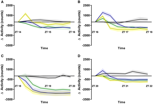
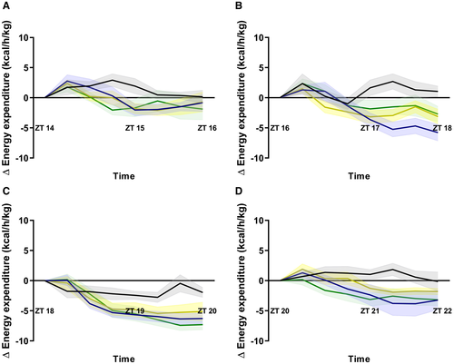
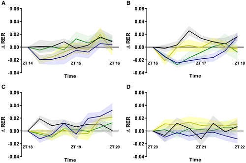
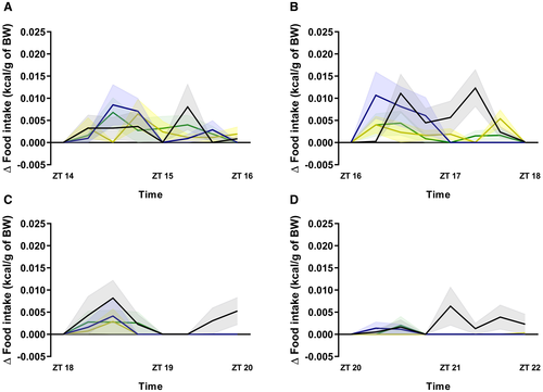
ALAN effects on locomotor activity showed significant effects of light (P < 0.001 to P < 0.0001) and interaction (P < 0.0001) for all four time points, although the most pronounced effects were seen in the second half of the dark period. All three wavelengths contributed significantly to these effects of light on locomotor activity (Figure 1). Before each light exposure, locomotor activity showed a clear daily rhythm entrained to the light/dark cycle (Supporting Information Figure S3). A significant effect of light on energy expenditure was only found at ZT16-18 (P = 0.0453), and significant interaction effects were found at ZT16-18 (P < 0.0001), ZT18-20 (P < 0.0001), and ZT20-22 (P = 0.0143). This effect was mostly caused by blue (P < 0.0001) and white (P = 0.0159) light at ZT16-18, blue (P = 0.0198) and green (P = 0.0245) light at ZT 18-20, and the green light (P = 0.0205) at ZT20-22 (Figure 2). For RER, a significant light effect was found at ZT16-18 (P = 0.0289) as well as significant interaction effects at ZT16-18 (P < 0.0001) and ZT18-20 (P < 0.0001). The effects on RER were mostly caused by the effects of blue (P = 0.0026) and green (P = 0.0427) light (Figure 3). Lastly, significant effects of light on food intake were found at ZT16-18 (P = 0.0344) and ZT18-20 (P = 0.0335). A significant interaction effect was found at ZT16-18 (P = 0.0045). The suppressive effects on food intake were mainly caused by the green (P = 0.0224) and white (P = 0.0360) light from ZT16-18 and the blue (P = 0.0481) and white (P = 0.0196) light from ZT18-20 (Figure 4). The detailed statistical analysis of these metabolic data are displayed in Supporting Information Tables S5-S12.
Surprisingly, with only three 2-hour nightly light pulses over a period of 6 weeks, we observed a significant difference in the percentage of body weight gain between light treatments, with the animals exposed to blue and green light showing a slightly lower weight gain over the course of the experiment (Supporting Information Figure S2, Supporting Information Tables S3-S4), which may be partially explained by the acute inhibitory effects of blue and green light described above.
Plasma measurements
Plasma glucose, insulin, and corticosterone concentrations were measured after the third 2-hour light exposure for each time point and wavelength. An overall significant effect of ALAN was observed in plasma glucose (P = 0.0019, Figure 5A), with a significant effect especially from blue light (P = 0.0312). No significant differences were found at any time point in the plasma insulin or corticosterone levels of light-exposed animals compared with dark controls, regardless of the wavelength used (Figures 5B-5C). A detailed statistical analysis of the changes in plasma values is shown in Supporting Information Tables S13-S15.

Clock and metabolic genes in the liver
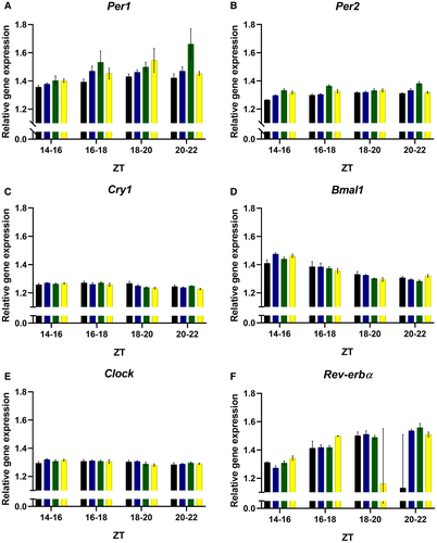
No significant ALAN-induced changes in the relative expression of the clock genes cryptochrome (Cry), brain and muscle Arnt-like protein-1 (Bmal1), circadian locomotor output cycles kaput (Clock), or Rev-erb (Figure 6) were found, regardless of timing and wavelength of the light exposure. Moreover, a significant increase in the expression of Per1 was found in the blue light–exposed group (P = 0.0155), whereas increases in Per2 were observed with green (P = 0.0004) and white light (P = 0.0004). The detailed statistical analysis of the changes in hepatic clock gene expression is shown in Supporting Information Tables S16-S18.
For the genes involved in carbohydrate metabolism, ALAN caused significant changes in the expression of phosphoenolpyruvate carboxykinase (Pepck) (P = 0.0072), glucokinase (Gck) (P = 0.0112), acetyl-CoA carboxylase 1 (Acc1) (P = 0.0005), fatty acid synthase (Fas) (P = 0.0002), and peroxisome proliferator-activated receptor alpha (Pparα) (P = 0.0003). Exposure to blue light caused a significant reduction in the expression of Pepck (P = 0.0404) (Figure 7A). Additionally, Gck expression was significantly increased with blue (P = 0.0107), green (P = 0.0002), and white (P = 0.0386) light (Figure 7B), while Pdk4 expression decreased with blue light exposure (P = 0.0306) (Figure 7D).
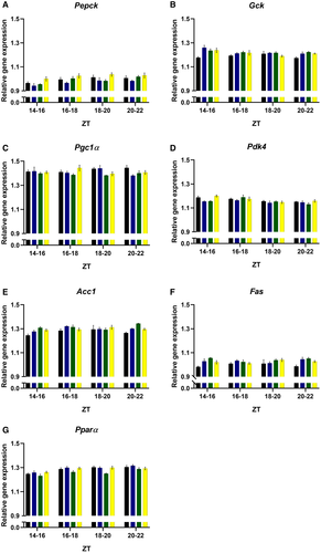
Among the genes involved in lipids metabolism, Acc1 significantly increased with all three wavelengths (P = 0.0128 for blue, P = 0.0076 for green, and P = 0.0195 for white) (Figure 7E), and such an effect was also observed for the expression of Fas (P = 0.0344 for blue, P < 0.0001 for green, and P = 0.0071 for white) (Figure 7F). Lastly, Pparα only showed a significant decrease with green light exposure (P = 0.0032) (Figure 7G). A detailed statistical analysis of the changes in metabolic gene expression is shown in Supporting Information Tables S19-S21.
cFos expression in SCN and PVN
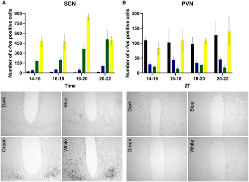
ALAN showed significant effects of light (P < 0.0001) and interaction (P = 0.0003) on c-Fos expression in the SCN as well as a significant effect of light (P = 0.0010) for c-Fos expression in the PVN. The number of c-Fos–positive cells in the SCN was significantly increased by all wavelengths compared with dark controls (P = 0.0132 for blue, P = 0.0004 for green, and P < 0.0001 for white), with the highest numbers observed after exposure to white light (Figure 8A). The effects of blue light were only significant at ZT20-22, whereas the effects of green and white light were apparent at all four time points. Moreover, in the PVN, we observed that the number of c-Fos–positive cells was significantly decreased in the blue (P = 0.0099) and green light (P = 0.0019) exposed animals. This inhibitory effect of ALAN was only observed at the beginning of the dark period. No differences were found for c-Fos expression in the PVN between dark controls and animals exposed to white light. A more detailed statistical analysis of the c-Fos results can be found in Supporting Information Tables S22-S24.
Discussion
Here, we show that 2 hours of ALAN acutely causes metabolic alterations and changes in the expression of clock and metabolic genes in the liver, and that these effects are dependent on both the timing and wavelength of the light exposure. In addition, we show that next to irradiance and photon flux, light-induced activation of c-Fos expression in the SCN also depends on wavelength. Remarkably, the effect of nocturnal light exposure on c-Fos expression in the PVN was completely opposite to that in the SCN. Contrary to the stimulation observed in the SCN, ALAN decreased c-Fos expression in the PVN. Moreover, inhibition of c-Fos expression in the PVN was only observed after blue and green light exposure, whereas white light was most effective in the SCN.
Our results show that ALAN in rats reduces locomotor activity regardless of the wavelength and time of exposure, which proves that in rats, negative masking ((19)) occurs independently of the wavelength and time of the dark phase when the light pulse is given. The robustness of this effect is clearly indicated by the fact that a similar analysis on the day before or after the light exposure did not reveal such a reduction. Remarkably, though, on some occasions, increased activity was also observed, especially during the first 15-minute period (that is, contrary to decades of negative masking data). The effect was observed mostly during the first half of the dark phase and especially with white and blue light. We do not have a straightforward explanation, although changes in the relative contribution of photoreceptors shortly after the onset of light exposure and possible daily rhythms therein could be involved ((20, 21)); often, a short bout of increased activity was also observed after regular lights-on with polychromatic light at ZT0 (Supporting Information Figure S3). Therefore, we think it more resembles a rarely studied phenomenon in the context of chronobiology called light-induced locomotor activity/activation ((22-24)).
Interestingly, although the reduction of locomotor activity by ALAN was significant for all time points and wavelengths, a significant concomitant reduction in energy expenditure was observed only at ZT16-18 with blue and white light. This apparent discrepancy could be merely a technical issue, as a decreasing trend was observed after each light exposure, with changes in the locomotor activity being just easier to detect than changes in energy expenditure. Moreover, whereas a significant decrease in energy expenditure was only observed at ZT16-18, the largest decrease in locomotor activity was observed at ZT18-20, indicating light might also have an activity independent effect on energy expenditure.
At ZT16-18, a significant reduction in RER also was observed, both during blue and green light exposure. The decrease in RER (i.e., indicating a higher lipid metabolism) was accompanied by a reduction in food intake during green light exposure but not during blue light exposure. The discrepancy between feeding behavior and peripheral metabolism indicates that light may also have independent effects on these parameters.
Surprisingly, white and green light–exposed animals showed no significant changes in plasma glucose values. Previously, our laboratory showed that ALAN, especially white and green light, acutely impaired glucose tolerance in rats ((12)) and we observed a similar effect with blue light in the diurnal rodent Arvicanthis ansorgei ((25)). However, in both these experiments, a glucose tolerance test was used to investigate glucose metabolism, which prevents a direct comparison with the current results. Moreover, animals were fasted for a few hours before the glucose tolerance tests, and the fasting state not only changes the glucose response but also changes SCN activity ((26, 27)). Moreover, when the Arvicanthis were not fasted ((25)) we did not observe any significant effects of ALAN on basal plasma glucose concentrations, whereas in the current experiment, we found that blue light reduced blood glucose. The difference between the effects of blue light on nonfasted blood glucose in these two experiments could be because Arvicanthis are diurnal rodents, whereas here we used nocturnal rats. Additionally, the setup and characteristics of the light exposure equipment differed slightly between the two experiments.
Although previous animal studies have shown that ALAN acutely increases the sympathetic and decreases the parasympathetic tone of the autonomic nerves that innervate peripheral organs such as the pancreas, adrenals, and liver ((3, 28, 29)), we did not find any significant changes in plasma insulin or corticosterone levels. In the existing literature, conflicting results have been reported regarding changes in corticosterone levels in response to acute LAN. Direct changes in absolute levels have not always been reported, and even opposite changes can be found ((3, 11, 12, 25, 30, 31)). These inconsistent findings may be related to differences among studies including the intensity, duration, wavelength, and circadian phase of the light exposure as well as sex and species involved. Moreover, our basal corticosterone levels are rather high (150-250 ng/mL); therefore, we cannot exclude these values are affected by the stress of the pentobarbital anesthesia, thoracotomy, and cardiac puncture, and it should be considered a limitation of our study. Nonetheless, even if our experimental conditions affected the baseline concentrations of corticosterone in all groups (dark controls included), we did not find any differences among the groups because of the different light conditions. We also did not observe a significant increase in the liver expression of Pepck as was previously reported with white light ((11)). Moreover, we did observe an unexpected decrease in the expression of this gene with blue light, which matches the observed reduction in plasma glucose but goes against previous evidence that exposure to blue-enriched white light at the beginning of the resting phase (i.e., ZT14) has a detrimental effect on plasma glucose in mice ((32)). However, we measured plasma glucose immediately after a 2-hour light pulse, whereas Nagai et al. ((32)) reported significant changes in plasma glucose 48 hours after the light pulse. Second, the Nagai et al. ((32)) study was performed using C57BL/6J mice, indicating again that the effects of ALAN may be species dependent.
The results of the current study did confirm the light-induced increased expression of Per2 in the liver observed before ((11, 32)). In addition, we found an increased expression of Gck with all the wavelengths and a decreased expression of Pgc1α and Pdk4 with green and blue light exposure, respectively, providing further evidence for light-induced changes in carbohydrate metabolism. Likewise, the increased expression of Acc1 and Fas caused by ALAN indicates that light at the wrong time of the day can also acutely affect hepatic lipid metabolism regardless of the wavelength. In fact, the increased lipid metabolism indicated by the changes in gene expression nicely fits with the decrease in RER observed. Unfortunately, plasma fatty acids and triglycerides were not measured, but the current findings support the changes in liver lipids observed in zebra finches ((33)) and lipid metabolism in rats ((34)) and men ((13)) reported previously.
Although we did find lower energy expenditure and changes in oxygen consumption with blue light as was reported in humans before ((35)), contrary to what was expected, green and not blue light was better mimicking the detrimental effects of white light overall. Thus, in accordance with our previous study on glucose tolerance ((12)), we show that green light causes more metabolic changes than blue light. Our results with green light once more indicate a putative role for middle-wavelength cones in the effects induced by ALAN. Because both blue and green light are able to suppress melatonin ((36)), it is not likely that the marked difference between the metabolic effects of green and blue light are melatonin dependent.
The intrinsically photosensitive retinal ganglion cells (ipRGCs) have been described extensively before in humans ((37)), mice ((38-40)), rats ((41)) and diurnal rodents ((42)). These ipRGCs are known to contain melanopsin, a photosensitive pigment with a peak of action at short-wavelength light (465-485 nm, i.e., the blue-appearing portion of the visible spectrum). In addition to their intrinsic photosensitivity, ipRGCs also receive light information via the rods and cones ((16)). Our data suggest that M-cones, which are sensitive to wavelengths of ~520 nm (green light), might be playing an important role in the metabolic effects of ALAN. Although only few studies have reported on the possible physiological effects of green light, a study in mice ((4)) did report effects on sleep/arousal and corticosterone release. In that study, it was proposed that blue light mainly stimulates the SCN via the M1 subtype of ipRGCs, whereas green light stimulates another type of ipRGCs that mainly project to the ventrolateral preoptic area ((4, 43, 44)). However, according to our immunohistochemical results, in rats, green light activates the SCN more effectively than blue light. We did not study the ventrolateral preoptic area, but in the PVN, we found an inhibition of c-Fos expression with blue and green light but not with white light. Thus, although these rat and mice results are not identical, both studies clearly indicate that different wavelengths may affect the SCN and other brain areas differently. It is highly unlikely that S-cones play a major role in our observations because they were barely stimulated with any of the light conditions we used (Supporting Information Table S1). Moreover, in our setup, the green and white light stimulated the rods much more effectively than the blue light, which in turn may change the information that ipRGCs send to the SCN. However, it is important to mention that although we equated the photon flux between the three light conditions, this does not mean that the α-opic irradiance and illuminance is the same for all wavelengths used, which makes comparisons among the different light conditions difficult. For example, the blue light used activated melanopsin to a greater extend (Supporting Information Table S1), but it also activated rods and cones, which in turn influences the firing rate of the ipRGCs ((16)). However, the green and white light activated the same photoreceptors, albeit to a different degree. Nevertheless, because one of the properties of photoreceptors is the principle of univariance (i.e., it cannot distinguish between changes in intensity and in wavelength), in principle, all lights could be equally effective in activating them, which can be considered a limitation of our study. A better approach for the future will be to study the role of ipRGCs in the effects of ALAN on metabolism by using the method of silent substitution ((45)).
The inhibitory effects of nocturnal light on c-fos expression in the PVN were to be expected in view of the abundant presence of gamma-Aminobutyric acid (GABA)-containing neurons in the SCN and the strong stimulation of c-fos activity in the SCN by nocturnal light. In fact, previously, we have shown that SCN-mediated GABA release in the PVN is essential for the inhibitory effect of nocturnal light on melatonin release ((2, 46)). However, the differential effects of wavelength on c-Fos expression in the SCN and PVN indicate that the inhibitory effects of light on the PVN may also be mediated via direct projections of the ganglion cells to the PVN and not indirectly via the SCN. Additionally, ipRGCs also project to the lateral hypothalamic area where the major population of orexin neurons is located; because these orexin neurons, either glutamatergic or GABAergic, among others, project to spinally projecting PVN neurons ((47, 48)), this may be another pathway for light to reach preautonomic neurons in the PVN and hence affect peripheral metabolism. Indeed, recent publications clearly indicated alternative pathways not involving the SCN for light to affect peripheral clocks and metabolism ((49-51)), nicely fitting with the existence of separate populations of ipRGCs projecting to the SCN and to non-SCN target areas ((52, 53)).
The increased gene expression in the liver (Per2, Gck, Acc1, Fas) nicely correlates with the effects of light on c-Fos expression in the SCN, whereas the decreased gene expression in the liver (Pgc1α, Pdk4) correlates with the wavelength-dependent effects of light on c-Fos expression in the PVN. The selective inhibition of neural activity in the PVN with blue and green light makes it unlikely that the metabolic effects of ALAN, particularly those of white light, are only due to changes in ANS activity, which supports a recent observation from our group on ALAN-induced changes in the liver transcriptome ((54)).
Even though the particular effects of each wavelength may differ from one study to the other, all the evidence points toward the fact that exposure to ALAN acutely disturbs energy metabolism, which with repeated exposure, in the end, may lead to metabolic diseases such as obesity and diabetes, even in low intensities ((7, 34, 55, 56)). We are aware that the sample sizes for our immunohistochemical analysis and gene measurements were small (that is, n = 4 per time point). Also, the interpretation and translation of our current results in nocturnal rats to human health must be done cautiously; nonetheless, they go hand in hand with the detrimental effects of light on energy metabolism in humans reported before ((13, 57)). Together with all the evidence of ALAN affecting energy metabolism in both an acute and chronic way ((6, 11, 12, 25, 32, 54, 55)), this results in several recommendations being made regarding the spectrum of light we should use ((58)). The current study provides further evidence to aid our understanding of ALAN, shiftwork, and metabolic disorders. However, additional studies involving other species, genetically modified animals, and humans are needed to further elucidate the metabolic effects of different wavelengths and the neural pathways involved.
Conclusion
We demonstrated in rats that exposure to ALAN causes changes in energy expenditure, RER, and clock and metabolic gene expression in the liver. All these effects were dependent on the time of day, which suggests an interaction with the circadian timing system. Moreover, the effects observed were also dependent on wavelength, suggesting involvement of ipRGCs, M-cones, and rods. Our work warrants further precaution in the use of electronic devices that emit low- and middle-length wavelengths of light at night in order to prevent negative metabolic consequences among users.
Acknowledgments
We thank Nikita L. Korpel for measuring the RIN values and Eva van Sambeek for her assistance with part of the immunohistochemistry.
Funding agencies
This work was supported by a doctoral fellowship from the “NeuroTime” Erasmus + Program (AMV) and the University of Amsterdam (AK). JM is supported by the French National Research Agency (ANR-14-CE13-0002-01 ADDiCLOCK JCJC) and the Institut Danone France, Fondation pour la Recherche Médicale.
Disclosure
The authors declared no conflict of interest.
Author contributions
AMV designed research, executed all experiments, analyzed samples, performed statistical analysis, created the figures, and wrote the manuscript. WIGRR assisted during experiments and revised the manuscript. JM advised on experimental design and revised the manuscript. AK designed research and wrote the manuscript. AK is the guarantor of this work and, as such, has full access to all the data in the study and takes responsibility for the integrity of the data and the accuracy of the data analysis.


