Mild Exercise Does Not Prevent Atherosclerosis in APOE*3-Leiden.CETP Mice or Improve Lipoprotein Profile of Men with Obesity
Abstract
Objective
Exercise has been shown to improve cardiometabolic health, yet neither the molecular connection nor the effects of exercise timing have been elucidated. The aim of this study was to investigate whether ad libitum or time-restricted mild exercise reduces atherosclerosis development in atherosclerosis-prone dyslipidemic APOE*3-Leiden.CETP mice and whether mild exercise training in men with obesity affects lipoprotein levels.
Methods
Mice were group-housed and subjected to ad libitum or time-restricted (first or last 6 hours of the active phase) voluntary wheel running for 16 weeks while on a cholesterol-rich diet, after which atherosclerosis development was assessed in the aortic root. Furthermore, nine men with obesity followed a 12-week mild exercise training program. Lipoprotein levels were measured by nuclear magnetic resonance spectroscopy in plasma collected pre and post exercise training.
Results
Wheel running did not affect plasma lipid levels, uptake of triglyceride-derived fatty acids by tissues, and aortic atherosclerotic lesion size or severity. Markers of training status were unaltered. Exercise training in men with obesity did not alter lipoprotein levels.
Conclusions
Mild exercise training does not reduce dyslipidemia or atherosclerosis development in APOE*3-Leiden.CETP mice or affect lipoprotein levels in humans. Future research on the effects of (time-restricted) exercise on atherosclerosis or lipid metabolism should consider more vigorous exercise protocols.
Study Importance
What is already known?
- ► Exercise improves cardiometabolic health in obesity.
- ► Improving peripheral lipolysis could reduce atherosclerosis development.
- ► Exercise at different times of day modulates cellular metabolism differentially.
What does this study add?
- Mild exercise does not prevent atherosclerosis in APOE*3-Leiden.CETP mice.
- Mild exercise over 3 months does not improve the lipoprotein profile of men with obesity.
How might these results change the direction of research or the focus of clinical practice?
High-intensity exercise rather than mild exercise should be advised to support lipid-lowering drug treatment in individuals with obesity. Future research on timed exercise should consider more vigorous exercise protocols.
Introduction
Cardiovascular diseases (CVD) are the leading mortality cause worldwide, as 30% of all deaths can be attributed to CVD events ((1)). The main pathology underlying CVD is atherosclerosis, a progressive thickening of arterial walls as a consequence of the accumulation of cholesterol, macrophages, and cell debris, resulting in narrowing of the arteries. One of the risk factors for the development of atherosclerosis is dyslipidemia, characterized by high plasma levels of cholesterol and triglycerides (TG), which is often associated with obesity ((2)). Atherosclerosis development is initiated by the influx of atherogenic lipoprotein particles, including low-density lipoprotein (LDL) and TG-rich lipoprotein remnants, which are oxidized and aggregate. Retention of these particles causes inflammation resulting in an influx of monocytes. Within the artery wall, these monocytes mature into macrophages that engulf modified particles to become cholesterol-laden foam cells. These cells can die, leading to acceleration of local inflammation and plaque growth ((3)). Numerous lipid-lowering compounds have been developed in order to treat CVD. However, even the most effective ones, statins and proprotein convertase subtilisin/kexin type 9 inhibitors, are not able to prevent more than 25% to 45% of cardiovascular events ((4, 5)).
In addition to medication, lifestyle interventions, among which exercise, have shown to further increase treatment effectiveness ((6)). Exercise is furthermore beneficial for the primary prevention of cardiometabolic disease in humans ((7, 8)) and mice ((9-11)). Yet, the molecular connection between exercise and atherosclerosis development is not clear. Several studies have linked lowered inflammatory markers to reduced atherosclerotic lesion development in apolipoprotein E–deficient (Apoe-/-) mice subjected to voluntary exercise ((9, 12-14)). Besides inflammatory modulation, exercise possibly affects lipid metabolism, as it has been shown to induce lipid uptake and oxidation by skeletal muscle ((15)). Exercise-induced TG-derived fatty acid (FA) uptake by skeletal muscle could be accompanied by accelerated hepatic lipoprotein remnant clearance, thereby reducing plasma cholesterol levels and atherosclerosis development. Because ApoE is essential for the interaction with the LDL receptor, exercise studies using Apoe-/- mice are unsuitable to investigate this pathway and may underestimate the beneficial effects of exercise. Unlike Apoe-/- mice, APOE*3-Leiden.CETP mice have an intact ApoE-LDL receptor pathway, allowing for the investigation of the role of TG-rich lipoprotein remnant clearance in the context of atherosclerosis development ((16)). We thus hypothesize that exercise induces TG-derived FA uptake by skeletal muscles accompanied by hepatic uptake of cholesterol-enriched lipoprotein remnants, thereby reducing hypercholesterolemia and atherosclerosis development in APOE*3-Leiden.CETP mice. Low compliance to lifestyle interventions in disease prevention remains an issue. If exercise at lower intensities, i.e., mild exercise, is able to improve cardiometabolic health, compliance to exercise could be higher as compared with vigorous exercise protocols.
It is noteworthy that there is a tight interplay between the circadian clock and exercise metabolism. Two studies that compared early active phase (AP) to early rest phase treadmill running in mice showed distinct time-dependent metabolic responses ((17, 18)), underlining that timing of exercise can dictate the amplitude of the exercise response. Applying ideal exercise timing could optimize the effectiveness of exercise and may be an additional strategy to improve cardiometabolic health for individuals with a low compliance. In a small cohort of patients with type 2 diabetes mellitus, morning exercise resulted in greater glycemic disturbance than afternoon exercise, strengthening the importance of the timing of exercise in health management ((19)). However, the influence of the timing of exercise on CVD and other health outcomes is unknown. Morning exercise has been shown to result in greater weight loss in overweight individuals compared with afternoon exercise ((20)). In contrast, evening exercise may improve sleep quality and thereby health if performed > 1 hour before bedtime ((21)). Moreover, voluntary wheel running during the late AP reduced diet-induced obesity in mice more efficiently than wheel running during the early AP ((22)). As studies on optimal exercise timing for cardiometabolic health are currently lacking, no advice can be given.
In this study, we investigated whether ad libitum (AL) voluntary wheel running in group-housed APOE*3-Leiden.CETP mice fed a cholesterol-rich Western-type diet, as a mild form of exercise, improves lipid metabolism and reduces atherosclerosis development. Simultaneously, we investigated whether wheel running access during the first or last 6 hours of the AP is more beneficial with regard to metabolic health and atherosclerosis development. In a translational approach, men with obesity participated in a 12-week mild training program to investigate the effects of mild exercise training on lipoprotein metabolism.
Methods
Animal study
Female APOE*3-Leiden.CETP mice were used, as specifically the females develop hypercholesterolemia and atherosclerosis when fed a cholesterol-rich Western-type diet ((23, 24)). To obtain APOE*3-Leiden.CETP mice, heterozygous APOE*3-Leiden mice were crossbred with homozygous human cholesteryl ester transfer protein (CETP) transgenic mice (both on a C57Bl/6J background) ((25)). Mice (8-12 weeks old) were group-housed (n = 4/cage) in light-tight cabinets at 21°C under standard 12/12-hour light-dark conditions. The cabinets were illuminated with white fluorescent light (intensity: ca. 85 µW/cm2). Experiments that took place during darkness were performed under red light. Mice were fed a Western-type diet (containing 16% fat and 0.15% cholesterol; Diet T; Altromin, Lage, Germany) AL. After a dietary run-in period of 3 weeks, mice were block-randomized over four groups based on body weight, fat mass, lean mass (both measured by EchoMRI 100-Analyzer; EchoMRI, Houston, Texas), plasma TG, and total plasma cholesterol (TC). Control mice were compared with three intervention groups (with an average lesion size of 1.50 × 10⁵ ± 0.4 × 105 µm, with 16 mice, we will be able to detect a one-third reduction in lesion size with a power of 80% and type 1 error rate of 5%) with running wheel access during either the first or last 6 hours of the AP or AL (24 hours). Time-restricted exercise groups were provided with running wheels with a diameter of 10.5 cm (TSE Systems Inc., Bad Homburg v.d.H., Germany) and an automated breaking system. Wheel rotations were automatically monitored, and running distance was calculated (TSE PhenoMaster v6.5.0; TSE Systems Inc.). The AL exercise group was provided with running wheels with a diameter of 23 cm. Wheel rotations were automatically monitored (ClockLab v3.11; Actimetrics, Wilmette, Illinois). Plasma lipid levels, body weight, and body composition were measured every 4 weeks. After 8 weeks, blood was collected from the tail vein at four time points (Zeitgeber time [ZT] 0, 6, 12, 18) to determine circadian rhythmicity of plasma lipids and glucose. After 16 weeks of wheel running, TG-derived FA and cholesteryl ester uptake from intravenously injected very LDL (VLDL)-like particles were assessed at lights on (ZT0) and lights off (ZT12) before the mice (27-31 weeks old) were sacrificed. All animal experiments were performed in accordance with the Institute for Laboratory Animal Research Guide for the Care and Use of Laboratory Animals and were approved by the National Committee for Animal experiments by the Ethics Committee on Animal Care and Experimentation of the Leiden University Medical Center.
Plasma lipids and blood glucose
Plasma TG, TC, high-density lipoprotein cholesterol (HDL-C), non–HDL-C, and blood glucose were measured as described in Supporting Information Methods 1. TC exposure was calculated by multiplying the average plasma TC concentrations (millimolar) between four consecutive weeks by the number of study weeks. Total area under the curve of diurnal TG/TC/glucose at week 8 was calculated by multiplying the average plasma concentration (millimolar) between four consecutive time points by the number of hours.
TG-derived FA and cholesteryl ester and uptake by organs
VLDL-like TG-rich particles (80 nm diameter) double-labeled with glycerol tri[3H]oleate and [14C]cholesteryl oleate were prepared as previously described ((26)). At the end of the 16-week exercise intervention period, after a t = 0 blood collection, mice were injected with VLDL-like particles (1 mg TG diluted in 200 µL PBS per mouse) to measure the tissue uptake of TG-derived FA and cholesteryl esters. After 15 minutes, animals were euthanized by CO2 inhalation and perfused with ice-cold PBS for 5 minutes before tissues were collected for further analyses. The hearts were isolated for atherosclerosis quantification. Processing of the materials for radioactivity measurement is described in Supporting Information Methods 2.
Atherosclerosis quantification
After sacrifice, hearts were fixated in 4% paraformaldehyde for 24 hours and stored in 70% ethanol before they were embedded in paraffin and cross-sectioned using a microtome (5 µm). Cross sections were stained with haematoxylin-phloxine. Atherosclerotic lesion sizes of four cross sections throughout the aortic root with a distance of 50 µm, starting at open aortic valves, were quantified with imaging software (ImageJ version 1.50i; National Institutes of Health, Bethesda, Maryland). Atherosclerotic plaques were scored subjectively for lesion severity (Mild: type I-III lesions; Severe: type IV-V lesions), according to the guidelines of the American Heart Association adapted for mice, as previously described ((23)).
Protein quantification
Protein abundance of mitochondrial oxidative phosphorylation complexes I-V was measured by Western blotting, and protein abundance of citrate synthase was determined by automated Western blot with Wes (ProteinSimple, San Jose, California) in the tibialis anterior (TA) muscle as described in Supporting Information Methods 3.
Cytokine measurement
Interleukin (IL) 6, monocyte chemoattractant protein 1, IL-1β, IL-10, keratinocyte chemoattractant, and tumor necrosis factor alpha were measured in freshly frozen plasma samples collected at week 8 using a customized six-plex U-Plex Biomarker Group 1 (mouse) Kit (Meso Scale Discovery; Rockville, Maryland) according to the manufacturer’s protocol.
Lipidomics in human plasma
Men with overweight or obesity (n = 9, 40-70 years old) with metabolic syndrome were subjected to a mild exercise training protocol for 12 weeks, as described ((27)). In short, two aerobic sessions and one resistance exercise session were performed per week. Aerobic cycling was performed on a stationary bike for 30 minutes at 70% of the maximal workload. Resistance exercise training consisted of three sets of 10 repetitions at 60% of the maximal voluntary contraction of the following exercises: leg press, leg extension, chest press, lateral pull down, horizontal row, triceps extension, biceps curl, and abdominal crunch. Maximal workload and maximal voluntary contraction were assessed prior to the exercise regime and every 4 weeks for training schedule adjustment. Fasted plasma samples were collected before and after the 12-week period, after which lipoprotein profile was measured by nuclear magnetic resonance (NMR) spectroscopy. The elaborate NMR spectroscopy protocol is described in Supporting Information Methods 4. The local ethics committee approved the study, which was performed following the declaration of Helsinki principles.
Statistical analysis
Statistical analyses between groups were performed with one-way ANOVA. Two-way ANOVA was used to compare multiple time points between groups. Paired t tests were used to compare lipoprotein markers pre and post exercise training in the human data. Data sets were tested for normality. In case of non-normal distributions, nonparametric tests were performed (Friedman test for repeated measures followed by Kruskal-Wallis test for the different comparison of all groups at different time points). Data are presented as means (SEM). Differences at P ≤ 0.05 were considered statistically significant. Statistical analyses were performed with GraphPad Prism software, version 8.0 (GraphPad, La Jolla, California). Statistical outliers were removed after identification by the Grubbs test.
Results
Wheel running distance is proportional to duration of wheel access during active phase
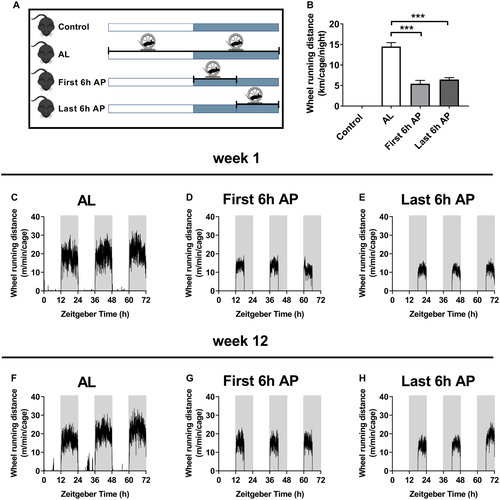
Mice housed in groups of four had access to running wheels either AL, during the first 6 or the last 6 hours of the AP, or no access (control) for 16 weeks (Figure 1A). Mice with wheel access during the first and last 6 hours of the AP ran similar distances (5.4± 0.8 and 6.4± 0.5 km per cage per night, respectively), while mice with AL access ran more than twice as far (14.5± 1.0 km per cage per night; Figure 1B). In kilometers per mouse per night, distances were 1.4± 0.2, 1.6± 0.1, and 3.6± 0.2, respectively. Running wheel activity was proportional to the duration of wheel access during the AP in all three exercise groups, during both week 1 and 12 (Figures 1C-1H). Even though mice with AL access were able to run during the rest (light) phase, wheel running activity during that phase was negligible.
Mild exercise reduces fat mass without affecting food intake in mice
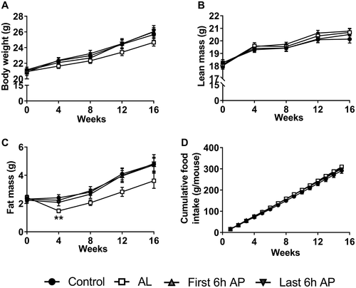
Body weight (gain) and lean mass did not differ between the groups (Figures 2A-2B). Mice with AL wheel access showed an initial decrease in fat mass compared with the control group, which stabilized later on (Figure 2C). In contrast, this was not seen in mice with wheel running access during the first or last 6 hours of the AP. Cumulative food intake did not differ between the groups (Figure 2D).
Mild exercise does not reduce plasma lipid levels in mice
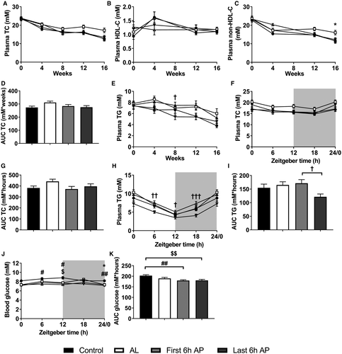
To investigate the effects of voluntary wheel running on plasma lipid metabolism, blood was collected regularly. Surprisingly, voluntary wheel running did not reduce total plasma cholesterol (Figure 3A). In fact, the AL group showed higher plasma TC compared with the control group, although this effect did not reach significance (P = 0.076 and P = 0.075 for week 12 and 16, respectively). This was attributed to an increase in non–HDL-C rather than HDL-C, which was significantly higher at week 16 (Figures 3B-3C). Nonetheless, TC exposure throughout the study did not differ between the groups (Figure 3D). Plasma TG was only lower in the last 6 hours of AP group as compared with the first 6 hours of AP group at week 8 but besides that was unaltered (Figure 3E). At week 8 of the intervention, diurnal plasma samples were collected, and TC did not differ between groups (Figures 3F-3G). Diurnal plasma TG was lower in the last 6 hours of AP group compared with the first 6 hours of AP group (Figures 3H-3I), but this may not have been consistent throughout the study, as plasma TG only differed between these groups in week 8 (Figure 3E). Diurnal non-fasted blood glucose was significantly lower in the first and last 6 hours of AP groups compared with control (Figures 3J-3K), but this effect may not have persisted, as the groups did not differ at ZT12 during week 16 (data not shown).
Mild exercise does not increase TG-derived FA uptake by skeletal muscle or cholesteryl ester uptake by liver in mice
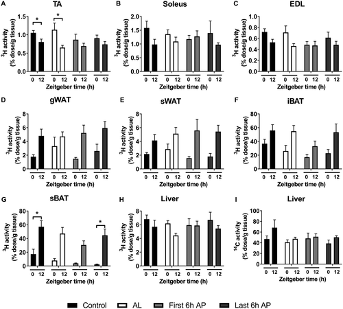
We hypothesized that exercise increases TG-derived FA uptake by muscle and thereby accelerates hepatic clearance of cholesterol-rich lipoprotein remnants. We, therefore, injected mice with VLDL-like TG-rich particles double-labeled with glycerol tri[3H]oleate and [14C]cholesteryl oleate. As certain tissues combust, store, or handle lipids more than others, relative uptake of these labels differs between tissues, hence the wide range of 3H-activity uptake as displayed in Figure 4. As circadian rhythms dictate fluctuations in metabolic activity of tissues, we included time points ZT0 (lights on) and ZT12 (lights off), as differences in tracer uptake are generally large between these time points. Several tissues showed a significant difference between ZT0 and ZT12 (Figures 4A, 4G). However, the exercise groups did not show higher [3H]oleate uptake by the hind limb muscles TA, soleus, and extensor digitorum longus compared with the control group (Figures 4A-4C). Similarly, [3H]oleate uptake by gonadal white adipose tissue, subcutaneous white adipose tissue, interscapular brown adipose tissue, subscapular brown adipose tissue, and liver did not differ between the groups (Figures 4D-4H). In line with this, there was no difference between the groups in [14C]cholesteryl oleate uptake by the liver, indicating that uptake of delipidated core remnants by the liver was unaffected by mild exercise (Figure 4I).
Mild exercise does not increase markers of training status in mice
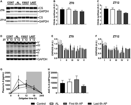
Because mild exercise did not affect lipid metabolism, we next investigated whether the exercise stimulus, i.e., sharing a running wheel in a group cage of four mice, was sufficient to induce markers of training status. Protein abundance of citrate synthase (Figures 5A-5C) and the mitochondrial oxidative phosphorylation complexes I-V in TA (Figures 5D-5F), as this mixed muscle is generally regarded as responsive to running exercise, as well as plasma levels of the cytokines IL-6 (Figures 5G-5H), monocyte chemoattractant protein 1, IL-1β, IL-10, keratinocyte chemoattractant, and tumor necrosis factor alpha (Supporting Information Figures S1A-S1E) were not altered significantly in any exercise group compared with the control.
Mild exercise does not reduce atherosclerotic lesion area and severity in mice
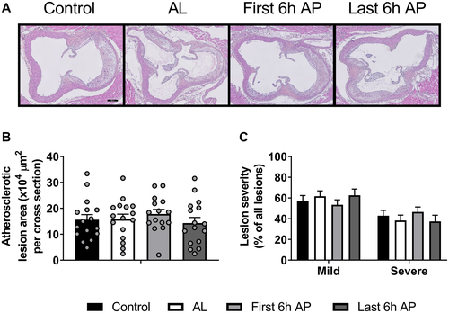
To determine the effect of voluntary wheel running on atherosclerosis development, we analyzed atherosclerotic lesion area and severity in the aortic root after 16 weeks of voluntary wheel running. Neither atherosclerotic lesion area (Figures 6A-6B) nor lesion severity (Figure 6C) were different between the groups.
Mild exercise training does not improve lipoprotein profile in men with obesity
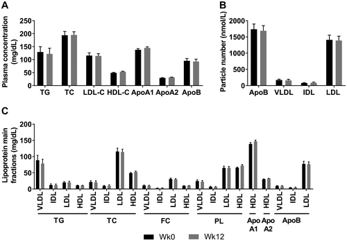
To investigate the effects on lipoprotein metabolism in humans, nine men with obesity followed a mild exercise protocol for 12 weeks. We performed NMR spectroscopy on plasma samples collected before and after the 12-week exercise training regime to measure their plasma lipoprotein profile. Mild exercise training did not affect the concentration and composition of the main lipoprotein fractions VLDL, intermediate-density lipoproteins, LDL, and HDL (Figures 7A-7C) or their subfractions (Supporting Information Figures S2A-S2C) despite a mild but significant reduction of body fat from 29.8% (3.0%) to 28.7% (3.0%) (−1.1%; P = 0.0021).
Discussion
Exercise has long been recognized as an effective strategy to increase treatment effectiveness in CVD. Despite the fact that exercise performance is higher in the late compared with the early AP in humans ((28, 29)), the influence of the timing of exercise on metabolic health orchestrated by circadian rhythms is largely unknown. In this study, we therefore aimed to investigate the effects of timed voluntary exercise on lipid metabolism in relation to atherosclerosis development. We subjected group-housed female APOE*3-Leiden.CETP mice to either AL, first 6 hours or last 6 hours of the AP, or no voluntary wheel running for 16 weeks. Surprisingly, we did not find a reduction in dyslipidemia or atherosclerosis development in any of the exercise groups. AL but not timed exercise caused a significant initial reduction in fat mass compared with the other groups. This may be explained by the differences in running distances between the groups. Wheel running failed to increase lipoprotein-TG–derived FA uptake by skeletal muscle or uptake of cholesterol-enriched lipoprotein remnant particles by liver and did not reduce plasma lipid levels. In fact, plasma non–HDL-C was increased in the AL group at the end of the study. This may have been caused by small, nonsignificant elevations in intake of food and, thus, cholesterol. Similarly, in a translational setting, nine men with obesity followed a 12-week mild exercise training program. Although there was a small significant reduction in fat mass, we did not observe changes in lipoprotein levels and composition of VLDL, intermediate-density lipoproteins, LDL, and HDL and their subfractions. Prolonging the study duration may have potentially altered the lipoprotein profile. Exercise was not specifically prescribed to a time of day. However, participants voluntarily trained consistently at the same time of day, either in the morning, afternoon, or evening. Exercise timing did not affect the outcome. According to the World Health Organization, average TG, TC, and HDL-C levels of participants were acceptable but not ideal, and LDL cholesterol levels were above the acceptable range ((30)).
Several studies investigating voluntary wheel running in single-housed mice have found either reduced atherosclerosis development or lesion regression ((9, 12, 13, 31)). Compared with those studies reporting running distances of 7 to 15 km per mouse per night ((9, 12, 13, 31)), wheel running distances observed here were markedly shorter. It is possible that the exercise stimulus was too mild to trigger an exercise response. Accordingly, we observed no changes in the abundance of citrate synthase and mitochondrial proteins involved in energy metabolism in muscle or plasma markers of exercise response such as IL-6. While only few of the other studies looking at voluntary wheel running and atherosclerosis quantified these markers with inconsistent results ((9, 13)), IL-6 has widely been recognized as an acute exercise response marker, increasing with endurance exercise ((32, 33)). Accordingly, we did not observe decreased plasma lipid levels in the exercise groups or in the men with obesity after 12 weeks of exercise training. Previous reports on the effects of exercise training on plasma lipids are rather inconsistent, as some studies have reported decreases in plasma TC and TG levels in mice ((9, 12, 31, 34)) and humans ((35)), while other studies have observed no changes in mice ((10, 11)) or humans ((36)). One variable in these studies that possibly explains the differences is the exercise intensity. In our study, mice were group housed, which resulted in limited wheel accessibility and milder training compared with single housing. Additionally, single housing female mice induces stress and excessive food intake ((37)), which might have resulted in elevated cholesterol exposure in this study and could thus be a confounder.
Comparing exercise studies proves to be difficult. Human studies have shown large variation in exercise protocols and thereby intensity as well as in compliance and heterogeneity. Animal studies have often shown variation in sex, mouse models, diet, and likely wheel circumference and resistance, of which the latter two are seldom reported. Differences in wheel resistance between or even within studies could affect exercise intensity, potentially causing differences in training effects on glucose or lipid metabolism. In this study, wheel circumference differed between the AL and AP groups. This may have affected wheel resistance, although wheel running was proportional to the duration of access during the night in all groups. Group housing may have introduced variability in wheel running activity between individual mice, possibly caused by cage hierarchies and social stress, but this was not quantified. Besides voluntary wheel running, different exercise protocols such as forced treadmill running or forced swimming have shown to reduce atherosclerosis development in mice ((10, 11, 38, 39)). These exercise protocols are more vigorous, albeit linked to more stress ((40, 41)). As time-restricted voluntary wheel running in group-housed female APOE*3-Leiden.CETP mice does not reduce atherosclerosis, studying the effects of early versus late AP exercise on atherosclerosis with a more vigorous exercise protocol could elucidate if exercise timing is important for cardiovascular health. Future preclinical and clinical studies in this line could help to establish patient guidelines regarding exercise timing in disease prevention. However, rather than “one size fits all,” any recommendations will possibly require personalization, as several studies have shown that chronotype is tightly linked to exercise performance at different times of day. With early chronotypes performing better early during the day and late chronotypes later during the day ((28, 42)), chronotype may similarly dictate optimal exercise timing for health benefits.
Previously, we observed that increased TG-derived FA uptake from TG-rich lipoproteins by pharmacologically activated brown adipose tissue accelerated the hepatic clearance of cholesterol-enriched lipoprotein remnants and reduced hypercholesterolemia and atherosclerosis development in APOE*3-Leiden.CETP mice ((26)). We hypothesized that voluntary wheel running similarly induces the uptake of TG-derived FA by skeletal muscle, consequently increasing hepatic lipoprotein remnant clearance and reducing plasma cholesterol. Evidently, we did not observe this, likely because the exercise was too mild. Alternatively, the type of exercise may be important, as in humans, resistance but not aerobic exercise training was able to reduce lipoprotein remnant particles ((43)). Different types of exercise may thus differently affect metabolism and health, possibly determining the ideal exercise timing. Whereas late-afternoon aerobic exercise has been shown to be superior in improving endurance compared with morning aerobic exercise ((44)), increases in strength and hypertrophy in response to resistance exercise are similar irrespective of time of day ((45)). Future studies on exercise timing and health should thus take the type of exercise into account, as a combination of resistance and aerobic exercise potentially offers the widest range of benefits.
In conclusion, we showed that mild exercise training does neither reduce dyslipidemia or atherosclerosis in female mice with a humanized lipoprotein metabolism nor alter the lipoprotein profile in men with obesity. The limitations lie hereby in the comparability of exercise intensities and possible sex differences between the preclinical and clinical studies. Future research should consider more vigorous exercise protocols to study the effects of (time-restricted) exercise on cardiometabolic health and further investigate long-term health benefits of mild exercise training. The focus should therefore lie on the deeper understanding of exercise-modulated lipoprotein and immunometabolism, which impacts the development and progression of atherosclerosis.
Acknowledgments
The authors thank Lianne van der Wee-Pals, Isabel M. Mol, Hetty C.M. Sips, and Sereh L.S.A. Winter for excellent technical support; Professor Johanna H. Meijer for providing us with running wheels; and all participants for participating in this study.
Funding agencies
This study was financed by grants from the Novo Nordisk Foundation (NNF18OC0032394), the Netherlands Cardiovascular Research Initiative: an initiative with support of the Dutch Heart Foundation (CVON2014-02 ENERGISE), and the Dutch Diabetes Research Foundation (2009.60.003). SK is supported by Dutch Heart Foundation Grant (2017T016), and PCNR is Established Investigator of the Dutch Heart Foundation.
Disclosure
The authors declared no conflict of interest.


