Pattern of muscle strength improvement after intravenous immunoglobulin therapy in multifocal motor neuropathy
Abstract
Introduction
In multifocal motor neuropathy (MMN), knowledge about the pattern of treatment response in a wide spectrum of muscle groups, distal as well as proximal, after intravenous immunoglobulin (IVIg) initiation is lacking.
Methods
Hand-held dynamometry data of 11 upper and lower limb muscles, from 47 patients with MMN was reviewed. Linear mixed models were used to determine the treatment response after IVIg initiation and its relationship with initial muscle weakness.
Results
All muscle groups showed a positive treatment response after IVIg initiation. Changes in SD scores ranged from +0.1 to +0.95. A strong association between weakness at baseline and the magnitude of the treatment response was found.
Discussion
Improved muscle strength in response to IVIg appears not only in distal, but to a similar degree also in proximal muscle groups in MMN, with the largest response in muscle groups that show the greatest initial weakness.
Abbreviations
-
- Abd
-
- abduction
-
- CI
-
- confidence interval
-
- CFB
-
- change from baseline
-
- EFNS
-
- European Federation of Neurological Societies
-
- Ext
-
- extension
-
- Fl
-
- flexion
-
- HHD
-
- hand-held dynamometer
-
- IVIg
-
- intravenous immunoglobulin
-
- MMN
-
- multifocal motor neuropathy
-
- MRC
-
- Medical Research Council
-
- PNS
-
- Peripheral Nerve Society
-
- UMCU
-
- University Medical Center Utrecht
1 INTRODUCTION
Multifocal motor neuropathy (MMN) is a rare motor neuropathy, characterized by progressive, asymmetric, and predominantly distal arm and leg weakness.1 Intravenous immunoglobulin (IVIg) is the first-line treatment in MMN, and its efficacy in improving muscle strength has been confirmed repeatedly.2-6
Muscle involvement and treatment response are often based on muscle strength assessments. In MMN, the method most commonly used to quantify muscle strength is hand-held dynamometry.2-4, 6 However, trials that quantified muscle strength using a hand-held dynamometer (HHD) evaluated preselected distal muscle groups with preserved contraction, or calculated sum scores. Therefore, knowledge about the pattern of treatment response in individual muscle groups, distal as well as proximal, after IVIg initiation is not supported by observational data. 7, 8
Insight into the course of muscle strength in both distal and proximal muscle groups after IVIg initiation could contribute to optimization of therapeutic strategies.9 This study aims to determine the pattern of treatment response at a group level in a wide spectrum of individual muscle groups after IVIg initiation in treatment-naive MMN patients, and to explore the relationship between initial muscle weakness and treatment benefit.
2 MATERIALS
2.1 Study design
In this cohort study, we reviewed data from the electronic medical records of all consecutive treatment-naïve patients, diagnosed with probable or definite MMN according the diagnostic criteria of the European Federation of Neurological Societies (EFNS) and Peripheral Nerve Society (PNS),8 who received their first treatment with IVIg (Gammagard; Hyland Baxter, Glendale, CA), and who underwent muscle strength assessments in the University Medical Center Utrecht (UMCU, The Netherlands) between 2012 and 2018. Subjects were excluded if they had been treated with other immunosuppressive drugs in the 6 mo before initial measurements or had comorbidities that had a direct effect on muscle strength.
The Medical Ethics Committee of the UMCU (protocol number 17–832) approved the study protocol, and concluded that no informed consent was required, provided that the data were anonymized before analysis.
2.2 Procedures
2.2.1 Testing procedure
During each objective muscle strength evaluation using HHD, 11 muscle groups were tested bilaterally. Hand-grip strength was tested with a hydraulic HHD (Jamar, Sammons & Preston, Bolingbrook, IL, USA). Pinch- and key-grip strength were tested with a hydraulic pinch gauge (Baseline, Fabrication Enterprises, White Plains NY, USA). The other eight muscle groups (wrist extension [Ext], elbow flexion [Fl] and Ext, shoulder abduction [Abd], ankle dorsiflexion, knee Ext, hip Abd and Fl) were manually tested with an HHD (MicroFET2, Hogan Health Industries, Salt Lake City UT, USA) using the break method. With this technique, the examiner pushes the HHD against the subject's limb until the subject's maximal effort is overcome, and the joint gives way. To reduce the risk of measurement errors, objective muscle strength evaluations were performed according to standard procedures described elsewhere, including test positions, placement of the HHD, duration of contraction, and verbal encouragements. 10, 11 All muscle strength evaluations with an HHD were conducted by the same physical therapist (J.N.E.B.).
Each measurement was repeated twice. If the value of the second measurement deviated by more than 10% from the first, the measurement was repeated until the difference between two individual scores was less than 10%. In this case, generally, a third or fourth measurement was sufficient. Of these two scores, the higher value was noted. The entire testing protocol took approximately 40 min per patient.
Muscle strength assessments of the second treatment were not available for all patients. Between 2012 and 2015, according to the local protocol, strength evaluation using HHD was done only after the first IVIg treatment. After 2015, muscle strength using HDD was evaluated both after first and second treatment. Pre-treatment measurements with HHD were carried out within 1 wk prior to or during the first 3 days of hospitalization for IVIg treatment. According to the local protocol, the follow-up (post-treatment) measurements were obtained between 21 and 35 days after the first IVIg treatment course, and (if performed) between 21 and 35 days after the second IVIg treatment course. Datasheets with muscle strength values were added to the electronic patient file.
2.2.2 Data collection
All data were collected prospectively from the time of diagnosis. Demographic data were retrieved, and objective muscle strength evaluations were collected at the first and, if available, second follow-up. After data collection (by J.N.E.B), the datasheet was anonymized by the data manager of the research institute of the UMCU). This was then provided to the researchers for statistical analysis.
2.3 Analysis
Baseline data were summarized by calculating the mean and the SD for normally distributed data, and the median for non-normally distributed data. Muscle strength data were standardized by sex- and age.12, 13 The standardized scores, or z-scores, indicate by how much the patient's values deviate (measured in number of SDs) from the average muscle strength of a control population of similar age and sex. To illustrate, a standardized score of −2 indicates that the patient is 2 SDs below the expected muscle strength for his or her age.
Due to the repeated measurements per patient, we used a linear mixed model to estimate the mean change in standardized muscle strength from baseline at the first and second follow-up visits. The measurement prior to IVIg initiation was taken as baseline and incorporated in the model as a fixed covariate, in addition to follow-up visit as factor (ie, visit 1 or 2). Each model was fitted per muscle group and incorporated a random intercept for subject number. In order to estimate the average, pooled effect, all muscle groups were combined into a single model, accounting for clustering within muscle groups and patients by specifying two random intercepts for each clustering variable. To compare the average treatment response between distal and proximal muscle groups, muscle groups were clustered accordingly and an additional indicator variable was added to the model as a fixed factor.
3 RESULTS
3.1 Patient characteristics
Data were available for 47 MMN patients who fulfilled the inclusion criteria, and comprised a total of 2574 isometric muscle strength measurements. Their patient characteristics are summarized in Table 1.
| Total number of included patients | 47 | |
|---|---|---|
| No. of patients with second follow-up | 20 | |
| Age, y | 52 | (11) |
| Male/female | 40/7 | |
| Disease durationa, mo | 25 | (2–161) |
| Treatment delayb, mo | 1 | (0–52) |
- Abbreviations: Age, age at diagnosis (SD).
- a Disease duration = time from first weakness to start of treatment (median and range).
- b Treatment delay = time from diagnosis to start of treatment with IVIg (median and range).
3.2 Treatment response of individual muscle groups
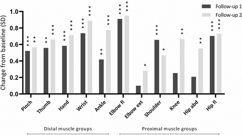
Figure 1 shows the average treatment response at a group level of individual muscle groups over time, when compared with baseline measurements (muscle strength prior to IVIg initiation). The change from baseline (CFB), expressed as number of SDs, indicated a statistically significant treatment response in most muscle groups at the first follow-up, and in all muscle groups at the second follow-up (see Supporting Information Table S1, which is available online).

The average, pooled effect over all muscle groups was 0.56 (95% confidence interval [CI], 0.37–0.75, P < .001). Changes in SD-scores ranged from 0.42 to 0.89 in the distal muscle groups (ie, pinch, thumb, hand, wrist and ankle), and 0.1 to 0.95 in the proximal muscle groups (ie, elbow, shoulder, knee and hip). Pairwise comparison between distal and proximal muscle strength groups showed no difference in muscle strength gain (P = .77). Compared to baseline, this treatment response was significant for both the first and the second follow-up, except for the first follow-up of elbow Ext, knee Ext, and hip Abd. In the upper extremity, elbow Fl and wrist Ext showed the largest CFB scores. Hip Fl and ankle dorsiflexion showed the largest change in the lower extremity.
The treatment response increased during the second follow-up in almost all individual muscle groups, except for shoulder Abd. Pairwise comparison showed a significant additional increase in muscle strength between first and second follow-up for ankle Fl, knee Ext and hip Abd (all P < .05).
3.3 Relationship between initial muscle strength and treatment response
Table 2 shows the average CFB for each individual muscle group compared to its baseline muscle strength. Despite variability between individual patients, most analyzed muscle groups showed a strong association between initial weakness at baseline and the magnitude of the treatment response, with the largest CFB in the weakest muscle groups. Regression coefficients ranged from −0.06 (SE 0.04) to −0.42 (SE 0.08), and were significant in all muscle groups, except for hand grip. The coefficient indicates that for each SD loss at baseline, patients gained up to an additional 0.42 SD after IVIg initiation. Especially in the more proximal muscle groups, that is the shoulder (−0.33), hip (−0.40) and knee (−0.42), the magnitude of the treatment response depended more strongly on muscle strength at baseline as compared to the more distal muscle groups, such as those for hand grip (−0.06) or ankle dorsiflexion (−0.17). Supporting Information Figure S1 provides additional supporting data.
| Slope (SE) | P value | |
|---|---|---|
| Pinch | −0.22 (0.03) | <.001 |
| Thumb | −0.17 (0.03) | <.001 |
| Hand | −0.06 (0.04) | .158 |
| Wrist | −0.14 (0.04) | <.001 |
| Elbow fl | −0.24 (0.05) | <.001 |
| Elbow ext | −0.20 (0.05) | <.001 |
| Shoulder | −0.33 (0.08) | <.001 |
| Ankle | −0.17 (0.05) | .001 |
| Knee ext | −0.42 (0.08) | <.001 |
| Hip abd | −0.40 (0.08) | <.001 |
| Hip fl | −0.28 (0.07) | <.001 |
4 DISCUSSION
The current study showed: (1) an overall positive treatment response in both distal and proximal muscle groups after IVIg initiation, and (2), an increase in treatment response as initial muscle strength (prior to IVIg initiation) decreased.
Despite variation on a group level, proximal muscle groups responded at a similar level as distal muscle groups during IVIg initiation. The similarity in treatment response between proximal and distal muscle groups was also noted in by previous research.14
When the first and second follow-up with IVIg were compared, the second follow-up caused an overall, additional treatment response in all muscle groups. However, within the wide range of tested muscle groups, this additional effect was only significant (P < .05) in muscles around the ankle, knee, and hip; muscle groups of the lower extremity. Since symptoms in the lower extremity initially occur in only 34% of MMN patients, 1 this finding is surprising, and might suggest that muscle groups in the lower extremity responded more slowly to IVIg initiation than muscle groups in the upper extremity. This delay in treatment response may have led to an underestimation of the effectiveness of IVIg in the lower extremity.
The positive treatment response in more proximal muscle groups of arms and legs, may not have been detected previously due the use of the Medical Research Council (MRC) scale both in the clinical and experimental setting. The use of the MRC scale for the quantification of muscle strength is controversial because of its subjectivity.15 Moreover, its insensitivity for detecting muscle weakness results in a risk of classifying muscle groups that are actually weakened as MRC 5.16, 17 This insensitivity may cause structural underestimation of muscle weakness, in particular in large muscle groups that initially exhibited mild muscle weakness. Although distal arm and/or leg weakness is distinct in MMN, it is possible that MMN also causes a mild loss of strength in the proximal muscles relative to the level before disease onset. This possibly more widespread loss of muscle function could explain the fact that MMN patients report that, in addition to primary loss of manual dexterity, they also have fatigue and loss of walking ability.18
Finally, the current study found that muscle groups that were very weak (<−4 SD) prior to IVIg initiation still benefitted from IVIg. In fact, the weaker the initial muscle strength, the greater the treatment response in SD. This finding suggests that severe weakness in the early phase of MMN may be related to reversible damage, such as focal demyelination, or inflammatory processes involving of the nodes of Ranvier, rather than irreversible axonal damage.19 However, it is important to note that this finding was the result of a group analysis, and that on an individual level there were also patients who experienced prolonged weakness or paralysis in some muscle groups (also see Supporting Information Figure S1).
Although objective muscle strength measurements using HHD can provide a more reliable assessment of muscle strength than MRC scores, they can also be prone to measurement errors. The reliability of HHD depends on the technique and strength of the examiner. 20, 21 A ceiling effect can occur when it is difficult for an examiner to provide sufficient resistance at high forces (commonly around 200 to 300 Newton) in strong muscle groups such as the quadriceps. 22 In this study, the impact of these errors was reduced as much as possible by using a standardized protocol applied by the same experienced evaluator. Since the treatment response increased consistently during consecutive measurements, a learning effect may also have had an impact on the results. To minimize this potential learning effect in the current study, patients were familiarized with the test. In addition, the results of this study show that the treatment response continued to increase during the second follow-up, making a significant influence of a learning effect less likely. The difference in the magnitude of the treatment response between elbow Fl and elbow Ext may have been caused by the normative values that were used, although a true difference in treatment responsiveness cannot be excluded. Furthermore, it cannot be ruled out that part of the initial muscle weakness in the proximal muscle groups in arms and legs can be explained by deconditioning. Also the delayed response in proximal lower extremity muscles could be explained by better physical fitness in patients who are more active when mobility is improved due to stronger distal muscles. Placebo effect could also have played a role, and though rare, some patients probably have had a conduction block in proximal muscles. Due to the relatively short follow-up period of our study, it is not possible to accurately discriminate between true or placebo effects. To gain more insight in the mechanisms that underlie the treatment response, a longer follow-up study with repeated electrodiagnostic evaluation is required.
The results of this study revealed an overall positive treatment response after IVIg initiation, demonstrated in proximal as well as distal muscle groups. In addition, weaker muscle groups benefitted most from IVIg treatment, even if there initially was complete paralysis at baseline.
ACKNOWLEDGMENTS
We thank Veerle van Dongen and Leo Swart for their help with the data-collection. The authors received no financial support for the research, authorship, and/or publication of this article.
CONFLICTS OF INTEREST
None of the authors has any conflict of interest to disclose.
ETHICAL PUBLICATION STATEMENT
We confirm that we have read the Journal's position on issues involved in ethical publication and affirm that this report is consistent with those guidelines.
Open Research
DATA AVAILABILITY STATEMENT
The corresponding author is able to provide the anonymized data of this study upon reasonable request from qualified investigators.


