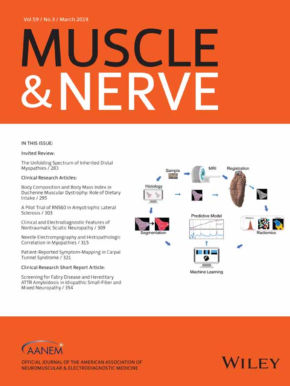
Texture as an imaging biomarker for disease severity in golden retriever muscular dystrophy
Aydin Eresen PhD
Department of Electrical and Computer Engineering, Texas A&M University, College Station, Texas, USA
Search for more papers by this authorLejla Alic PhD
Department of Electrical and Computer Engineering, Texas A&M University at Qatar, Doha, Qatar
Search for more papers by this authorSharla M. Birch DVM, PhD
College of Veterinary Medicine & Biomedical Sciences, Texas A&M University, College Station, Texas, USA
Search for more papers by this authorWade Friedeck MS
College of Veterinary Medicine & Biomedical Sciences, Texas A&M University, College Station, Texas, USA
Search for more papers by this authorJohn F. Griffin IV DVM, PhD
College of Veterinary Medicine & Biomedical Sciences, Texas A&M University, College Station, Texas, USA
Search for more papers by this authorJoe N. Kornegay DVM, PhD
College of Veterinary Medicine & Biomedical Sciences, Texas A&M University, College Station, Texas, USA
Search for more papers by this authorCorresponding Author
Jim X. JI PhD
Department of Electrical and Computer Engineering, Texas A&M University, College Station, Texas, USA
Department of Electrical and Computer Engineering, Texas A&M University at Qatar, Doha, Qatar
Correspondence to: J. X. Ji; e-mail: [email protected]Search for more papers by this authorAydin Eresen PhD
Department of Electrical and Computer Engineering, Texas A&M University, College Station, Texas, USA
Search for more papers by this authorLejla Alic PhD
Department of Electrical and Computer Engineering, Texas A&M University at Qatar, Doha, Qatar
Search for more papers by this authorSharla M. Birch DVM, PhD
College of Veterinary Medicine & Biomedical Sciences, Texas A&M University, College Station, Texas, USA
Search for more papers by this authorWade Friedeck MS
College of Veterinary Medicine & Biomedical Sciences, Texas A&M University, College Station, Texas, USA
Search for more papers by this authorJohn F. Griffin IV DVM, PhD
College of Veterinary Medicine & Biomedical Sciences, Texas A&M University, College Station, Texas, USA
Search for more papers by this authorJoe N. Kornegay DVM, PhD
College of Veterinary Medicine & Biomedical Sciences, Texas A&M University, College Station, Texas, USA
Search for more papers by this authorCorresponding Author
Jim X. JI PhD
Department of Electrical and Computer Engineering, Texas A&M University, College Station, Texas, USA
Department of Electrical and Computer Engineering, Texas A&M University at Qatar, Doha, Qatar
Correspondence to: J. X. Ji; e-mail: [email protected]Search for more papers by this authorABSTRACT
Introduction: Golden retriever muscular dystrophy (GRMD), an X-linked recessive disorder, causes similar phenotypic features to Duchenne muscular dystrophy (DMD). There is currently a need for a quantitative and reproducible monitoring of disease progression for GRMD and DMD. Methods: To assess severity in the GRMD, we analyzed texture features extracted from multi-parametric MRI (T1w, T2w, T1m, T2m, and Dixon images) using 5 feature extraction methods and classified using support vector machines. Results: A single feature from qualitative images can provide 89% maximal accuracy. Furthermore, 2 features from T1w, T2m, or Dixon images provided highest accuracy. When considering a tradeoff between scan-time and computational complexity, T2m images provided good accuracy at a lower acquisition and processing time and effort. Conclusions: The combination of MRI texture features improved the classification accuracy for assessment of disease progression in GRMD with evaluation of the heterogenous nature of skeletal muscles as reflection of the histopathological changes. Muscle Nerve 59:380–386, 2019
Supporting Information
| Filename | Description |
|---|---|
| mus26386-sup-0001-FigureS1.docxWord 2007 document , 403.8 KB | Supplementary Figure S1. The framework represents the overall process of texture analysis to select MRI modality and textural features. Qualitative and quantitative middle slice MRI images are acquired from a 15 months-old GRMD dog acquired with a 3T clinical scanner: T1w (a), T2w (b), T1m (c), T2m (d), DWf (e) and DFf (f) images. |
| mus26386-sup-0002-TableS1.docxWord 2007 document , 31.2 KB | Supplementary Table S1. MRI acquisition parameters on 3T MRI scanner |
| mus26386-sup-0003-TableS2.docxWord 2007 document , 17.1 KB | Supplementary Table S2. List of textural features |
| mus26386-sup-0004-TableS3.docxWord 2007 document , 65.5 KB | Supplementary Table S3. List of features provided maximized accuracy for T1w and T2w images. |
| mus26386-sup-0005-TableS4.docxWord 2007 document , 24.6 KB | Supplementary Table S4. List of features provided maximized accuracy for T1m and T2m images. |
| mus26386-sup-0006-TableS5.docxWord 2007 document , 36.6 KB | Supplementary Table S5. List of features provided maximized accuracy for DWf and DFf images. |
Please note: The publisher is not responsible for the content or functionality of any supporting information supplied by the authors. Any queries (other than missing content) should be directed to the corresponding author for the article.
REFERENCES
- 1Cheung JY, Bonventre JV, Malis CD, Leaf A. Calcium and ischemic injury. N Engl J Med 1986; 314: 1670–1676.
- 2Mendell JR, Shilling C, Leslie ND, Flanigan KM, al-Dahhak R, Gastier-Foster J, et al. Evidence-based path to newborn screening for Duchenne muscular dystrophy. Ann Neurol 2012; 71: 304–313.
- 3Brinkmeyer-Langford C, Kornegay JN. Comparative genomics of X-linked muscular dystrophies: the golden retriever model. Curr Genomics 2013; 14: 330–342.
- 4US National Library of Medicine, MedlinePlus. Duchenne muscular dystrophy. Available at https://medlineplus.gov/ency/article/000705.htm. Accessed December 7, 2018
- 5Liu JMK, Okamura CS, Bogan DJ, Bogan JR, Childers MK, Kornegay JN. Effects of prednisone in canine muscular dystrophy. Muscle Nerve 2004; 30: 767–773.
- 6Kornegay JN, Spurney CF, Nghiem PP, Brinkmeyer-Langford CL, Hoffman EP, Nagaraju K. Pharmacologic management of Duchenne muscular dystrophy: target identification and preclinical trials. ILAR J 2014; 55: 119–149.
- 7Vulin A, Barthélémy I, Goyenvalle A, Thibaud J-L, Beley C, Griffith G, et al. Muscle function recovery in golden retriever muscular dystrophy after AAV1-U7 exon skipping. Mol Ther 2012; 20: 2120–2133.
- 8Barthélémy I, Uriarte A, Drougard C, Unterfinger Y, Thibaud J-L, Blot S. Effects of an immunosuppressive treatment in the GRMD dog model of Duchenne muscular dystrophy. PLoS One 2012; 7: e48478.
- 9Yang G, Lalande V, Chen L, Azzabou N, Larcher T, de Certaines JD, et al. MRI texture analysis of GRMD dogs using orthogonal moments: a preliminary study. IRBM 2015; 36: 213–219.
- 10Lerario A, Bonfiglio S, Sormani M, Tettamanti A, Marktel S, Napolitano S, et al. Quantitative muscle strength assessment in Duchenne muscular dystrophy: longitudinal study and correlation with functional measures. BMC Neurol 2012; 12: 1–8.
- 11Mcdonald CM, Henricson EK, Abresch RT, Florence JM, Eagle M, Gappmaier E, et al. The 6-minute walk test and other endpoints in Duchenne muscular dystrophy: longitudinal natural history observations over 48 weeks from a multicenter study. Muscle Nerve 2013; 48: 343–356.
- 12Martins-Bach AB, Malheiros J, Matot B, Martins PCM, Almeida CF, Caldeira W, et al. Quantitative T2 combined with texture analysis of nuclear magnetic resonance images identify different degrees of muscle involvement in three mouse models of muscle dystrophy: mdx, Largemyd and mdx/Largemyd. PLoS One 2015; 10: e0117835.
- 13Vohra RS, Mathur S, Bryant ND, Forbes SC, Vandenborne K, Walter GA. Age-related T2 changes in hindlimb muscles of mdx mice. Muscle Nerve 2016; 53: 84–90.
- 14Qin EC, Jugé L, Lambert SA, Paradis V, Sinkus R, Bilston LE. In vivo anisotropic mechanical properties of dystrophic skeletal muscles measured by anisotropic MR elastographic imaging: the mdx mouse model of muscular dystrophy. Radiology 2014; 273: 726–735.
- 15Fan Z, Wang J, Ahn M, Shiloh-Malawsky Y, Chahin N, Elmore S, et al. Characteristics of magnetic resonance imaging biomarkers in a natural history study of golden retriever muscular dystrophy. Neuromuscul Disord 2014; 24: 178–191.
- 16Park J, Wicki J, Knoblaugh SE, Chamberlain JS, Lee D. Multi-parametric MRI at 14T for muscular dystrophy mice treated with AAV vector-mediated gene therapy. PLoS One 2015; 10: e0124914.
- 17Yokota T, Lu Q-L, Partridge T, Kobayashi M, Nakamura A, Takeda S, et al. Efficacy of systemic morpholino exon-skipping in Duchenne dystrophy dogs. Ann Neurol 2009; 65: 667–676.
- 18Wokke BH, Van Den Bergen JC, Hooijmans MT, Verschuuren JJ, Niks EH, Kan HE. T2 relaxation times are increased in skeletal muscle of DMD but not BMD patients. Muscle Nerve 2016; 53: 38–43.
- 19Hooijmans MT, Doorenweerd N, Baligand C, Verschuuren JJGM, Ronen I, Niks EH, et al. Spatially localized phosphorous metabolism of skeletal muscle in Duchenne muscular dystrophy patients: 24–month follow-up. PLoS One 2017; 12: e0182086.
- 20Willcocks RJ, Arpan IA, Forbes SC, Lott DJ, Senesac CR, Senesac E, et al. Longitudinal measurements of MRI-T2 in boys with Duchenne muscular dystrophy: effects of age and disease progression. Neuromuscul Disord 2014; 24: 393–401.
- 21Arpan I, Willcocks RJ, Forbes SC, Finkel RS, Lott DJ, Rooney WD, et al. Examination of effects of corticosteroids on skeletal muscles of boys with DMD using MRI and MRS. Neurology 2014; 83: 974–980.
- 22Godi C, Ambrosi A, Nicastro F, Previtali SC, Santarosa C, Napolitano S, et al. Longitudinal MRI quantification of muscle degeneration in Duchenne muscular dystrophy. Ann Clin Transl Neurol 2016; 3: 607–622.
- 23Akima H, Lott D, Senesac C, Deol J, Germain S, Arpan I, et al. Relationships of thigh muscle contractile and non-contractile tissue with function, strength, and age in boys with Duchenne muscular dystrophy. Neuromuscul Disord 2012; 22: 16–25.
- 24Wren TAL, Bluml S, Tseng-Ong L, Gilsanz V. Three-point technique of fat quantification of muscle tissue as a marker of disease progression in Duchenne muscular dystrophy: preliminary study. AJR Am J Roentgenol 2008; 190: W8–W12.
- 25Arpan I, Forbes SC, Lott DJ, Senesac CR, Daniels MJ, Triplett WT, et al. T2 mapping provides multiple approaches for the characterization of muscle involvement in neuromuscular diseases: a cross-sectional study of lower leg muscles in 5-15-year-old boys with Duchenne muscular dystrophy. NMR Biomed 2013; 26: 320–328.
- 26Fischmann A, Hafner P, Gloor M, Schmid M, Klein A, Pohlman U, et al. Quantitative MRI and loss of free ambulation in Duchenne muscular dystrophy. J Neurol 2013; 260: 969–974.
- 27Forbes SC, Walter GA, Rooney WD, Wang D-J, DeVos S, Pollaro J, et al. Skeletal muscles of ambulant children with Duchenne Muscular Dystrophy: validation of multicenter study of evaluation with MR imaging and MR spectroscopy. Radiology 2013; 269: 198–207.
- 28Hollingsworth KG, Garrood P, Eagle M, Bushby K, Straub V. Magnetic resonance imaging in Duchenne muscular dystrophy: longitudinal assessment of natural history over 18 months. Muscle Nerve 2013; 48: 586–588.
- 29Kim HK, Laor T, Horn PS, Racadio JM, Wong B, Dardzinski BJ. T2 mapping in Duchenne muscular dystrophy: distribution of disease activity and correlation with clinical assessments. Radiology 2010; 255: 899–908.
- 30Vohra R, Accorsi A, Kumar A, Walter G, Girgenrath M. Magnetic resonance imaging is sensitive to pathological amelioration in a model for laminin-deficient Congenital muscular dystrophy (MDC1A). PLoS One 2015; 10: e0138254.
- 31Heier CR, Guerron AD, Korotcov A, Lin S, Gordish-Dressman H, Fricke S, et al. Non-invasive MRI and spectroscopy of mdx mice reveal temporal changes in dystrophic muscle imaging and in energy deficits. PLoS One 2014; 9: e112477.
- 32Wang J, Fan Z, Vandenborne K, Walter G, Shiloh-Malawsky Y, An H, et al. A computerized MRI biomarker quantification scheme for a canine model of Duchenne muscular dystrophy. Int J Comput Assist Radiol Surg 2013; 8: 763–774.
- 33Bish LT, Sleeper MM, Forbes SC, Morine KJ, Reynolds C, Singletary GE, et al. Long-term systemic myostatin inhibition via liver-targeted gene transfer in golden retriever muscular dystrophy. Hum Gene Ther 2011; 22: 1499–1509.
- 34Vohra RS, Lott D, Mathur S, Senesac C, Deol J, Germain S, et al. Magnetic resonance assessment of hypertrophic and pseudo-hypertrophic changes in lower leg muscles of boys with Duchenne muscular dystrophy and their relationship to functional measurements. PLoS One 2015; 10: e0128915.
- 35Wokke BH, van den Bergen JC, Versluis MJ, Niks EH, Milles J, Webb AG, et al. Quantitative MRI and strength measurements in the assessment of muscle quality in Duchenne muscular dystrophy. Neuromuscul Disord 2014; 24: 409–416.
- 36Mathur S, Lott DJ, Senesac C, Germain SA, Vohra RS, Sweeney HL, et al. Age-related differences in lower-limb muscle cross-sectional area and torque production in boys with Duchenne muscular mystrophy. Arch Phys Med Rehabil 2010; 91: 1051–1058.
- 37Richards P, Saywell WR, Heywood P. Pseudohypertrophy of the temporalis muscle in Xp21 muscular dystrophy. Dev Med Child Neurol 2000; 42: 786–787.
- 38Roberts TC, Blomberg KEM, McClorey G, Andaloussi SEL, Godfrey C, Betts C, et al. Expression analysis in multiple muscle groups and serum reveals complexity in the microRNA transcriptome of the mdx mouse with implications for therapy. Mol Ther Nucleic Acids 2012; 1: 1–9.
- 39Ricotti V, Evans MRB, Sinclair CDJ, Butler JW, Ridout DA, Hogrel J-Y, et al. Upper limb evaluation in Duchenne muscular dystrophy: fat-water quantification by MRI, muscle force and function define endpoints for clinical trials. PLoS One 2016; 11: e0162542.
- 40Burakiewicz J, Sinclair CDJ, Fischer D, Walter GA, Kan HE, Hollingsworth KG. Quantifying fat replacement of muscle by quantitative MRI in muscular dystrophy. J Neurol 2017; 264: 2053–2067.
- 41Valentine BA, Cooper BJ, Cummings JF, Lahunta A. Canine X-linked muscular dystrophy: morphologic lesions. J Neurol Sci 1990; 97: 1–23.
- 42Ponrartana S, Ramos-Platt L, Wren TAL, Hu HH, Perkins TG, Chia JM, et al. Effectiveness of diffusion tensor imaging in assessing disease severity in Duchenne muscular dystrophy: preliminary study. Pediatr Radiol 2015; 45: 582–589.
- 43Hooijmans MT, Damon BM, Froeling M, Versluis MJ, Burakiewicz J, Verschuuren JJGM, et al. Evaluation of skeletal muscle DTI in patients with duchenne muscular dystrophy. NMR Biomed 2015; 28: 1589–1597.
- 44Li GD, Liang YY, Xu P, Ling J, Chen YM. Diffusion tensor imaging of thigh muscles in Duchenne muscular dystrophy: correlation of apparent diffusion coefficient and fractional anisotropy values with fatty infiltration. AJR Am J Roentgenol 2016; 206: 867–870.
- 45Thibaud J-L, Monnet A, Bertoldi D, Barthélémy I, Blot S, Carlier PG. Characterization of dystrophic muscle in golden retriever muscular dystrophy dogs by nuclear magnetic resonance imaging. Neuromuscul Disord 2007; 17: 575–584.
- 46Willcocks RJ, Rooney WD, Triplett WT, Forbes SC, Lott DJ, Senesac CR, et al. Multicenter prospective longitudinal study of magnetic resonance biomarkers in a large duchenne muscular dystrophy cohort. Ann Neurol 2016; 79: 535–547.
- 47Kornegay JN. The golden retriever model of Duchenne muscular dystrophy. Skeletal Muscle 2017; 7: 1–21.
- 48Kinali M, Arechavala-Gomeza V, Cirak S, Glover A, Guglieri M, Feng L, et al. Muscle histology vs MRI in Duchenne muscular dystrophy. Neurology 2011; 76: 346–353.
- 49Thibaud J-L, Azzabou N, Barthelemy I, Fleury S, Cabrol L, Blot S, et al. Comprehensive longitudinal characterization of canine muscular dystrophy by serial NMR imaging of GRMD dogs. Neuromuscul Disord 2012; 22(Suppl 2): S85–S99.
- 50Zhang M-H, Ma J-S, Shen Y, Chen Y. Optimal classification for the diagnosis of duchenne muscular dystrophy images using support vector machines. Int J Comput Assist Radiol Surg 2016; 11: 1755–1763.
- 51 Committee for the Update of the Guide for the Care and Use of Laboratory Animals. Guide for the care and use of laboratory animals. Washington, DC: National Academy Press; 2011.
10.17226/25801 Google Scholar
- 52Eresen A, Birch SM, Alic A, Griffin JF IV, Kornegay JN, Ji JX. New similarity metric for registration of MRI to histology: golden retriever Muscular Dystrophy Imaging. IEEE Trans Biomed Eng 2018. doi: https://doi.org/10.1109/TBME.2018.2870711.
- 53Tustison NJ, Avants BB, Cook PA, Zheng Y, Egan A, Yushkevich PA, et al. N4ITK: improved N3 bias correction. IEEE Trans Med Imaging 2010; 29: 1310–1320.
- 54Klein S, Staring M, Murphy K, Viergever MA, Pluim JPW. Elastix: a toolbox for intensity-based medical image registration. IEEE Trans Med Imaging 2010; 29: 196–205.
- 55Kass M, Witkin A, Terzopoulos D. Snakes: active contour models. Int J Comput Vis 1988; 1: 321–331.
- 56Deoni SCL, Rutt BK, Peters TM. Rapid combined T1 and T2 mapping using gradient recalled acquisition in the steady state. Magn Reson Med 2003; 49: 515–526.
- 57Glover GH, Schneider E. Three-point Dixon technique for true water/fat decomposition with B0 inhomogeneity correction. Magn Reson Med 1991; 18: 371–383.
- 58Haralick RM, Shanmugam K, Dinstein IH. Textural features for image classification. IEEE Trans Syst Man Cybern B Cybern 1973; SMC-3: 610–621.
10.1109/TSMC.1973.4309314 Google Scholar
- 59Galloway MM. Texture analysis using gray level run lengths. Comput Graph Image Process 1975; 4: 172–179.
10.1016/S0146-664X(75)80008-6 Google Scholar
- 60Ojala T, Pietikäinen M, Harwood D. A comparative study of texture measures with classification based on featured distributions. Pattern Recognit 1996; 29: 51–59.
- 61Burges CJC. A tutorial on support vector machines for pattern recognition. Data Min Knowl Discov 1998; 2: 121–167.
- 62Scholkopf B, Smola AJ. Learning with kernels: support vector machines, regularization, optimization, and beyond. Cambridge, MA: MIT Press; 2001.
10.7551/mitpress/4175.001.0001 Google Scholar
- 63Zhu W, Zeng NF, Wang N. Sensitivity, specificity, accuracy, associated confidence interval and ROC analysis with practical SAS implementations. NESUG Proceedings: Health Care and Life Sciences; Baltimore, Maryland. Available at http://www.nesug.org/Proceedings/nesug10/hl/hl07.pdf. Accessed December 7, 2018.
- 64Maroco J, Silva D, Rodrigues A, Guerreiro M, Santana I, de Mendonça A. Data mining methods in the prediction of Dementia: a real-data comparison of the accuracy, sensitivity and specificity of linear discriminant analysis, logistic regression, neural networks, support vector machines, classification trees and random forests. BMC Res Notes 2011; 4: 1–14.
- 65Armbrustmacher VW. Pathology of the muscular dystrophies and the congenital nonprogressive myopathies. Pathol Annu 1980; 15: 301–333.
- 66Cullen MJ, Mastaglia FL. Morphological changes in dystrophic muscle. Br Med Bull 1980; 36: 145–166.
- 67Duda D, Kretowski M, Azzabou N, de Certaines JD. MRI texture analysis for differentiation between healthy and golden retriever muscular dystrophy dogs at different phases of disease evolution. Proceedings of the IFIP International Conference on Computer Information Systems and Industrial Management, September 24–26, 2015, Warsaw, Poland.


