On the RF heating of coronary stents at 7.0 Tesla MRI
Corresponding Author
Lukas Winter
Berlin Ultrahigh Field Facility (B.U.F.F.), Max-Delbrueck Center for Molecular Medicine, Berlin, Germany
Correspondence to: Lukas Winter, Dr.rer.nat., Berlin Ultrahigh Field Facility (B.U.F.F.), Max-Delbrueck Center for Molecular Medicine, Robert-Roessle-Strasse 10, 13125 Berlin, Germany. E-mail: [email protected]Search for more papers by this authorEva Oberacker
Berlin Ultrahigh Field Facility (B.U.F.F.), Max-Delbrueck Center for Molecular Medicine, Berlin, Germany
Search for more papers by this authorCelal Özerdem
Berlin Ultrahigh Field Facility (B.U.F.F.), Max-Delbrueck Center for Molecular Medicine, Berlin, Germany
Search for more papers by this authorYiyi Ji
Berlin Ultrahigh Field Facility (B.U.F.F.), Max-Delbrueck Center for Molecular Medicine, Berlin, Germany
Search for more papers by this authorFlorian von Knobelsdorff-Brenkenhoff
Berlin Ultrahigh Field Facility (B.U.F.F.), Max-Delbrueck Center for Molecular Medicine, Berlin, Germany
Experimental and Clinical Research Center (ECRC), a joint cooperation between the Charité Medical Faculty and the Max-Delbrueck Center for Molecular Medicine, Berlin, Germany
Search for more papers by this authorGerd Weidemann
Physikalisch Technische Bundesanstalt (PTB), Braunschweig and Berlin, Germany
Search for more papers by this authorBernd Ittermann
Physikalisch Technische Bundesanstalt (PTB), Braunschweig and Berlin, Germany
Search for more papers by this authorFrank Seifert
Physikalisch Technische Bundesanstalt (PTB), Braunschweig and Berlin, Germany
Search for more papers by this authorThoralf Niendorf
Berlin Ultrahigh Field Facility (B.U.F.F.), Max-Delbrueck Center for Molecular Medicine, Berlin, Germany
Experimental and Clinical Research Center (ECRC), a joint cooperation between the Charité Medical Faculty and the Max-Delbrueck Center for Molecular Medicine, Berlin, Germany
Search for more papers by this authorCorresponding Author
Lukas Winter
Berlin Ultrahigh Field Facility (B.U.F.F.), Max-Delbrueck Center for Molecular Medicine, Berlin, Germany
Correspondence to: Lukas Winter, Dr.rer.nat., Berlin Ultrahigh Field Facility (B.U.F.F.), Max-Delbrueck Center for Molecular Medicine, Robert-Roessle-Strasse 10, 13125 Berlin, Germany. E-mail: [email protected]Search for more papers by this authorEva Oberacker
Berlin Ultrahigh Field Facility (B.U.F.F.), Max-Delbrueck Center for Molecular Medicine, Berlin, Germany
Search for more papers by this authorCelal Özerdem
Berlin Ultrahigh Field Facility (B.U.F.F.), Max-Delbrueck Center for Molecular Medicine, Berlin, Germany
Search for more papers by this authorYiyi Ji
Berlin Ultrahigh Field Facility (B.U.F.F.), Max-Delbrueck Center for Molecular Medicine, Berlin, Germany
Search for more papers by this authorFlorian von Knobelsdorff-Brenkenhoff
Berlin Ultrahigh Field Facility (B.U.F.F.), Max-Delbrueck Center for Molecular Medicine, Berlin, Germany
Experimental and Clinical Research Center (ECRC), a joint cooperation between the Charité Medical Faculty and the Max-Delbrueck Center for Molecular Medicine, Berlin, Germany
Search for more papers by this authorGerd Weidemann
Physikalisch Technische Bundesanstalt (PTB), Braunschweig and Berlin, Germany
Search for more papers by this authorBernd Ittermann
Physikalisch Technische Bundesanstalt (PTB), Braunschweig and Berlin, Germany
Search for more papers by this authorFrank Seifert
Physikalisch Technische Bundesanstalt (PTB), Braunschweig and Berlin, Germany
Search for more papers by this authorThoralf Niendorf
Berlin Ultrahigh Field Facility (B.U.F.F.), Max-Delbrueck Center for Molecular Medicine, Berlin, Germany
Experimental and Clinical Research Center (ECRC), a joint cooperation between the Charité Medical Faculty and the Max-Delbrueck Center for Molecular Medicine, Berlin, Germany
Search for more papers by this authorAbstract
Purpose
Examine radiofrequency (RF) induced heating of coronary stents at 7.0 Tesla (T) to derive an analytical approach which supports RF heating assessment of arbitrary stent geometries and RF coils.
Methods
Simulations are performed to detail electromagnetic fields (EMF), local specific absorption rates (SAR) and temperature changes. For validation E-field measurements and RF heating experiments are conducted. To progress to clinical setups RF coils tailored for cardiac MRI at 7.0T and coronary stents are incorporated into EMF simulations using a human voxel model.
Results
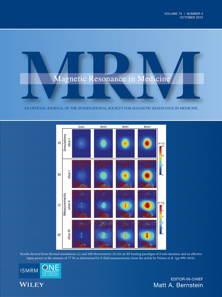
Our simulations of coronary stents at 297 MHz were confirmed by E-field and temperature measurements. An analytical solution which describes SAR(1g tissue voxel) induced by an arbitrary coronary stent interfering with E-fields generated by an arbitrary RF coil was derived. The analytical approach yielded a conservative estimation of induced SAR(1g tissue voxel) maxima without the need for integrating the stent into EMF simulations of the human voxel model.
Conclusion
The proposed analytical approach can be applied for any patient, coronary stent type, RF coil configuration and RF transmission regime. The generalized approach is of value for RF heating assessment of other passive electrically conductive implants and provides a novel design criterion for RF coils. Magn Reson Med 74:999–1010, 2015. © 2014 Wiley Periodicals, Inc.
REFERENCES
- 1Ladd ME. High-field-strength magnetic resonance: potential and limits. Top Magn Reson Imaging 2007; 18: 139–152.
- 2Regatte RR, Schweitzer ME. Ultra-high-field MRI of the musculoskeletal system at 7.0T. J Magn Reson Imaging 2007; 25: 262–269.
- 3Niendorf T, Sodickson DK, Krombach GA, Schulz-Menger J. Toward cardiovascular MRI at 7 T: clinical needs, technical solutions and research promises. Eur Radiol 2010; 20: 2806–2816.
- 4Ugurbil K. The road to functional imaging and ultrahigh fields. Neuroimage 2012; 62: 726–735.
- 5Moser E, Stahlberg F, Ladd ME, Trattnig S. 7-T MR–from research to clinical applications? NMR Biomed 2012; 25: 695–716.
- 6Trattnig S, Zbýň Š, Schmitt B, Friedrich K, Juras V, Szomolanyi P, Bogner W. Advanced MR methods at ultra-high field (7 Tesla) for clinical musculoskeletal applications. Eur Radiol 2012; 22: 2338–2346.
- 7van der Kolk AG, Hendrikse J, Zwanenburg JJ, Visser F, Luijten PR. Clinical applications of 7 T MRI in the brain. Eur J Radiol 2013; 82: 708–718.
- 8Niendorf T, Graessl A, Thalhammer C, et al. Progress and promises of human cardiac magnetic resonance at ultrahigh fields: a physics perspective. J Magn Reson 2013; 229: 208–222.
- 9Kraff O, Fischer A, Nagel AM, Mönninghoff C, Ladd ME. MRI at 7 Tesla and above: demonstrated and potential capabilities. J Magn Reson Imaging 2015; 41: 13–33.
- 10Ugurbil K. Magnetic resonance imaging at ultrahigh fields. IEEE Trans Biomed Eng 2014; 61: 1364–1379.
- 11Shellock FG. The Reference Manual for Magnetic Resonance Safety. Implants and Devices. Los Angeles, CA: Biomedical Research Publishing Company; 2014. 744 p.
- 12IEC. 60601-2-33 Medical electrical equipment - Part 2–33: particular requirements for the basic safety and essential performance of magnetic resonance equipment for medical diagnosis. Edition 3.0. 2010.
- 13 ICNIRP. ICNIRP guidelines - for limiting exposure to time-varying electric, magnetic and electromagnetic fields (up to 300GHz). Health Phys 1998; 74: 494–522.
- 14Rauschenberg J, Nagel AM, Ladd SC, et al. Multicenter study of subjective acceptance during magnetic resonance imaging at 7 and 9.4 T. Invest Radiol 2014; 49: 249–259.
- 15van der Kolk AG, Zwanenburg JJ, Brundel M, Biessels GJ, Visser F, Luijten PR, Hendrikse J. Intracranial vessel wall imaging at 7.0-T MRI. Stroke 2011; 42: 2478–2484.
- 16Madai VI, von Samson-Himmelstjerna FC, Bauer M, Stengl KL, Mutke MA, Tovar-Martinez E, Wuerfel J, Endres M, Niendorf T, Sobesky J. Ultrahigh-field MRI in human ischemic stroke–a 7 tesla study. PLoS One 2012; 7: e37631.
- 17Sammet CL, Yang X, Wassenaar PA, Bourekas EC, Yuh BA, Shellock F, Sammet S, Knopp MV. RF-related heating assessment of extracranial neurosurgical implants at 7T. Magn Reson Imaging 2013; 31: 1029–1034.
- 18Kraff O, Wrede KH, Schoemberg T, Dammann P, Noureddine Y, Orzada S, Ladd ME, Bitz AK. MR safety assessment of potential RF heating from cranial fixation plates at 7 T. Med Phys 2013; 40: 042302.
- 19Dula AN, Virostko J, Shellock FG. Assessment of MRI issues at 7 T for 28 implants and other objects. AJR Am J Roentgenol 2014; 202: 401–405.
- 20van Rijn GA, Mourik JE, Teeuwisse WM, Luyten GP, Webb AG. Magnetic resonance compatibility of intraocular lenses measured at 7 Tesla. Invest Ophthalmol Vis Sci 2012; 53: 3449–3453.
- 21Schrom T, Thelen A, Asbach P, Bauknecht HC. Effect of 7.0 Tesla MRI on upper eyelid implants. Ophthal Plast Reconstr Surg 2006; 22: 480–482.
- 22Wezel J, Kooij BJ, Webb AG. Assessing the MR compatibility of dental retainer wires at 7 Tesla. Magn Reson in Med 2014; 72: 1191–1198.
- 23Ansems J, Kolk A, Kroeze H, Van den Berg NAT, Borst G, Luijten PR, Webb AG, Renema WKJ, Klomp DWJ. MR imaging of patients with stents is safe at 7.0 Tesla. In Proceedings of the 20th Annual Meeting of ISMRM, Melbourne, Australia, 2012. Abstract 2764.
- 24Montalescot G, Sechtem U, Achenbach S, Andreotti F, Arden C, Budaj A, Bugiardini R, Crea F, Cuisset T, Di Mario C. 2013 ESC guidelines on the management of stable coronary artery disease. The Task Force on the Management of Stable Coronary Artery Disease of the European Society of Cardiology. Eur Heart J 2013; 34: 2949–3003.
- 25Muhlberger V, Kobel C, Kaltenbach L, Pachinger O. Austrian National CathLab Registry (ANCALAR): cardiac catheterization, coronary angiography (CA), and percutaneous coronary intervention (PCI) in Austria during the year 2011 (Registry Data with Audit including 2012). Wien Klin Wochenschr 2013; 125: 736–749.
- 26Ulrich MR, Brock DM, Ziskind AA. Analysis of trends in coronary artery bypass grafting and percutaneous coronary intervention rates in Washington state from 1987 to 2001. Am J Cardiol 2003; 92: 836–839.
- 27Rawson NS, Chu R, Ismaila AS, Terres JA. The aging Canadian population and hospitalizations for acute myocardial infarction: projection to 2020. BMC Cardiovasc Disord 2012; 12: 25.
- 28Go AS, Mozaffarian D, Roger VL, Benjamin EJ, Berry JD, Blaha MJ, Dai S, Ford ES, Fox CS, Franco S. Heart disease and stroke statistics–2014 update: a report from the American Heart Association. Circulation 2014; 129: e28.
- 29Sharpe RE Jr, Levin DC, Parker L, Rao VM. The recent reversal of the growth trend in MRI: a harbinger of the future? J Am Coll Radiol 2013; 10: 599–602.
- 30Thalhammer C, Renz W, Winter L, et al. Two-dimensional sixteen channel transmit/receive coil array for cardiac MRI at 7.0 T: design, evaluation, and application. J Magn Reson Imaging 2012; 36: 847–857.
- 31Graessl A, Renz W, Hezel F, et al. Modular 32-channel transceiver coil array for cardiac MRI at 7.0T. Magn Reson Med 2014; 72: 276–290.
- 32Vaughan J, Adriany G, Snyder C, Tian J, Thiel T, Bolinger L, Liu H, DelaBarre L, Ugurbil K. Efficient high-frequency body coil for high-field MRI. Magn Reson Med 2004; 52: 851–859.
- 33Brunner DO, De Zanche N, Fröhlich J, Paska J, Pruessmann KP. Travelling-wave nuclear magnetic resonance. Nature 2009; 457: 994–998.
- 34Jin J-M. Theory and computation of electromagnetic fields: New York: John Wiley & Sons; 2010. 572 p.
10.1002/9780470874257 Google Scholar
- 35Virtanen H, Huttunen J, Toropainen A, Lappalainen R. Interaction of mobile phones with superficial passive metallic implants. Phys Med Biol 2005; 50: 2689.
- 36Kastrati A, Mehilli J, Dirschinger J, Pache J, Ulm K, Schühlen H, Seyfarth M, Schmitt C, Blasini R, Neumann F-J. Restenosis after coronary placement of various stent types. Am J Cardiol 2001; 87: 34–39.
- 37Bakhai A, Booth J, Delahunty N, Nugara F, Clayton T, McNeill J, Davies S, Cumberland D, Stables R. The SV stent study: a prospective, multicentre, angiographic evaluation of the BiodivYsio phosphorylcholine coated small vessel stent in small coronary vessels. Int J Cardiol 2005; 102: 95–102.
- 38Christiansen EH, Jensen LO, Thayssen P, Tilsted H-H, Krusell LR, Hansen KN, Kaltoft A, Maeng M, Kristensen SD, Bøtker HE. Biolimus-eluting biodegradable polymer-coated stent versus durable polymer-coated sirolimus-eluting stent in unselected patients receiving percutaneous coronary intervention (SORT OUT V): a randomised non-inferiority trial. Lancet 2013; 381: 661–669.
- 39Santoro D, Winter L, Müller A, Vogt J, Renz W, Özerdem C, Grässl A, Tkachenko V, Schulz-Menger J, Niendorf T. Detailing radio frequency heating induced by coronary stents: a 7.0 Tesla magnetic resonance study. PLoS One 2012; 7: e49963.
- 40Oberacker E, Winter L, Seifert F, Marek J, Weidemann G, Hoffmann E, Niendorf T. En route to ultrahigh field cardiac MR in patients: RF safety assessment of intracoronary stents at 7.0T using numerical simulations and E-field measurements. In Proceedings of the 22nd Annual Meeting of ISMRM, Milan, Italy, 2014. Abstract 0177.
- 41Christ A, Kainz W, Hahn EG, Honegger K, Zefferer M, Neufeld E, Rascher W, Janka R, Bautz W, Chen J. The virtual family—development of surface-based anatomical models of two adults and two children for dosimetric simulations. Phys Med Biol 2010; 55: N23.
- 42Clemens M, Weiland T. Discrete electromagnetism with the finite integration technique. Prog Electromagn Res 2001; 32: 65–87.
10.2528/PIER00080103 Google Scholar
- 43Winter L, Özerdem C, Hoffmann W, Santoro D, Müller A, Waiczies H, Seemann R, Graessl A, Wust P, Niendorf T. Design and evaluation of a hybrid radiofrequency applicator for magnetic resonance imaging and RF induced hyperthermia: electromagnetic field simulations up to 14.0 Tesla and proof-of-concept at 7.0 Tesla. PLoS One 2013; 8: e61661.
- 44 American Society for Testing and Materials International. Designation F2182-011: standard test method for measurement of radio frequency induced heating near passive implants during magnetic resonance imaging. West Conshohocken, PA: ASTM International; 2011.
- 45Weidemann G, Frese I, Seifert F, Cassara A, Hoffmann W, Ittermann B. A system for calibrated measurements of RF electromagnetic fields inside a clinical MR scanner. In Proceedings of the 22nd Annual Meeting of ISMRM, Milan, Italy, 2014. Abstract 1370.
- 46Klepsch T, Lindel T, Hoffmann W, Botterweck H, Ittermann B, Seifert F. Calibration of fibre-optic RF E/H-field probes using a magnetic reso-nance (MR) compatible TEM cell and dedicated MR measurement techniques. Biomed Tech 2012; 57: 1.
10.1515/bmt-2012-4428 Google Scholar
- 47Hasgall P, Neufeld E, Gosselin MC, Klingenböck A, Kuster N. IT'IS database for thermal and electromagnetic parameters of biological tissues. http://www.itis.ethz.ch/database. Version 2.4, Published July 30, 2013. Accessed January 1, 2014.
- 48Ishihara Y, Calderon A, Watanabe H, Okamoto K, Suzuki Y, Kuroda K. A precise and fast temperature mapping using water proton chemical shift. Magn Reson Med 1995; 34: 814–823.
- 49Wonneberger U, Schnackenburg B, Wlodarczyk W, Walter T, Streitparth F, Rump J, Teichgräber UKM. Intradiscal temperature monitoring using double gradient-echo pulse sequences at 1.0 T. J Magn Reson Imaging 2010; 31: 1499–1503.
- 50Kuroda K, Oshio K, Chung AH, Hynynen K, Jolesz FA. Temperature mapping using the water proton chemical shift: a chemical shift selective phase mapping method. Magn Reson Med 1997; 38: 845–851.
- 51Katscher U, Voigt T, Findeklee C, Vernickel P, Nehrke K, Dossel O. Determination of electric conductivity and local SAR via B1 mapping. IEEE Trans Med Imaging 2009; 28: 1365–1374.
- 52van Lier AL, Brunner DO, Pruessmann KP, Klomp DWJ, Luijten PR, Lagendijk JJW, van den Berg CAT. B1(+) Phase mapping at 7 T and its application for in vivo electrical conductivity mapping. Magn Reson Med 2012; 67: 552–561.
- 53Peters RD, Henkelman RM. Proton-resonance frequency shift MR thermometry is affected by changes in the electrical conductivity of tissue. Magn Reson Med 2000; 43: 62–71.
10.1002/(SICI)1522-2594(200001)43:1<62::AID-MRM8>3.0.CO;2-1 CAS PubMed Web of Science® Google Scholar
- 54Gellermann J, Wlodarczyk W, Feussner A, Fähling H, Nadobny J, Hildebrandt B, Felix R, Wust P. Methods and potentials of magnetic resonance imaging for monitoring radiofrequency hyperthermia in a hybrid system. Int J Hyperthermia 2005; 21: 497–513.
- 55Dieringer MA, Renz W, Lindel T, et al. Design and application of a four-channel transmit/receive surface coil for functional cardiac imaging at 7T. J Magn Reson Imaging 2011; 33: 736–741.
- 56Winter L, Kellman P, Renz W, Gräßl A, Hezel F, Thalhammer C, Von Knobelsdorff Brenkenhoff F, Tkachenko V, Schulz-Menger J, Niendorf T. Comparison of three multichannel transmit/receive radiofrequency coil configurations for anatomic and functional cardiac MRI at 7.0T: implications for clinical imaging. Eur Radiol 2012; 22: 2211–2220.
- 57King RWP, Smith GS, Owens M, Wu TT. Antennas in matter: fundamentals, theory, and applications. Cambridge, MA: MIT Press; 1981. 868 p.
- 58Eichfelder G, Gebhardt M. Local specific absorption rate control for parallel transmission by virtual observation points. Magn Reson Med 2011; 66: 1468–1476.
- 59Graesslin I, Homann H, Biederer S, Bornert P, Nehrke K, Vernickel P, Mens G, Harvey P, Katscher U. A specific absorption rate prediction concept for parallel transmission MR. Magn Reson Med 2012; 68: 1664–1674.
- 60Metzger GJ, Snyder C, Akgun C, Vaughan T, Ugurbil K, Van de Moortele PF. Local B1+ shimming for prostate imaging with transceiver arrays at 7T based on subject-dependent transmit phase measurements. Magn Reson Med 2008; 59: 396–409.
- 61Schmitter S, DelaBarre L, Wu X, Greiser A, Wang D, Auerbach EJ, Vaughan JT, Ugurbil K, Van de Moortele PF. Cardiac imaging at 7 Tesla: single- and two-spoke radiofrequency pulse design with 16-channel parallel excitation. Magn Reson Med 2013; 70: 1210–1219.
- 62Katscher U, Bornert P, Leussler C, van den Brink JS. Transmit SENSE. Magn Reson Med 2003; 49: 144–150.
- 63Zhu Y. Parallel excitation with an array of transmit coils. Magn Reson Med 2004; 51: 775–784.
- 64Yarmolenko PS, Moon EJ, Landon C, Manzoor A, Hochman DW, Viglianti BL, Dewhirst MW. Thresholds for thermal damage to normal tissues: an update. Int J Hyperthermia 2011; 27: 320–343.
- 65Murbach M, Neufeld E, Capstick M, Kainz W, Brunner DO, Samaras T, Pruessmann KP, Kuster N. Thermal tissue damage model analyzed for different whole-body SAR and scan durations for standard MR body coils. Magn Reson Med 2013; 71: 421–431.