

Sprayed Cells and Polyelectrolyte Films for Biomaterial Functionalization: The Influence of Physical PLL–PGA Film Treatments on Dental Pulp Cell Behavior
Corresponding Author
Ivan V. Panayotov
EA4203 Laboratoire de Bio-santé et Nano-science, Université Montpellier 1, Montpellier, France
Search for more papers by this authorPierre-Yves Collart-Dutilleul
EA4203 Laboratoire de Bio-santé et Nano-science, Université Montpellier 1, Montpellier, France
Search for more papers by this authorHamideh Salehi
EA4203 Laboratoire de Bio-santé et Nano-science, Université Montpellier 1, Montpellier, France
Search for more papers by this authorMarta Martin
Laboratoire Charles Coulomb UMR 5221, Université Montpellier 2, Montpellier, France
Laboratoire Charles Coulomb UMR 5221, CNRS, Montpellier, France
Search for more papers by this authorAttila Végh
Biological Research Centre of the Hungarian Academy of Sciences, Institute of Biophysics, Szeged, Hungary
Search for more papers by this authorJacques Yachouh
EA4203 Laboratoire de Bio-santé et Nano-science, Université Montpellier 1, Montpellier, France
Search for more papers by this authorBoyan Vladimirov
Department of Maxillofacial Surgery, Medical University, Plovdiv, Bulgaria
Search for more papers by this authorPéter Sipos
Department of Pharmaceutical Technology, University of Szeged, Szeged, Hungary
Search for more papers by this authorBalázs Szalontai
Biological Research Centre of the Hungarian Academy of Sciences, Institute of Biophysics, Szeged, Hungary
Search for more papers by this authorCsilla Gergely
Laboratoire Charles Coulomb UMR 5221, Université Montpellier 2, Montpellier, France
Laboratoire Charles Coulomb UMR 5221, CNRS, Montpellier, France
Search for more papers by this authorFrédéric J. G. Cuisinier
EA4203 Laboratoire de Bio-santé et Nano-science, Université Montpellier 1, Montpellier, France
Search for more papers by this authorCorresponding Author
Ivan V. Panayotov
EA4203 Laboratoire de Bio-santé et Nano-science, Université Montpellier 1, Montpellier, France
Search for more papers by this authorPierre-Yves Collart-Dutilleul
EA4203 Laboratoire de Bio-santé et Nano-science, Université Montpellier 1, Montpellier, France
Search for more papers by this authorHamideh Salehi
EA4203 Laboratoire de Bio-santé et Nano-science, Université Montpellier 1, Montpellier, France
Search for more papers by this authorMarta Martin
Laboratoire Charles Coulomb UMR 5221, Université Montpellier 2, Montpellier, France
Laboratoire Charles Coulomb UMR 5221, CNRS, Montpellier, France
Search for more papers by this authorAttila Végh
Biological Research Centre of the Hungarian Academy of Sciences, Institute of Biophysics, Szeged, Hungary
Search for more papers by this authorJacques Yachouh
EA4203 Laboratoire de Bio-santé et Nano-science, Université Montpellier 1, Montpellier, France
Search for more papers by this authorBoyan Vladimirov
Department of Maxillofacial Surgery, Medical University, Plovdiv, Bulgaria
Search for more papers by this authorPéter Sipos
Department of Pharmaceutical Technology, University of Szeged, Szeged, Hungary
Search for more papers by this authorBalázs Szalontai
Biological Research Centre of the Hungarian Academy of Sciences, Institute of Biophysics, Szeged, Hungary
Search for more papers by this authorCsilla Gergely
Laboratoire Charles Coulomb UMR 5221, Université Montpellier 2, Montpellier, France
Laboratoire Charles Coulomb UMR 5221, CNRS, Montpellier, France
Search for more papers by this authorFrédéric J. G. Cuisinier
EA4203 Laboratoire de Bio-santé et Nano-science, Université Montpellier 1, Montpellier, France
Search for more papers by this authorAbstract
Further development of biomaterials is expected as advanced therapeutic products must be compliant to good manufacturing practice regulations. A spraying method for building-up polyelectrolyte films followed by the deposition of dental pulp cells by spraying is presented. Physical treatments of UV irradiation and a drying/wetting process are applied to the system. Structural changes and elasticity modifications of the obtained coatings are revealed by atomic force microscopy and by Raman spectroscopy. This procedure results in thicker, rougher and stiffer film. The initially ordered structure composed of mainly α helices is transformed into random/β-structures. The treatment enhanced dental pulp cell adhesion and proliferation, suggesting that this system is promising for medical applications.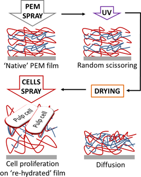
Supporting Information
As a service to our authors and readers, this journal provides supporting information supplied by the authors. Such materials are peer reviewed and may be re-organized for online delivery, but are not copy-edited or typeset. Technical support issues arising from supporting information (other than missing files) should be addressed to the authors.
| Filename | Description |
|---|---|
| mabi201400256-sm-0001-SuppFig-S1.docx229.7 KB | Figure S1. (Spray deposition of polyelectrolyte multilayers; (A): Quantity of solution deposited by spraying; (B): Volume of sprayed solution deposited on samples for 0.57ml (four spraying) and 1.27 ml (eight spraying) total deposited volume; (C): Total spray image (St: diameter = 7 cm and the disk surface (Ss: diameter = 1.4 cm). Contact angle measurements have been performed to characterize the surface of PEM's. A sessile drop of the reference liquid (water in our case) was applied with a syringe onto a specimen, which was placed on a plate capable of moving in three dimensions. Specimens were dried for 5 min in air at room temperature before the drop deposition. Water drop profiles were recorded with a CCD camera connected to a video-acquisition card. Contact angles were measured and recorded with ImageJ software (NIH, Bethesda, MD, USA, http://rsb.info.nih.gov/ij/). The contact angle of a water drop was measured after each layer deposition. Contact angles were determined by averaging data obtained at five different spots (one measurement per spot) after each polyelectrolyte adsorption step. Contact angle measurements during (PLL-PGA)5-PLL construction showed an increase of hydrophobicity after PLL adsorption, and of hydrophilicity after PGA deposition (Figure S2). There is no statistically significant difference (p = 0.156) between the contact angle of glass surface and that of the (PLLPGA)5PLL films (PLL 6 on Figure S2). The mean contact angle of a water drop was 64°±3° on the PEM surface, while it was 57° ± 3° on the bare glass surface. Figure S2. Contact angle changes during (PLL-PGA)5-PLL film build up; Five measurements have been done for each PEM deposition on five different glass disks. PLL increased the hydrophobicity, in contrast, PGA increased the hydrophilicity of the film surface.) Contact angle measurements showed alternating changes according to the nature of the last-adsorbed polyelectrolyte. Multilayers having PLL as the outermost layer were more hydrophobic than those of terminated by PGA, most probably due to the lower polarity of the PLL head-group, similarly as in the case of polyallilamine/polystyrenesulfonate (PAH/PSS) multilayers. The difference between the contacts angles became more expressed as the number of layers grew, i.e. as the structure of the PLL/PGA film became more perfect. |
Please note: The publisher is not responsible for the content or functionality of any supporting information supplied by the authors. Any queries (other than missing content) should be directed to the corresponding author for the article.
References
- 1 a) G. Decher, Science 1997, 277, 1232; b) G. H. Decher, J. D. Schmitt, J. Thin Solid Films 1992, 210/211, 831.
- 2 G. Decher, in: Multilayer Thin Films, Wiley-VCH Verlag GmbH & Co, KGaA 2003, p. 1.
- 3 a) G. Ladam, C. Gergely, B. Senger, G. Decher, J. C. Voegel, P. Schaaf, F. J. Cuisinier, Biomacromolecules 2000, 1, 674; b) G. Ladam, P. Schaaf, F. J. G. Cuisinier, G. Decher, J.-C. Voegel, Langmuir 2000, 17, 878. c) F. Tristan, G. Palestino, J. L. Menchaca, E. Perez, H. Atmani, F. Cuisinier, G. Ladam, Biomacromolecules 2009, 10, 2275.
- 4 a) C. Picart, Ph. Lavalle, P. Hubert, F. J. G. Cuisinier, G. Decher, P. Schaaf, J.-C. Voegel, Langmuir 2001, 23, 7414. b) C. Picart, C. Gergely, Y. Arntz, J. C. Voegel, P. Schaaf, F. J. Cuisinier, B. Senger, Biosens. Bioelectron 2004, 20, 553.
- 5 P. Lavalle, C. Gergely, F. J. G. Cuisinier, G. Decher, P. Schaaf, J. C. Voegel, C. Picart, Macromolecules 2002, 35, 4458.
- 6 C. P. Vazquez, T. Boudou, V. Dulong, C. Nicolas, C. Picart, K. Glinel, Langmuir 2009, 25, 3556.
- 7 L. Richert, F. Boulmedais, P. Lavalle, J. Mutterer, E. Ferreux, G. Decher, P. Schaaf, J. C. Voegel, C. Picart, Biomacromolecules 2004, 5, 284.
- 8 J. B. Schlenoff, S. T. Dubas, T. Farhat, Langmuir 2000, 16, 9968.
- 9 C. Porcel, P. Lavalle, G. Decher, B. Senger, J. C. Voegel, P. Schaaf, Langmuir 2007, 23, 1898.
- 10 a) G. Cado, H. Kerdjoudj, A. Chassepot, M. Lefort, K. Benmlih, J. Hemmerle, J. C. Voegel, L. Jierry, P. Schaaf, Y. Frere, F. Boulmedais, Langmuir 2012, 28, 8470; b) P. Schaaf, J. C. Voegel, L. Jierry, F. Boulmedais, Adv. Mater. 2012, 24, 1001.
- 11 M. Cohen, A. Bahoric, H. M. Clarke, Plast. Reconstr. Surg. 2001, 107, 1208.
- 12
H. A. R. Bahoric, A
H. M. Clarke,
R. M. Zuker,
Can. J. Plast. Surg.
1997,
5, 153.
10.1177/229255039700500301 Google Scholar
- 13 F. A. Navarro, M. L. Stoner, C. S. Park, J. C. Huertas, H. B. Lee, F. M. Wood, D. P. Orgill, J. Burn Care Rehabil. 2000, 21, 513.
- 14 A. T. Hafez, D. J. Bagli, A. Bahoric, K. Aitken, C. R. Smith, D. Herz, A. E. Khoury, J. Urol. 2003, 169, 2316.
- 15 A. Roberts, B. E. Wyslouzil, L. Bonassar, Biotechnol. Bioeng. 2005, 91, 801.
- 16 J. T. Seil, T. J. Webster, Int. J. Nanomed. 2011, 6, 1095.
- 17 a) S. Facca, P. Gillet, J. F. Stoltz, P. Netter, D. Mainard, J. C. Voegel, N. Benkirane-Jessel, Bio-med. Mater. Eng. 2008, 18, 231; b) J. R. Tritz, R. De Isla, N. Charif, N. Pinzano, A. Mainard, D. Bensoussan, D. Netter, P. Stoltz, J. F. Benkirane-Jessel, N. Huselstein, C., Soft Matter 2010, 6, 5165.
- 18 G. Papaccio, A. Graziano, R. d'Aquino, M. F. Graziano, G. Pirozzi, D. Menditti, A. De Rosa, F. Carinci, G. Laino, J. Cell. Physiol. 2006, 208, 319.
- 19 R. d'Aquino, A. Graziano, M. Sampaolesi, G. Laino, G. Pirozzi, A. De Rosa, G. Papaccio, Cell Death Differ. 2007, 14, 1162.
- 20 G. T. Huang, S. Gronthos, S. Shi, J. Dent. Res. 2009, 88, 792.
- 21 A. Arthur, S. Shi, A. C. Zannettino, N. Fujii, S. Gronthos, S. A. Koblar, Stem Cells 2009, 27, 2229.
- 22 J. L. Arrondo, A. Muga, J. Castresana, F. M. Goni, Progr. Biophys. Mol. Biol. 1993, 59, 23.
- 23 L. Richert, A. Schneider, D. Vautier, C. Vodouhe, N. Jessel, E. Payan, P. Schaaf, J. C. Voegel, C. Picart, Cell Biochem. Biophys. 2006, 44, 273.
- 24 P. G. Lavalle, C. Gergely, F. J. G. Cuisinier, G. Decher, P. Schaaf, J. C. Voegel, C. Picart, Macromolecules 2002, 35, 4458.
- 25 M. Kolasinska, R. Krastev, P. Warszynski, J. Colloid Interface Sci. 2007, 305, 46.
- 26 C. Brunot, L. Ponsonnet, C. Lagneau, P. Farge, C. Picart, B. Grosgogeat, Biomaterials 2007, 28, 632.
- 27 E. Biazar, M. Heidari, A. Asefnejad, N. Montazeri, Int. J. Nanomed. 2011, 6, 631.
- 28 C. Porcel, P. Lavalle, V. Ball, G. Decher, B. Senger, J. C. Voegel, P. Schaaf, Langmuir 2006, 22, 4376.
- 29 S. E. Burke, C. J. Barrett, Biomacromolecules 2005, 6, 1419.
- 30 F. S. Boulmedais, P. Schwinté, C. Gergely, J.-C. Voegel, P. Schaaf, Langmuir 2002, 18, 4523.
- 31 F. B. Boulmedais, M. Bozonnet, P. Schwinté, J.-C. Voegel, P. Schaaf, Langmuir 2003, 19, 9873.
- 32 L. Richert, P. Lavalle, D. Vautier, B. Senger, J. F. Stoltz, P. Schaaf, J. C. Voegel, C. Picart, Biomacromolecules 2002, 3, 1170.
- 33 a) A. Schneider, G. Francius, R. Obeid, P. Schwinte, J. Hemmerle, B. Frisch, P. Schaaf, J. C. Voegel, B. Senger, C. Picart, Langmuir 2006, 22, 1193; b) K. Ren, T. Crouzier, C. Roy, C. Picart, Adv. Funct. Mater. 2008, 18, 1378.
- 34 L. Kocgozlu, P. Lavalle, G. Koenig, B. Senger, Y. Haikel, P. Schaaf, J. C. Voegel, H. Tenenbaum, D. Vautier, J. Cell. Sci. 2010, 123, 29.
- 35 L. Richert, A. J. Engler, D. E. Discher, C. Picart, Biomacromolecules 2004, 5, 1908.
- 36 H. Hertz, Reine Angew. Math. 1981, 156.
- 37 M. Martin, O. Benzina, V. Szabo, A. G. Vegh, O. Lucas, T. Cloitre, F. Scamps, C. Gergely, PLoS ONE 2013, 8, e56286.
- 38 M. B. Saab, N. Bec, M. Martin, E. Estephan, F. Cuisinier, C. Larroque, C. Gergely, Cell Biochem. Biophys. 2013, 3, 399.
- 39 R. E. Mahaffy, C. K. Shih, F. C. MacKintosh, J. Kas, Physical review letters 2000, 85, 880.
- 40 M. J. Rosenbluth, W. A. Lam, D. A. Fletcher, Biophys. J. 2006, 90, 2994.
- 41 G. T. Hermanson, Bioconjugate Techniques, Academic Press, New York 1996, p. 303.
- 42 K. H. Jones, J. A. Senft, J. Histochem. Cytochem. 1985, 33, 77.

