Corticostriatal plasticity in the nucleus accumbens core
Funding information
This work was supported by NIH NS060803
Abstract
Small fluctuations in striatal glutamate and dopamine are required to establish goal-directed behaviors and motor learning, while large changes appear to underlie many neuropsychological disorders, including drug dependence and Parkinson's disease. A better understanding of how variations in neurotransmitter availability can modify striatal circuitry will lead to new therapeutic targets for these disorders. Here, we examined dopamine-induced plasticity in prefrontal cortical projections to the nucleus accumbens (NAc) core. We combined behavioral measures of male mice, presynaptic optical studies of glutamate release kinetics from prefrontal cortical projections, and postsynaptic electrophysiological recordings of spiny projection neurons within the NAc core. Our data show that repeated amphetamine promotes long-lasting but reversible changes along the corticoaccumbal pathway. In saline-treated mice, coincident cortical stimulation and dopamine release promoted presynaptic filtering by depressing exocytosis from glutamatergic boutons with a low-probability of release. The repeated use of amphetamine caused a frequency-dependent, progressive, and long-lasting depression in corticoaccumbal activity during withdrawal. This chronic presynaptic depression was relieved by a drug challenge which potentiated glutamate release from synapses with a low-probability of release. D1 receptors generated this synaptic potentiation, which corresponded with the degree of locomotor sensitization in individual mice. By reversing the synaptic depression, drug reinstatement may promote allostasis by returning corticoaccumbal activity to a more stable and normalized state. Therefore, dopamine-induced synaptic filtering of excitatory signals entering the NAc core in novice mice and paradoxical excitation of the corticoaccumbal pathway during drug reinstatement may encode motor learning, habit formation, and dependence.
Abbreviations
-
- D1R
-
- D1-type dopamine receptors
-
- D2R
-
- D2-type dopamine receptors
-
- eEPSCs
-
- evoked excitatory postsynaptic currents
-
- mEPSC
-
- miniature excitatory postsynaptic currents
-
- NAc
-
- nucleus accumbens
-
- PFC
-
- prefrontal cortex
-
- PPR
-
- paired pulse ratio
-
- rm
-
- repeated measures
-
- SEM
-
- mean ± standard error
-
- SPN
-
- spiny projection neuron
-
- TTX
-
- tetrodotoxin
-
- VTA
-
- ventral tegmental area
-
- WD
-
- withdrawal day
Significance
The striatum is a subcortical structure within the brain that helps to execute learned movements and goal-directed behaviors. Drug addiction, Parkinson's disease, and other neuropsychiatric disorders are produced by transient or permanent changes in the neurotransmitter dopamine. These investigations seek new targets and alternative pharmacological treatments for drug dependence by showing how the repeated use of the psychostimulant amphetamine can promote long-lasting alterations in striatal function which might encode habit learning and addictive behaviors.
1 INTRODUCTION
Addiction is a chronic condition characterized by drug seeking behavior and relapse following long periods of withdrawal. Psychostimulant drugs have a high potential for abuse as they acutely increase brain dopamine levels (Sulzer, 2011) and can produce long-lasting changes at glutamatergic synapses in the striatum. These synaptic changes are critical for the expression of behavioral and motoric responses (Day & Carelli, 2008; Pessiglione, Seymour, Flandin, Dolan, & Frith, 2006). Understanding how altered corticostriatal activity can lead to dependency is required to identify new therapeutic targets (Dackis, 2004).
The striatum represents the main input for cortical signals entering the corticostriatal-basal ganglia-thalamocortical feedback loop (Albin, Young, & Penney, 1989; Parent & Hazrati, 1995). The striatum is a continuum of largely similar neural networks and is broadly partitioned into anatomical regions that are defined by both function and anatomy of glutamatergic inputs from the cortex and dopamine inputs from the midbrain. The dorsal striatum participates in motoric control and motor learning and contributes to decision-making, action selection, and initiation (Balleine, Delgado, & Hikosaka, 2007; Bamford & Bamford, 2019; Darvas & Palmiter, 2009, 2011; Wang et al., 2013). The ventral striatum, which includes the nucleus accumbens (NAc) core and a surrounding shell, is commonly associated with rewarding behaviors (Haber, 2011). The NAc shell responds to a broad variety of physiological and pharmacological stimuli, including psychostimulant drugs, whereas the NAc core may encode the reinstatement of drug seeking behavior (Kalivas & Volkow, 2005; Zahm, 1999). However, there is significant functional overlap between these regions, and reward responses and habits appear to be encoded by plasticity that is generated throughout the striatum (Bamford & Bamford, 2019; Haber, 2011).
The NAc consists of a tripartite network where excitatory glutamatergic signals from the prefrontal cortex (PFC) and modulatory dopamine inputs from the ventral tegmental area (VTA) form synapses on spiny projection neurons (SPNs) (Nirenberg et al., 1997). The NAc also contains a relatively low number of modulatory cholinergic and GABAergic interneurons that participate in synaptic function and plasticity (Bamford & Cepeda, 2009; Cepeda, Bamford, Andre, & Levine, 2010).
While the modulation of cortical signals by dopamine contributes to motor learning and cue-dependent behaviors (Darvas & Palmiter, 2009, 2011; Wang et al., 2013), long-lasting plasticity in corticostriatal activity generated by too much or too little dopamine contributes to the signs and symptoms of many neuropsychological diseases, including addiction, Parkinson's disease, and schizophrenia (Bamford, Wightman, & Sulzer, 2018; Cepeda et al., 2010; Goto & Grace, 2007; Kalivas & Volkow, 2005). The repeated use of psychostimulants in rodents has been shown to reduce corticoaccumbal activity (McFarland & Kalivas, 2001; McFarland, Lapish, & Kalivas, 2003), suggesting that long-lasting alterations in glutamate transmission within the NAc core may participate in locomotor sensitization.
In the present study, we combined electrophysiological recordings in SPNs and optical recordings from individual cortical boutons within the NAc core of male mice to determine if repeated amphetamine in a regimen that produces robust locomotor sensitization (Beutler et al., 2011) can modify corticoaccumbal neurotransmission. We found that the repeated non-contingent use of amphetamine promoted a long-lasting, frequency-dependent plasticity in corticoaccumbal neurotransmission during withdrawal. Glutamate release from presynaptic boutons in the NAc core was enhanced at low frequencies and reduced at higher frequencies. This high-frequency chronic presynaptic depression was reversed by a frequency-dependent presynaptic potentiation during drug reinstatement that was mediated through D1-type dopamine receptors (D1Rs). The synaptic potentiation developed through a specific subset of cortical boutons and was proportional to the expression of locomotor sensitization in individual animals. These data suggest that this long-lasting presynaptic plasticity may contribute to addictive behaviors by promoting sensitized responses and allostatic normalization during relapse (Ahmed & Koob, 2005).
2 MATERIALS AND METHODS
2.1 Animals
All experimental procedures were approved by the Institutional Animal Care and Use Committee at the University of Washington and Yale University. C57BL/6 male mice (n = 96), aged 30–60 days, were obtained from Jackson Labs (Bar Harbor, Maine) and housed together in a modified specific pathogen-free vivarium with a normal 12-hr light/dark cycle. Male mice were used to avoid potential confounds that might be associated with ovulation. Mice were provided access to food and water ad libitum, except during locomotor recording. For terminal procedures, mice were anesthetized using Beuthanasia (270 mg/kg i.p.) or Ketamine (650 mg/kg i.p.) with Xylazine (44 mg/kg i.p.) prior to sacrifice.
2.2 Experimental design
Mice were randomly assigned to amphetamine and saline experimental groups. For the electrophysiological experiments, mice received either amphetamine or saline for 5 days and were sacrificed on withdrawal day (WD) 10 or WD 21. The data collecting time points for the saline-treated control mice were the same as those for the amphetamine-treated mice. For experiments used to compare results from WD 10 and WD 21, the data obtained from saline-treated mice sacrificed on WD 10 or WD 21 were compared and the results were pooled together, if similar. For the optical experiments, mice received either amphetamine or saline for 5 days. They received additional amphetamine or saline treatments on experiment days corresponding to WD 10 and WD 21 and were sacrificed on WD 50. When possible, the experimenters were blind to group assignment and outcome. Sample-size estimates were determined by a power analysis. Data Handling: The study included 96 male mice. All mice used in the study were included in the data set. During the electrophysiology experiments, “outliers” were defined as cells demonstrating >20% change in holding current or evidence of polysynaptic activity (see below). For the optical studies, outliers were defined by slice movement that could not be compensated by automated software (see Methods). Data that were defined as an outlier were deemed failed experiments and were removed from all subsequent analyses. Replicates: All physiological recordings were replicated in four or more mice. The number of experimental repetitions is indicated in the results section. Results were pooled and averaged together as one “n.”
2.3 Behavior
Locomotor responses were determined using animal activity monitor cages (San Diego Instruments, CA). Four infrared beams, separated by 8.8 cm that cross the width of each chamber were connected to an IBM computer, which recorded the number of times each beam was broken. Locomotor activity was measured in ambulations (two consecutive beam interruptions) summated over 5 min intervals. On each test day, animals were acclimated to individual activity chambers for 90 min to allow the animal to become accustomed to its behavioral cage before subsequent injections of either amphetamine or saline. Following the injection, each animal was placed back into their respective activity chamber and ambulations were recorded over 90 min. To separate the effects of novelty from the pharmacological effects of the drug, animals were injected with saline, and acclimated to the locomotor chambers for 2 hr in the 2 days prior to measuring locomotor activity.
2.4 Electrophysiology
Evoked (e) and miniature (m) excitatory postsynaptic currents (EPSCs) were recorded in 185 SPNs from 65 mice. Standard techniques were used to prepare 300 µm slices for electrophysiology (Joshi et al., 2009). Sagittal sections containing the PFC and NAc core were cut at an interaural distance range of 0.72–1.44 mm from midline (Paxinos & Franklin, 2005). Brains were dissected, submerged in ice-cold, carbogenated (95% O2, 5% CO2) cutting solution containing (in mM): KCl 2.8, NaHCO3 26, NaH2PO4 1.25, MgSO4 2, MgCl2 1, CaCl2 1, sucrose 206, and glucose 10 (pH 7.2–7.4, 290–310 mOsm). Slices were prepared on a vibratome then transferred to an incubating chamber containing carbogenated artificial cerebrospinal fluid solution (aCSF) containing (in mM): NaCl 124, KCl 5, NaHCO3 26, NaH2PO4 1.25, MgCl2 2, CaCl2 2, and glucose 10 (pH 7.2–7.4, 290–310 mOsm) at room temperature. After 1 hr, slices were placed on the stage of an upright Zeiss Axioskop FS or an Olympus BX51WI microscope and submerged in continuously flowing carbogenated aCSF (3 ml/min), warmed to 35°C.
Whole-cell patch clamp recordings in voltage clamp mode were obtained from SPNs in the NAc core. Cells were visualized in slices with the aid of infrared videomicroscopy coupled with differential interference contrast optics. SPNs were identified by size (~8–12 µm) and typical passive membrane properties (Joshi et al., 2009). Electrophysiological properties were monitored throughout the recording and cells were removed from the analysis if the series resistance changed by >20%. Passive membrane properties of SPNs were determined in voltage clamp mode by applying a depolarizing step voltage command (10 mV) and using the membrane test function integrated in the pClamp software. For voltage clamp recordings, the patch pipette (4–6 MΩ) contained the following internal solution (in mM): Cs-methanesulfonate 125, KCl 3, NaCl 4, MgCl2 1, MgATP 5, EGTA 5, HEPES 8, Tris-GTP 1, Di-sodium phosphocreatine 10, leupeptin 0.1, and N-(2,6-Dimethylphenylcarbamoylmethyl) triethylammonium bromide 4 (QX-314; pH 7.2–7.3, 270–280 mOsm). eEPSCs were isolated by blocking GABAA receptors with bicuculline (10 µM). Cells were held at –70 mV to minimize further the contribution of GABAA-mediated events and that of voltage-gated conductances. Amphetamine was used to elevate striatal dopamine concentrations to ~3 µM after superfusion for 5–10 min (Bamford, Zhang, et al., 2004b), via a reversal of the dopamine transporter (Sulzer, 2011).
Synaptic currents were evoked by electrical stimulation of the deep cortical layers at strengths adjusted to 1.5× threshold. A twisted tungsten bipolar electrode (Plastics One, Roanoke, VA) was placed over the dorsal PFC, which preferentially projects to the NAc core (Gorelova & Yang, 1997). Single or paired 20 Hz current pulses (200 µs duration) were presented every 30 s. Each cell received 10 paired pulses in background aCSF before amphetamine or receptor ligands were added to the bath. eEPSC amplitudes and the paired pulse ratios (PPR) were averaged and compared 2.5 min before and 5–7.5 min after bath application of amphetamine or a receptor ligand. The PPR was determined by dividing the amplitude of the second pulse by the first pulse and then multiplying by 100. Cells demonstrating eEPSCs with variable latencies or prolonged durations suggesting polysynaptic responses were rejected from all analyses. Currents were Bessel filtered at 2 kHz and digitized at 50 µs using an IBM computer equipped with Digidata 1440A data acquisition and pClamp10.2 software (Molecular Devices). Peak currents were measured by subtracting the baseline from the eEPSC peak.
mEPSCs were recorded in gap-free mode with the Na+ channel blocker, tetrodotoxin, for 90 s, both before and 10 min following bath application of amphetamine and/or a receptor ligand. The membrane current was filtered at 1 kHz and digitized at 100 µs using Clampfit 10.1 (Molecular Devices, Inc., Foster City, CA). Spontaneous synaptic events were analyzed off-line using the Mini Analysis Program (Jaejin Software, Leonia, NJ). The threshold amplitude for the detection of an event was adjusted to at least two times above root mean square noise level (~2–3 pA at −70 mV). Synaptic events could be prevented by adding NBQX (10 µM) to the bath solution, indicating that they arose from activation of glutamatergic receptors.
2.5 Optical recordings
Optical recordings of cortical afferents in the NAc core were obtained from 151 slices from 31 mice, as described (Bamford, Zhang, et al., 2004b). Sagittal sections were prepared, as for the electrophysiology studies, and recovered for 1 hr in carbogenated aCSF containing (in mM): NaCl 109, KCl 5, NaHCO3 35, NaH2PO4 1.25, HEPES 20, MgCl2 1.2, CaCl2 2, and Glucose 10 (pH 7.3–7.4, 295–305 mOsm) at RT. During the experiment, slices were held in a RC-27L incubation chamber (56 µl/mm; Warner Instruments, Hamden, CT) and were perfused at 3 ml/min with the aCSF warmed to 35°C. To ensure equilibrium, sections were exposed to pharmacological agents for 10 min before stimulation-mediated unloading.
The endocytic tracer FM1-43 (N-[3-(triethylammonio)propyl]-4-(4-dibutylaminostyryl) pyridinium dibromide; Molecular Probes; 8 µM in aCSF) was loaded into presynaptic boutons by stimulating cortical layers V-VI at 10 Hz for 10 min with 400 µA, 200 µs pulses. This loading method ensures the absence of provoked striatal dopamine release and that changes in the release of FM1-43 are not due to inadequate FM1-43 loading of the recycling synaptic vesicle pool (Bamford, Robinson, et al., 2004a; Bamford, Zhang, et al., 2004b; Joshi et al., 2009). The stimulating electrode was of the same type and was placed in the same location as the one used for the electrophysiology studies. Similarly, the recording regions were the same as those in the electrophysiological experiments. Following loading, slices were superfused in ADVESEP-7 (1 mM in aCSF; CyDex, Overland Park, KS) for 2 min to remove adventitious staining. For stimulation-dependent destaining, pulse trains were again delivered to the cortex. During unloading, aCSF was supplemented with ADVASEP-7 (100 µM) to prevent recurrent endocytosis of dye into synaptic boutons. Electrical stimulation was provided by a Grass Stimulator (West Warwick, RI) through a stimulation isolator (AMPI, Jerusalem, Israel) and monitored by a Tektronix TDS 3014B digital oscilloscope (Beaverton, OR).
Fluorescent boutons in the NAc core were visualized using an LSM 510 NLO multiphoton laser-scanning microscope equipped with a titanium-sapphire laser (excitation 810 nm/emission 625 nm) and a 40× inverted oil objective (Zeiss). Images were captured in 8-bit, 123 × 123 µm regions of interest (ROI) at 512 × 512 pixel resolution and acquired at 22.5 s intervals using Zeiss LSM 510 software. To compensate for z-axis shift, a z-series of five images, separated by 1 µm in the z-axis, was obtained for each imaging period. The time series of images was analyzed for changes in fluorescence using Image J (National Institutes of Health, Rockville, MD) and custom software written in Interactive Data Language (Research Systems, Boulder, CO) (Bamford, Zhang, et al., 2004b). The software identified fluorescent puncta measuring 0.5–1.5 µm in diameter. The criteria for punctum inclusion were (a) spherical in shape, (b) fluorescence two standard deviations above the background, and (c) stimulation-dependent destaining. The IDL software aligned and combined the five-image z-series for each time interval and the overall intensity of the FM1-43 fluorescence was measured over the course of the time series. Image J was used to subtract background fluorescence of the tissue from the fluorescence intensity of each individual punctum. The results were then normalized by the maximal puncta fluorescence just prior to application of destaining stimulation. The half-time of fluorescence intensity decay during destaining (t1/2) was determined using a software algorithm written on SigmaPlot software (SPSS, Chicago, IL).
2.6 Drug administration
Bicuculline methiodide and NBQX were dissolved in dimethylsulfoxide and diluted to final concentrations in the aCSF. Amphetamine sulfate was dissolved in 0.9% normal saline (0.2 mg/ml) and injected i.p. Unless specified in the text, chemical and drugs were obtained from either Sigma (St. Louis, MO) or Abcam Biochemicals (Cambridge, MA).
2.7 Statistical analysis
Values given in the text and in the figures are mean ± standard error (SEM). “n” represents the number of mice, cells, or puncta indicated in the text. Differences in mean values were assessed by two-tailed Student's t tests (two groups), or repeated measures ANOVAs (multiple groups). Bonferroni t tests were used for data requiring multiple comparisons. Cumulative probability data was compared using the non-parametric Kolmogorov–Smirnov test. The non-parametric Mann–Whitney test was used to validate differences between cases where the data may have failed to meet the assumptions of parametric testing. Testing for normally distributed data in the optical studies was determined graphically using normal probability plots. When individual half-times are presented in a normal probability plot, a straight line was used to indicate a normally distributed population (Bamford, Robinson, et al., 2004a). To correlate electrophysiological results with behavioral data, numbers obtained in multiple cells from a single animal were averaged and treated as one “n” to minimize the possibility of obtaining a type I error caused by individual differences. Statistical analyses were performed with Statistica 6.1 (StatSoft, Tulsa, OK) and differences were considered significant if p < 0.05.
3 RESULTS
3.1 The repeated use of amphetamine promotes a depression in corticoaccumbal activity during withdrawal
We examined the effect of repeated amphetamine use on corticoaccumbal activity by performing whole-cell voltage clamp recordings of SPNs within the NAc core. C57B/6 mice, aged 4–8 weeks, were treated with amphetamine (2 mg/kg/d; i.p.) or an equivalent volume of 0.9% saline (10 µl/g mouse) once per day for five consecutive days. Mice were then sacrificed on WD 10 or WD 21 and acute sagittal slices encompassing the PFC and NAc core were prepared for electrophysiology (Figure 1a,b). Experiments in 4–8 week-old C57B/6 mice allowed comparisons with prior work in the NAc core (Wang et al., 2012) while the experimental time points of WD 10 and WD 21 allowed comparisons with earlier work in SPNs from the dorsal striatum following withdrawal from contingent and non-contingent use of psychostimulants (Bamford et al., 2008; Beutler et al., 2011; Storey et al., 2016; Wang et al., 2013). The passive cell membrane properties of SPNs from saline-treated mice (n = 13) were compared with those from amphetamine-treated mice (n = 26). The series resistance of each cell was <20 MΩ (11 ± 0.7 MΩ and 13 ± 0.6 MΩ for saline- and amphetamine-treated mice, respectively; mean ± SEM). Compared to SPNs from saline-treated mice, SPNs from amphetamine-treated mice demonstrated an increase in the membrane capacitance (93 ± 6 pF, n = 37 cells for saline vs. 122 ± 7 pF, n = 43 cells on WD 10; t(78) = 3.15, p = 0.002, two-tailed Student's t test) and in the time constant (1.1 ± 0.1 ms, n = 11 for saline vs. 2.4 ± 0.1 ms, n = 31 on WD 10; t(40) = 5.6, p < 0.001, two-tailed Student's t test). There was no change in resting membrane resistance (227 ± 31 MΩ, n = 37 cells for saline vs. 232 ± 23 MΩ, n = 43 cells for WD 10). The length of drug withdrawal had no effect on the membrane capacitance (121 ± 7 pF, n = 31 cells on WD 21) or resting membrane resistance (245 ± 28 MΩ, n = 19 on WD 21). However, the time constant was slightly higher on WD 10 than on WD 21 (1.8 ± 0.2 ms, n = 19; t(48) = 3.09, p = 0.003, two-tailed Student's t test). There was no difference in the passive membrane parameters of SPNs from saline-treated controls that were sacrificed on WD 10 or WD 21, so the results were pooled together. Thus, withdrawal from repeated amphetamine modifies SPNs by increasing their membrane capacitance and the time needed for cells to reach their resting state.
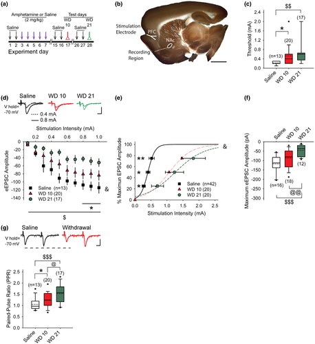
Prior experiments in mice and rats have demonstrated that repeated psychostimulants modify glutamatergic activity in the dorsal and ventral striatum (Bamford et al., 2008; McFarland et al., 2003; Pierce & Kalivas, 1997; Wang et al., 2013). To determine if withdrawal from repeated amphetamine can modify corticoaccumbal glutamatergic activity, we placed bipolar electrodes in the dorsal PFC and used single cortical pulses to elicit eEPSCs in SPNs within the NAc core. For these experiments, cortical stimulation was provided every 30 s and the stimulation intensity was progressively increased until an eEPSC was detected in the SPN. Compared to SPNs from saline-treated mice (0.22 ± 0.02 mA, n = 13 cells from five mice), the stimulation intensity required to evoke an eEPSC amplitude above 10 pA was 144% higher in cells from amphetamine-treated mice on WD 10 (0.53 ± 0.1 mA; n = 20 cells from six mice; t(31) = 2.48, p = 0.019, two-tailed Student's t test) and was 203% higher in SPNs from amphetamine-treated mice on WD 21 (0.65 ± 0.13 mA; n = 17 cells from four mice; t(28) = 2.92, p = 0.006, two-tailed Student's t test; Figure 1c). While the threshold was 24% higher on WD 21 compared to WD 10, the difference was not significant (t(35) = 0.79, p = 0.4, two-tailed Student's t test). The eEPSC threshold was similar in saline-treated mice sacrificed on WD 10 (0.22 ± 0.02 mA; n = 6 cells) and WD 21 (0.21 ± 0.03 mA; n = 7 cells) and the results were pooled together.
Cortical stimulation intensities above the firing threshold evoked larger eEPSC amplitudes in cells from saline-treated mice, compared to SPNs on WD 10 and on WD 21 (F(18, 333) = 1.98, p = 0.010; n = 50; 2-way ANOVA for interaction between stimulation intensity and treatment; Figure 1d). Additionally, a greater stimulation current was required to achieve a peak eEPSC amplitude following repeated amphetamine; the stimulation intensity (mA) required to obtain 25%, 50%, and 75% of maximum eEPSC amplitude (pA) in SPNs from saline-treated mice (0.27 ± 0.05 mA, 0.41 ± 0.06 mA, 0.52 ± 0.08 mA, respectively; n = 13 cells from five mice) was much lower than in cells from amphetamine-treated mice tested on WD 10 (0.71 ± 0.29 mA, 1.24 ± 0.49 mA, 1.82 ± 0.68 mA, respectively; n = 20 cells from nine mice) and on WD 21 (0.68 ± 0.09 mA, 1.07 ± 0.15 mA, 1.47 ± 0.24 mA, respectively; n = 19 cells from 10 mice; F(4, 98) = 3.48, p = 0.010, two-way ANOVA for interaction between stimulation intensity and treatment; Figure 1e). The stimulation intensity required to obtain 25%, 50%, and 75% of maximum eEPSC amplitude in SPNs from saline-treated mice sacrificed on WD 10 (0.25 ± 0.09 mA, 0.38 ± 0.1 mA, 0.5 ± 0.15 mA, respectively; n = 6 cells) was similar to saline-treated mice sacrificed on WD 21 (0.29 ± 0.06 mA, 0.44 ± 0.08 mA, 0.54 ± 0.09 mA, respectively; n = 7 cells), so the results were pooled together.
To assess synaptic activity, we measured the maximum achievable peak eEPSC amplitude in response to a single cortical stimulus. The average maximum peak eEPSC amplitude in SPNs from saline-treated mice (−118 ± 13 pA; n = 13 cells from four mice) was higher than the peak amplitudes obtained in SPNs from amphetamine-treated mice on WD 10 (−86 ± 12 pA; n = 19 cells from six mice), but the results were not significant (t(30) = 1.79, p = 0.080, two-tailed Student's t test; Figure 1f). The maximum peak eEPSC amplitude in saline-treated mice sacrificed on WD 10 (−119.34 ± 19; n = 6 cells) was similar to WD 21 (−116 ± 19; n = 7 cells), so the results were pooled together. The average peak eEPSC amplitude obtained in SPNs from amphetamine-treated mice on WD 21 (−46 ± 7 pA; n = 16 cells from four mice) was significantly lower when compared to saline (t(27) = 5.14, p < 0.001, two-tailed Student's t test) or WD 10 (t(33) = 2.83, p = 0.008, two-tailed Student's t test). Therefore, higher stimulation intensities were required to evoke and optimize EPSCs in SPNs from the NAc core during drug withdrawal, suggesting that a history of amphetamine exposure can cause a depression in corticoaccumbal activity.
Next, we used paired cortical pulses to determine if amphetamine can modify presynaptic corticoaccumbal activity (Mennerick & Zorumski, 1995). We measured the PPR (amplitude of the second eEPSC/amplitude of the first eEPSC) in response to cortical stimulation with 50 ms paired pulses applied every 30 s. Paired pulse depression can represent a Ca2+-dependent or Ca2+-independent reduction in presynaptic release and/or a postsynaptic release-dependent depression which may result from the unavailability of postsynaptic receptors (Kirischuk, Clements, & Grantyn, 2002).
Compared to SPNs from saline-treated mice (1.05 ± 0.07; n = 16 cells from seven mice), the PPR was 18% higher in cells from amphetamine-treated mice on WD 10 (1.25 ± 0.08; n = 18 cells from seven mice; t(32) = 2.2, p = 0.030, two-tailed Student's t test) and 45% higher in cells from amphetamine-treated mice on WD 21 (1.53 ± 0.12; n = 12 cells from seven mice; t(26) = 3.68, p < 0.001, two-tailed Student's t test; Figure 1g). The PPR was 22% higher on WD 21 compared to WD 10 (t(28) = 2.17, p = 0.023, two-tailed Student's t test). The PPR was similar in saline-treated mice sacrificed on WD 10 (1.07 ± 0.13; n = 7 cells) and WD 21 (1.04 ± 0.09; n = 9 cells) and the results were pooled together. Thus, the data suggested that repeated use of amphetamine in mice can promote an enduring and progressive synaptic depression along the corticoaccumbal pathway. This corticoaccumbal depression was characterized by a reduction in postsynaptic responsiveness to single and paired stimuli, as well as an increase in the PPR.
3.2 An amphetamine challenge in withdrawal reverses corticoaccumbal depression through D1-type dopamine receptors
Since reinstatement of drug-seeking behaviors can be generated by changes in glutamatergic activity within the NAc core (Baker et al., 2003; Bell, Duffy, & Kalivas, 2000; McFarland et al., 2003; Pierce, Bell, Duffy, & Kalivas, 1996) as well as within the dorsal striatum (Bamford et al., 2008; Storey et al., 2016; Wang et al., 2013), we tested whether an amphetamine challenge in withdrawal would reverse or block the synaptic depression observed in the NAc core. Mice were treated with saline or amphetamine for 5 days (Figure 1a). On WD 10, slices were prepared and 50 ms paired pulses were applied to the dorsal PFC every 30 s. In SPNs from saline-treated mice, amphetamine (10 µM) in vitro—which can elevate striatal dopamine concentrations to ~3 µM (Bamford, Zhang, et al., 2004b) via reversal of the dopamine transporter (Sulzer, 2011)—decreased the average amplitude of the first eEPSC of the pair by 16 ± 11% (−128 ± 26 pA in vehicle vs. −104 ± 23 pA with amphetamine; n = 11 cells from five mice; t(10) = 2.57, p = 0.027, paired t test) and increased the average PPR by 19 ± 6% (1.11 ± 0.09 in vehicle vs. 1.3 ± 0.13 in amphetamine; t(10) = 3.07, p = 0.012, paired t test; Figure 2a), indicating that novel exposure to amphetamine in vitro can reduce corticoaccumbal excitation. A higher concentration of amphetamine (20 µM) also reduced the probability of release from presynaptic boutons as the amplitude of the first eEPSC decreased (−43 ± 5%; −324 ± 76 pA in vehicle vs. −187 ± 42 pA with amphetamine; n = 5 cells from four mice; t(4) = 3.73, p = 0.020, paired t test) while the PPR increased (0.92 ± 0.05 in vehicle vs. 1.14 ± 0.08 in amphetamine; t(4) = 3.62, p = 0.022, paired t test; Figure 2b).
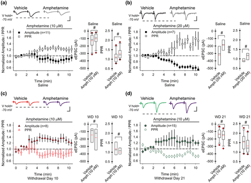
Next, we tested whether drug withdrawal would modify the inhibitory response of an amphetamine challenge. In SPNs from amphetamine-treated mice on WD 10, bath-applied amphetamine (10 µM) increased the amplitude of the first eEPSC by 30 ± 9% (−145 ± 41 pA in vehicle vs. −181 ± 49 pA with amphetamine; n = 8 cells from four mice; t(7) = 2.56, p = 0.037, paired t test) and reduced the PPR by 14 ± 6% (1.17 ± 0.12 in vehicle vs. 0.97 ± 0.1 with amphetamine; t(7) = 2.80, p = 0.026, paired t test; Figure 2c). On WD 21, amphetamine in vitro also increased the amplitude of the first eEPSC (22 ± 14%; −137 ± 20 pA in vehicle vs. −154 ± 22 pA with amphetamine; n = 12 cells from seven mice; t(11) = 2.92, p = 0.014, paired t test) and reduced the PPR (−12 ± 4%; 1.53 ± 0.12 in vehicle vs. 1.33 ± 0.12 with amphetamine; t(11) = 2.99, p = 0.012, paired t test; Figure 2d). Thus, in novice mice, acute amphetamine, with concurrent cortical stimulation, promoted a presynaptic depression within the NAc core that was characterized by a reduction in the eEPSC amplitude and an increase in the PPR. This depression was maintained in withdrawal and could be reversed by an amphetamine challenge.
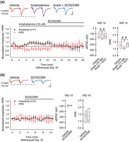
In a separate set of experiments, we determined if the synaptic potentiation that follows an amphetamine challenge in withdrawal might be generated through D1Rs, as occurs in the dorsal striatum (Bamford et al., 2008; Wang et al., 2013). In amphetamine-treated mice on WD 10, amphetamine (10 µM) in vitro increased the eEPSC amplitude (16 ± 6%; −144 ± 22 pA in vehicle vs. −163 ± 33 pA with amphetamine; n = 5 cells from three mice; t(4) = 4.76, p = 0.009, paired t test) and reduced the PPR (−12 ± 5%; 1.27 ± 0.11 in vehicle vs. 1.1 ± 0.18 with amphetamine; t(4) = 3.38, p = 0.028, paired t test; Figure 3a). The excitatory effect of an amphetamine challenge on corticoaccumbal activity was dependent on D1Rs, since the D1R antagonist SCH23390 (10 µM) blocked the increase in eEPSC amplitude (0.2 ± 13%, compared to vehicle; −141 ± 32 pA for amphetamine with SCH23390; t(4) = 0.15, p = 0.886, paired t test compared to vehicle) and prevented the decrease in the PPR (3 ± 9%, compared to vehicle; 1.28 ± 0.1 for amphetamine with SCH23390; t(4) = 0.13, p = 0.899, paired t test compared to vehicle). The D1R antagonist alone had no effect on the eEPSC amplitude (−0.7 ± 9%; −93 ± 10 pA in vehicle vs. −89 ± 15 pA in SCH23390; n = 5 cells from three mice; t(4) = 0.46, p = 0.670, paired t test) or the PPR (−2 ± 7%; 1.35 ± 0.2 in vehicle vs. 1.34 ± 0.2 in SCH23390; t(4) = 0.09, p = 0.934, paired t test) on WD 10 (Figure 3b), suggesting that the synaptic potentiation which follows a drug challenge in withdrawal is mediated through D1Rs.
3.3 Synaptic depression in withdrawal is dependent on cortical stimulation
To determine if this synaptic plasticity within the NAc core is dependent on cortical stimulation, we measured spontaneous mEPSCs in SPNs in the absence of stimulation. Alterations in the frequency of mEPSCs can also help to determine if changes are generated presynaptically while a change in the mEPSC amplitude may signify an alteration in postsynaptic responsiveness (Van der Kloot, 1991). The Na+ channel antagonist tetrodotoxin (TTX; 1 µM) was used to block spontaneous cortically derived action potentials and isolate presynaptic activity. Compared to mEPSCs measured in SPNs from saline-treated mice (1.13 ± 0.25; n = 13 cells from four mice), the frequency of mEPSCs was enhanced on WD 10 (3.14 ± 0.7; n = 7 cells from four mice; t(17) = 3.40, p = 0.003, two-tailed Student's t test), and there was a significant increase in the frequency–amplitude distribution (p < 0.05, Kolmogorov–Smirnov test; Figure 4a). Similarly, the average mEPSC amplitude was increased on WD 10 (10.8 ± 1 for saline vs.14.6 ± 1.5 for WD 10; t(17) = 2.22, p = 0.040, two-tailed Student's t test), and there was a significant change in the cumulative normalized amplitude–frequency distribution (p < 0.05, Kolmogorov–Smirnov test; Figure 4b). Thus, under non-stimulated conditions, the results are consistent with an increase in pre-and postsynaptic responsiveness following repeated amphetamine (Van der Kloot, 1991).
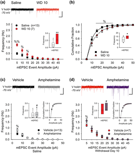
To test the effect of an amphetamine challenge under non-stimulated conditions, mEPSCs (with TTX) were observed in SPNs before and following amphetamine (10 µM) in vitro. In SPNs from saline-treated mice, amphetamine in vitro enhanced the frequency of mEPSC by 42 ± 13% (1.05 ± 0.26 Hz in vehicle vs. 1.3 ± 0.27 Hz in amphetamine; n = 13 cells from five mice; t(12) = 2.41, p = 0.034, paired t test) by selectively increasing high-frequency, low-amplitude inward currents (Figure 4c). Amphetamine had no effect on the average mEPSC amplitude (10.8 ± 0.9 pA in vehicle vs. 10.3 ± 0.6 pA in amphetamine; t(12) = 0.98, p = 0.347, paired t test) or the amplitude distribution (p > 0.1, Kolmogorov–Smirnov test). On WD 10, bath-applied amphetamine also increased the frequency of mEPSCs (63 ± 21%; 2.9 ± 0.69 Hz in vehicle vs. 3.8 ± 0.72 Hz in amphetamine; n = 7 cells from four mice; t(6) = 3.16, p = 0.019, paired t test), primarily by boosting high-frequency, low-amplitude currents (Figure 4d). An amphetamine challenge in withdrawal did not change the average mEPSC amplitude (14.6 ± 1.6 pA in vehicle vs. 13.5 ± 0.6 pA in amphetamine; t(6) = 0.88, p = 0.501, paired t test) or the amplitude distribution (p > 0.1, Kolmogorov–Smirnov test). Thus, under non-stimulated conditions, acute amphetamine had a small excitatory effect on presynaptic corticoaccumbal activity, with and without a history of psychostimulant exposure, and indicated that synaptic depression in drug withdrawal and the paradoxical potentiation that follows a drug challenge are dependent upon cortical stimulation.
3.4 Presynaptic depression and potentiation are frequency-dependent and modify a select subpopulation of presynaptic boutons in the NAc core
The data above indicate that synaptic depression was dependent on SPN activation. To investigate further, we directly measured the frequency dependence of presynaptic release by optical analysis using the endocytic tracer FM1-43 combined with multiphoton confocal microscopy (Bamford, Zhang, et al., 2004b; Wong, Sulzer, & Bamford, 2011). In these experiments, mice received saline or amphetamine (2 mg/kg/d; i.p.) for five consecutive days and were challenged with amphetamine on WD 10 and WD 21. To determine if presynaptic plasticity was long-lasting, optical recordings were performed on WD 50 (Figure 5a).
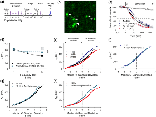
Stimulation of the dorsal PFC resulted in endocytosis of FM1-43 dye into recycling presynaptic vesicles in the NAc core, revealing fluorescent puncta that were distinctive of corticoaccumbal afferents (Bamford, Zhang, et al., 2004b; Wong et al., 2011) (Figure 5b). Following dye loading, cortical stimulation with a train of pulses produced exocytosis of FM1-43 dye from presynaptic boutons, characteristic of synaptic vesicle fusion (Bamford, Robinson, et al., 2004a). As FM1-43 destaining follows first-order kinetics (Joshi et al., 2009), corticostriatal release was characterized by the half-time of destaining (t1/2), defined as the time required for fluorescence to decay to half of its initial value (Figure 5c). In slices from saline-treated mice, an increase in cortical stimulation frequency at physiologically relative rates (Charpier, Mahon, & Deniau, 1999; Cowan & Wilson, 1994; Fellous, Rudolph, Destexhe, & Sejnowski, 2003; Kasper, Larkman, Lubke, & Blakemore, 1994; Stern, Kincaid, & Wilson, 1997) from 1 to 20 Hz produced a corresponding increase in FM1-43 release from the majority of boutons, which was reflected by a decrease in the average half-time of FM1-43 destaining (t1/2 = 305 ± 14 s at 1 Hz, n = 104 puncta; t1/2 = 246 ± 10 s at 10 Hz, n = 193 puncta; and t1/2 = 208 ± 6 s at 20 Hz, 330 puncta; F(2, 564) = 26.00, p = 0.001, repeated measures (rm)-ANOVA; Figure 5c,d). When amphetamine was bath-applied, an increase in the stimulation frequency also accelerated release (t1/2 = 309 ± 14 s at 1 Hz, n = 103 puncta; t1/2 = 269 ± 13 s at 10 Hz, n = 97 puncta; and t1/2 = 264 ± 10 s at 20 Hz, n = 193 puncta; F(2, 387) = 4.00, p = 0.020, rm-ANOVA) but to a lesser extent than in vehicle (F(2, 951) = 4.03, p = 0.029, 2-way ANOVA for interaction between frequency and amphetamine; Figure 5d). Compared to vehicle, amphetamine in vitro caused a progressive, frequency-dependent reduction in exocytosis so that presynaptic inhibition by amphetamine was observed at 20 Hz (p < 0.001, Mann–Whitney).
Prior investigations have shown the cortical boutons in the striatum have variable release kinetics (Bamford, Zhang, et al., 2004b). We used normal probability plots to determine if amphetamine differentially modifies a subset of corticoaccumbal boutons. When the half-time of FM-143 release is compared to the median ± standard deviation, a normally distributed population is reflected by a straight line (Bamford, Zhang, et al., 2004b). Increasing cortical stimulation frequencies from 1 to 20 Hz produced a corresponding increase in FM-143 release with a relatively greater effect on slower-releasing boutons (Figure 5e). Bath application of amphetamine had little effect at 1 and 10 Hz cortical stimulation. At 20 Hz, amphetamine inhibited corticoaccumbal release and produced a low-pass frequency filter with filtering applied specifically to a subset of boutons with a low probability of release (e.g., those with the highest t1/2; Figure 5f–h).
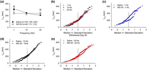
Next, we compared the release of FM-143 in brain slices from saline-treated mice with that obtained from amphetamine-treated mice on WD 50. In slices from amphetamine-treated mice on WD 50, an increase in cortical stimulation frequency produced little change in corticoaccumbal release (Figure 6a,b), suggesting that drug exposure caused a progressive frequency-dependent depression in the fractional release of FM1-43. Compared to saline-treated controls, corticoaccumbal release on WD 50 was augmented at 1 Hz (t1/2 = 247 ± 20 s, n = 46 puncta; p = 0.01, Mann–Whitney), unchanged at 10 Hz (t1/2 = 227 ± 13 s, n = 111 puncta) and depressed at 20 Hz (t1/2 = 242 ±12 s, n = 112 puncta; p = 0.01, Mann–Whitney; Figure 6a and c–e). Thus, under conditions of drug withdrawal, corticoaccumbal activity was enhanced during trains of low-frequency stimulation and was depressed at higher cortical frequencies. Evaluation of the kinetics of individual corticoaccumbal boutons showed that withdrawal from amphetamine increased release from boutons with intermediate kinetics at 1 Hz and from faster-releasing boutons at 10 Hz (Figure 6c,d). At 20 Hz, release was diminished only in those boutons with the lowest release probability (Figure 6e).
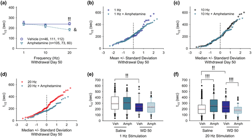
Next, we determined how an amphetamine challenge would modify frequency-dependent plasticity in corticoaccumbal release during withdrawal. Bath application of amphetamine in corticoaccumbal slices from amphetamine-treated mice on WD 50 caused a frequency-dependent increase in corticoaccumbal release (t1/2 = 237 ± 11 s at 1 Hz, n = 105 puncta; t1/2 = 219 ± 15 s at 10 Hz, n = 73 puncta; and t1/2 = 184 ± 12 s at 20 Hz, n = 60 puncta; F(2, 235) = 5.00, p = 0.010, rm-ANOVA; Figure 7a). Compared to untreated slices on WD 50, amphetamine in vitro produced a frequency-dependent increase in release (F(2, 951) = 4.00, p = 0.019, two-way ANOVA for interaction between frequency and treatment). At lower cortical stimulation frequencies of 1 and 10 Hz, amphetamine in vitro had little effect on presynaptic release in amphetamine-treated mice (Figure 7a–c). At higher stimulation frequencies (20 Hz), amphetamine boosted presynaptic release by assisting corticoaccumbal boutons with a low probability of release (Figure 7d). Therefore, the repeated use of amphetamine produced frequency-dependent plasticity in corticoaccumbal excitability. During withdrawal, presynaptic activity was enhanced at low frequencies (1 Hz) and depressed at high frequencies (20 Hz; Figure 7e,f). An amphetamine challenge in withdrawal had no effect on low-frequency potentiation but boosted corticoaccumbal activity at high frequencies to normalize synaptic function.
3.5 Synaptic potentiation correlates with locomotor sensitization
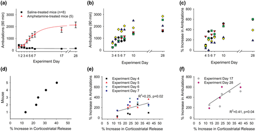
Evidence suggests that amphetamine-induced adaptations in glutamatergic signaling may underlie locomotor sensitization (Cornish, Duffy, & Kalivas, 1999; Ghasemzadeh, Permenter, Lake, Worley, & Kalivas, 2003; Knackstedt & Kalivas, 2009; Li et al., 1999; Storey et al., 2016; Wang et al., 2013). To investigate further, we treated mice with saline (n = 8) or amphetamine (n = 5) for five consecutive days and monitored their locomotor activity. Locomotor sensitization, defined as a progressive increase in locomotion with each treatment, was tested by an amphetamine challenge on challenge day 1 (corresponding to WD 10) and challenge day 2 (corresponding to WD 21; Figure 5a). Mice were sacrificed on experiment day 57 (WD 50) for optical measurements of corticoaccumbal release. We then compared locomotor ambulations in individual mice with the change in FM1-43 release following an amphetamine challenge in vitro.
Amphetamine in vivo produced locomotor sensitization with increasing ambulations following each amphetamine injection (Figure 8a). Locomotor ambulations varied widely between animals (Figure 8b) and generally increased following each treatment (292 ± 36%; range, 3%–603%; Figure 8c). On WD 50, the mice were sacrificed and we measured FM1-43 destaining in response to 20 Hz cortical stimulation. Amphetamine (10 µM) in vitro depressed FM1-43 release in saline-treated mice. In slices from amphetamine-treated mice, an amphetamine challenge in vitro increased FM1-43 release. The percent increase in corticoaccumbal release varied widely between amphetamine-treated mice (28.2 ± 3.3%; range, 22.5%–40.3%; Figure 8d). A linear regression comparison between the percent increase in corticostriatal release and the percent change in ambulations for each mouse revealed a significant correlation during the initial 5 days of treatment (R2 = 0.25, F(1, 18) = 6.08, p = 0.020, Figure 8e) and when challenged with amphetamine on challenge day 1 and challenge day 2 (R2 = 0.41, F(1, 8) = 5.72, p = 0.040; Figure 8f), indicating a correlation between the degree of presynaptic potentiation and sensitized locomotor responses.
4 DISCUSSION
Excitatory glutamatergic signals from the PFC and modulatory dopamine inputs from the VTA converge on SPNs in the NAc core, which initiate and direct signaling through “direct” and “indirect” pathways to generate go/no-go actions through striatal-thalamic-cortical pathways (Albin et al., 1989; Parent & Hazrati, 1995). While small changes in the availability of glutamate and dopamine are putatively required to establish goal-directed behaviors, motor learning, and habits through functional and structural alterations in striatal circuitry, large changes in these neurotransmitters appear to underlie many neuropsychological disorders, including drug dependence (Bamford et al., 2018; Kalivas & Volkow, 2005; Kalivas et al., 2003). Our data show that repeated psychostimulants, when delivered in a paradigm that releases dopamine (Bamford, Zhang, et al., 2004b), while allowing simultaneous measurements of behavior, promotes long-lasting, but reversible changes along the corticoaccumbal pathway (Figure 9).
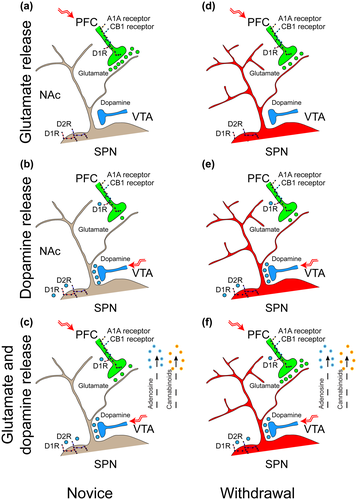
We combined presynaptic optical studies with postsynaptic electrophysiological recordings in the NAc core of male mice treated with repeated amphetamine. The results of these experiments show that under normal conditions, dopamine released by amphetamine promotes synaptic filtering by causing a frequency-dependent depression in a subset of corticoaccumbal boutons. The repeated use of amphetamine promoted a long-lasting and progressive depression in corticoaccumbal activity. This corticoaccumbal depression was characterized by activity- and frequency-dependent plasticity during withdrawal that was partially relieved by a drug challenge. The increase in glutamate release from excitatory corticoaccumbal boutons in response to the drug challenge was generated though D1Rs and corresponded with the degree of locomotor sensitization in individual animals. Thus, the boost in glutamate release during drug reinstatement promotes allostasis (Ahmed & Koob, 2005), an attempt to return corticoaccumbal activity to a more stable and normalized state.
These experiments were performed in young adolescent mice and at time points that corresponded to our earlier work in the dorsal and ventral striatum (Bamford et al., 2008; Beutler et al., 2011; Storey et al., 2016; Wang et al., 2013). Limitations of the study include the use of young mice rather than older rats that have been used extensively to analyse the effects of contingent psychostimulant use but see (Storey et al., 2016). The electrophysiology and optical data were collected in the slice preparation and may differ from plasticity that occurs in vivo, therefore presenting an avenue for future research. Another limitation is our exclusive use of male mice. While the data are not confounded by any possible effects of ovulation, sex-based comparisons were not available.
4.1 Dopamine promotes synaptic filtering
Our experiments in saline-treated control mice showed that novel exposure to amphetamine in vitro modulates corticoaccumbal activity. In the absence of stimulation, amphetamine provoked a small excitatory response in cortical boutons with a low probability of release, characterized by an increase in the frequency but not the amplitude of mEPSCs. This combination of changes in mEPSCs suggests that dopamine has a small excitatory effect on presynaptic boutons (Van der Kloot, 1991) in the NAc core and is consistent with the presence and function of D1Rs on corticoaccumbal axons (Dumartin, Doudnikoff, Gonon, & Bloch, 2007; Wang et al., 2012). The responses to amphetamine under non-stimulated conditions were similar in saline- and amphetamine-treated mice and suggested that any plasticity provoked by drug exposure had no effect on excitation provided by presynaptic D1Rs.
When SPNs from saline-treated mice became activated by stimulation of the PFC, amphetamine in vitro reduced corticoaccumbal activity. Corticoaccumbal depression was present at 20 Hz, but not at lower stimulation frequencies of 1 and 10 Hz. This frequency-dependent inhibition suggests the presence of presynaptic inhibition by the neuromodulators, adenosine and endocannabinoids. These neuromodulators are putatively released by SPNs in response to activation and provide retrograde inhibition of glutamate release (Wang et al., 2012). Frequency-dependent filtering of a subset of presynaptic excitatory afferents by dopamine also occurs in the dorsal striatum and may act as a mechanism to focus attention on meaningful information by boosting strong signals while inhibiting the weak (Bamford, Zhang, et al., 2004b; Dani & Zhou, 2004).
4.2 Stimulation-dependent synaptic depression increases during drug withdrawal and is generated though pre- and postsynaptic plasticity
We determined if the depression in corticoaccumbal activity provoked by amphetamine in novice, saline-treated mice might persist in mice exposed to several amphetamine treatments. The repeated use of amphetamine in mice promoted a synaptic depression along the corticoaccumbal pathway that lasted over 50 days. The depression was characterized by a reduction in postsynaptic responsiveness to single and paired (20 Hz) stimuli. Higher cortical stimulation intensities were required to evoke and optimize eEPSCs, and the maximal amplitude of eEPSCs progressively declined with the duration of withdrawal.
We found that the synaptic depression observed in amphetamine withdrawal was dependent on excitation of the PFC. Under non-stimulated conditions, SPNs from amphetamine-treated mice manifest an elevated frequency and a higher amplitude of mEPSCs compared to SPNs from saline-treated mice. This increase in frequency and amplitude suggests enhanced presynaptic release and postsynaptic responsiveness following repeated amphetamine (Van der Kloot, 1991). There was additional evidence of postsynaptic plasticity. Recordings in SPNs from amphetamine-treated mice demonstrated increased cellular capacitance and time constant, while the input resistance was unchanged. These findings are consistent with the increase in the dendritic length and spine density (Isokawa, 1997) of SPNs in the NAc core that follows repeated amphetamine (Robinson & Kolb, 1997). Therefore, changes in the membrane properties of SPNs likely represent a postsynaptic alteration in consequence of repeated exposure to the psychostimulant. Structural changes in the dendritic spines of SPNs in the NAc can persist for months after the last drug treatment (Li, Kolb, & Robinson, 2003) and may underlie or compensate for long-lasting alterations in glutamatergic and/or dopaminergic synaptic transmission (Garcia, Neely, & Deutch, 2010; Villalba & Smith, 2013).
There was also evidence of presynaptic depression, manifesting in an increase in the PPR (Mennerick & Zorumski, 1995). Although paired-pulse facilitation can also be generated by changes in postsynaptic receptors (Kirischuk et al., 2002), the results are similar to the presynaptic depression found in the dorsal striatum following contingent and non-contingent use of methamphetamine (Bamford et al., 2008) and amphetamine (Storey et al., 2016; Wang et al., 2013). The observed depression was not due to current spread, because (a) eEPSCs in SPNs required intact corticostriatal axons, (b) the resting membrane potential of SPNs was unchanged by cortical stimulation, and (c) similar methods produced opposing responses in saline-treated controls (Wong et al., 2015).
Neurotransmitter release from subpopulations of presynaptic boutons is reliant on neuronal firing frequencies (Bamford, Robinson, et al., 2004a; Bamford, Zhang, et al., 2004b; Sulzer, 2011), and this dependence is altered in disease (Bamford, Robinson, et al., 2004a; Joshi et al., 2009). Our optical recordings of FM1-43 release from presynaptic boutons in the NAc core validated the frequency-dependent plasticity found in post-synaptic recordings. By providing trains of cortical stimuli at physiologically relevant frequencies (Charpier et al., 1999; Cowan & Wilson, 1994; Fellous et al., 2003; Kasper et al., 1994; Stern et al., 1997), we measured the kinetics of vesicle fusion at presynaptic boutons in the NAc core. Compared to slices from saline-treated mice, FM1-43 release in slices from amphetamine-treated mice was elevated 1 Hz and became depressed at 20 Hz. Therefore, presynaptic corticoaccumbal activity in withdrawal was enhanced at no or low cortical stimulation frequencies and was depressed at higher cortical frequencies.
Different stimulation frequencies also differentially modulated certain subsets of corticoaccumbal boutons. In saline-treated controls, amphetamine had little effect at lower frequencies but inhibited boutons with a low probability of release at high frequencies. In amphetamine withdrawal, low-frequency stimulation enhanced exocytosis from boutons with high probability of release while higher frequencies depressed boutons with a low probability of release. These findings suggest that the synaptic filtering provided by dopamine in novice mice may persist during drug withdrawal.
4.3 Amphetamine reinstatement generates a frequency-dependent potentiation in corticoaccumbal activity
Synaptic potentiation likely plays a key role in the motivational circuitry underlying learning and dependence since supra-physiological glutamatergic drive promotes compulsive drug-seeking in addicts by decreasing the value of natural rewards (Kalivas & Volkow, 2005). In slices from saline-treated mice, acute amphetamine decreased the amplitude of the first eEPSC (of the pair) and increased in PPR, consistent with a decrease in glutamate release from corticoaccumbal boutons. Conversely, in amphetamine-treated mice, an amphetamine challenge in withdrawal increased the EPSC amplitude and decreased the PPR, suggesting that dopamine promotes synaptic filtering in novice mice but potentiates glutamate release in withdrawal.
The optical recordings confirmed a primary presynaptic mechanism of synaptic potentiation due to a drug challenge in withdrawal. In saline-treated mice, an amphetamine challenge in vitro produced a frequency-dependent decrease in release from boutons with a low probability of release. In drug withdrawal an amphetamine challenge produced a frequency-dependent boost in exocytosis from this same population of presynaptic boutons.
Synaptic potentiation in response to a drug challenge was not dependent on dopamine release kinetics (Bamford et al., 2008) and could not depend on changes in dopamine neuronal firing since it was measured in the striatal slice from which dopamine cell bodies were absent. The boost in presynaptic activity during a drug challenge appeared to be mediated though presynaptic mechanisms and was unchanged over the length of drug withdrawal. In comparison, the synaptic depression appears to be generated through both pre-and postsynaptic plasticity and worsened with the length of drug withdrawal, suggesting that drug reinstatement which attempts to normalize the synapse can become less effective over time.
4.4 Synaptic potentiation correlates with the degree of locomotor sensitization
Prior experiments in mice and rats have demonstrated that repeated psychostimulants modify glutamatergic activity in the dorsal and ventral striatum (Bamford et al., 2008; McFarland et al., 2003; Pierce & Kalivas, 1997; Wang et al., 2013). This plasticity modifies behavioral responses to subsequent drug treatments and appears to encode drug-seeking behavior (McFarland et al., 2003; Storey et al., 2016).
We tested whether the reversal of corticoaccumbal synaptic depression by an amphetamine challenge in withdrawal might encode sensitized locomotor responses. Comparisons between the percent increase in corticostriatal release and the percent change in ambulations for each mouse revealed a significant correlation between behavior and excitatory synaptic function. Therefore, drug reinstatement in withdrawal which moves synaptic activity from a depressed level to a potentiated state may be one mechanism involved in the initiation and maintenance of behavioral sensitization.
4.5 Mechanisms of synaptic filtering
SPNs exhibit a low tonic activity (~1 Hz) in vivo due to presynaptic inhibition of cortical inputs by tonic eCBs (Wang et al., 2012) and the prominent potassium currents found in mature SPNs (Wilson & Kawaguchi, 1996). Evidence indicates that both glutamate and dopamine need to be present simultaneously for the SPN to fire (Bamford et al., 2018). Our data suggests that under quiescent conditions when there is little cortical activity, dopamine can provoke a small excitatory response in SPNs which would bring the cells closer toward threshold. Once the PFC becomes active, dopamine provides a frequency-dependent suppression of corticoaccumbal boutons with a low probability of release. This filtering of cortical information by dopamine may then act to suppress extraneous information and provide a mechanism whereby the animal might focus attention on meaningful and relative information that leads to rewarding behaviors (Bamford & Bamford, 2019; Bamford, Zhang, et al., 2004b; Bamford et al., 2018; Wong et al., 2015). A similar filtering mechanism by dopamine has been described in the dorsal striatum (Bamford, Zhang, et al., 2004b) and has been more fully described within the NAc core (Wang et al., 2012). Published work demonstrates that synaptic filtering in the NAc core is mediated through presynaptic dopamine receptors as well as retrograde presynaptic inhibition that is generated via the stimulation-dependent release of adenosine and endocannabinoids (Wang et al., 2012). Adenosine receptors are present on D1- and D2-type (D2R) receptor expressing SPNs (Dunwiddie & Masino, 2001; Harvey & Lacey, 1997), while cannabinoid CB1 receptors may primarily regulate D2R-expressing SPNs (Grueter, Brasnjo, & Malenka, 2010; Wang et al., 2012). Adenosine and endocannabinoids are considered necessary for the establishment of reward and attention (Harvey & Lacey, 1996; Kalivas & Volkow, 2005; Wang et al., 2012). When dopamine and glutamate are released simultaneously, adenosine and endocannabinoids appear to focus excitatory signals toward D1-receptor expressing SPNs that drive behaviors and movements via the “direct” striatal pathway (Wang et al., 2012). This mechanism may persist in drug withdrawal, but excitation of the direct pathway would be significantly amplified by the potentiation of glutamate release that occurs in response to a drug challenge.
4.6 Potential mechanisms of synaptic plasticity
Evidence of synaptic depression during psychostimulant withdrawal has been found in the dorsal striatum (Bamford et al., 2008) and NAc (Bell et al., 2000; McFarland et al., 2003; Pierce et al., 1996), while potentiation at glutamatergic synapses during drug reinstatement has been found in several brain areas (Gass & Olive, 2008), including the NAc (Bell et al., 2000; McFarland et al., 2003; Pierce et al., 1996; Reid & Berger, 1996) and the dorsal striatum (Bamford et al., 2008; McKee & Meshul, 2005; Storey et al., 2016; Wang et al., 2013). The precise mechanism(s) underlying these changes remains unclear but may develop in part through plasticity generated in SPNs or striatal interneurons.
Infusion of AMPA into the NAc core induces reinstatement behavior (Ping, Xi, Prasad, Wang, & Kruzich, 2008) while PFC lesions, or blockade/inactivation of AMPA, NMDA, or metabotropic glutamate receptors in this region attenuate sensitization and cue-induced reinstatement (Backstrom & Hyytia, 2006; Beutler et al., 2011; Ghasemzadeh et al., 2003; Li et al., 1999). In the dorsal striatum, synaptic potentiation and locomotor sensitization can be blocked by D1R antagonists in withdrawal (Bamford et al., 2008; Kuribara, 1995; Wang et al., 2013).
Cholinergic interneurons have also been implicated in the mechanism of amphetamine-induced locomotor sensitization and drug seeking behavior. Experiments have revealed that both corticostriatal depression in withdrawal and subsequent potentiation by drug reinstatement rely on tonic excitation and inhibition by acetylcholine at nicotinic and muscarinic receptors located on corticostriatal axons (Bamford et al., 2008). Synaptic depression (Bamford et al., 2008) and locomotor sensitization (Kelly, Low, Rubinstein, & Phillips, 2008) are dependent on D2Rs, which act to reduce acetylcholine efflux (Bamford et al., 2008; Bickerdike & Abercrombie, 1997). Potentiation and locomotor sensitization are contingent on D1Rs (Bamford et al., 2008; Kuribara, 1995), which act to boost acetylcholine release (Bamford et al., 2008; Bickerdike & Abercrombie, 1997). These shifts in acetylcholine availability can then modulate corticostriatal activity by interacting with nicotinic receptors on corticostriatal axons (Bamford et al., 2008; Wang et al., 2013). In this way, the increase in glutamate might boost locomotor activity by activating SPNs that have a diminished capacity to respond (Gass & Olive, 2008) and promote an imbalance between striatal pathways by creating a net shift from a D2R-generated reduction motor activity along the “indirect” pathway to D1R-mediated excitation along the “direct” pathway (Bamford et al., 2018; Beutler et al., 2011; Wang et al., 2013).
Therapeutic approaches that target upstream interneurons might increase synaptic glutamate in withdrawal and facilitate extinction of drug-seeking behavior, while those that prevent potentiation would potentially act to disrupt the reinforcing effects of drugs (Gass & Olive, 2008). Since dopamine modifies corticoaccumbal activity by changing the kinetics of different subpopulations of excitatory boutons, our results may also explain how dopamine released by salient experiences might encode learned behaviors and movements and how disruption of corticoaccumbal filtering triggered by too much or too little dopamine would lead to habits and movement disorders like Parkinson's disease (Bamford & Cepeda, 2009; Cepeda et al., 2010).
DECLARATION OF TRANSPARENCY
The authors, reviewers, and editors affirm that in accordance to the policies set by the Journal of Neuroscience Research, this manuscript presents an accurate and transparent account of the study being reported and that all critical details describing the methods and results are present.
ACKNOWLEDGMENTS
The authors wish to thank Professor Michael S. Levine for his mentorship, support, and encouragement throughout the years.
CONFLICT OF INTEREST
The authors declare that they have no conflict of interest.
AUTHOR CONTRIBUTIONS
All authors had full access to all the data in the study and take responsibility for the integrity of the data and the accuracy of the data analysis. Conceptualization, N.S.B.; Methodology, N.S.B. and W.W.; Investigation, N.S.B. and W.W; Formal Analysis, N.S.B. and W.W; Resources: N.S.B.; Writing—Original Draft, N.S.B.; Writing—Review & Editing, N.S.B. and W.W; Visualization, N.S.B.; Supervision, N.S.B.; Funding Acquisition, N.S.B.


