Human papillomaviruses genotyping in plantar warts
Abstract
Plantar warts are caused by human papillomaviruses (HPVs) and have been associated with several HPV genotypes. However, there are few studies focused exclusively on plantar warts. In this work, we aim to identify the HPV genotypes of plantar warts and explore their relation to demographic and clinical characteristics of patients. A total of 72 patients diagnosed with plantar warts were recruited at the Laser unit at Podiatric Hospital, University of Barcelona, Spain. Inner hyperkeratosis laminar sections of warts were collected and DNA of samples were extracted. Amplification of a conserved region of the HPV L1 gene was performed with the SK-Polymerase chain reaction method. DNA amplicons were sequenced and HPV types identified. The most prevalent genotypes detected among the 105 analyzed plantar warts were HPV-57 (37.1%), HPV-27 (23.8%), HPV-1a (20.9%), HPV-2 (15.2%), and HPV-65 (2.8%). The majority of patients (78%) presented one single plantar wart, whereas multiple warts were detected in 22.2% of patients. One patient with multiple warts presented HPV types from two different genera, suggesting the spread of warts by self-inoculation as well as by de novo infection. No significant differences between the number of warts in toes, midfoot and heel were found. The most prevalent HPV types detected in all areas belonged to the alpha genus. This work provides new insight on plantar warts and their associated HPV genotypes, and evidences the usefulness and reliability of both the sample collection procedure and the PCR method used for HPV detection and typing. J. Med. Virol. 89:902–907, 2017. © 2016 Wiley Periodicals, Inc.
INTRODUCTION
Cutaneous warts are benign intraepidermal tumors of the skin with a variety of manifestations, including common warts (verrucae vulgaris), palmar and plantar warts (verrucae palmares et plantares), mosaic warts, flat warts (verrucae planae) and butcher's warts [Kirnbauer et al., 2004]. The most prevalent of these types are vulgar and plantar warts [Bruggink et al., 2012]. They are usually diagnosed by their clinical appearance [McCarthy, 1986] although definitive diagnosis depends upon histopathologic examination. Histologically, the lesions caused by the infection of keratinocytes are characterized by hyperkeratosis, parakeratosis, papillomatosis, and acanthosis [Cardoso and Calonje, 2011; Viennet et al., 2012]. These lesions are capable of resolving spontaneously by the action of the host immune system, or remain asymptomatic for long periods of time evolving ultimately to big tumors that persist for months or years [Yelverton, 2007; Androphy and Lowy, 2009].
Warts are caused by human papillomaviruses (HPVs) [Burns, 1992; Kirnbauer et al., 2004; Androphy and Lowy, 2009]. Currently, over 120 HPV types have been fully characterized and classified into five genera (alpha, beta, gamma, mu, and nu) and 16 species [Strauss et al., 1949; Bernard et al., 2010; Bruggink et al., 2012]. The most frequently HPV types associated with cutaneous warts are HPV-2, -3, -7, -10, -27, and -57 from the alpha genus, HPV-4, -60, and -65 from the gamma genus, and HPV-1, -63 from the mu genus [Bruggink et al., 2012; Al Bdour et al., 2013].
Among the cutaneous HPV types, HPV-2, -27, -57, and HPV-1 have been associated with the development of plantar warts [Sasagawa and Mitsuishi, 2012; Al Bdour et al., 2013]. However, there are few studies focused exclusively on plantar warts, and more studies are needed to yield insights regarding their associated HPV types.
In this work, we aim to identify the HPV genotypes present in a sample of Spanish patients with plantar warts, and to explore the relation between HPV types and demographic and clinical parameters.
MATERIALS AND METHODS
Study Design and Participants
A total of 72 patients diagnosed with plantar warts were recruited at the Laser unit at Podiatric Hospital, University of Barcelona, Spain.
Subjects were male or female patients, who were not under topical treatment for at least 3 months before the recruitment. Patients with neuropathy and/or vascular diseases were excluded from the study. Eligible patients agreed to participate and provided informed consent. The study was performed in accordance with the recommendations of the Declaration of Helsinki and approved by the University of Barcelona Bioethics Committee.
Patients Characteristics
We recorded the following characteristics: sex; age (4–11 years vs. 12–20 years vs. older than 21 years); location of plantar warts (right foot vs. left foot, heel-internal vs. external-, midfoot-internal vs. external-, barefoot-first ray vs. second ray vs. third ray vs. fourth ray vs. fifth ray-, toes-great toe vs. second toe vs. third toe vs. fourth toe vs. fifth toe); time to development of warts (<6 months, 6–12 months, >12 months, unknown); and number of warts per patient.
Sample Collection
Before sample collection, plantar warts were recorded on standard forms and pictures of each of them were taken. Then, disinfection with ethanol—was performed. Superficial hyperkeratosis of warts were cut in laminar sections by using a blade and discarded afterwards. A new blade was used to collect the inner hyperkeratosis laminar sections, which were introduced in an eppendorf tube containing 0.5 ml physiological serum and frozen at −80°C until further analysis.
HPV Identification
DNA of samples was extracted using a previously described phenol-chloroform-isoamyl alcohol method [Green and Sambrook, 2012]. DNA purity and concentration were determined spectrophotometrically using a NanoDrop Spectrophotometer ND-1000.
We used the SK-Polymerase Chain Reaction (SK-PCR) method described by Sasagawa and Mitsuishi [2012], which detects most of the common HPV types causing cutaneous warts. Briefly, this method uses two pairs of degenerated primers (primers SKF1 and F2 and SKR1 and R2) to amplify a conserved region of the HPV L1 gene DNA. The resulting fragment is about 210–238 base pairs length [Sasagawa and Mitsuishi, 2012]. PCR amplicons were purified using the Wizard SV Gel and PCR Clean-Up System (Promega, Madison, WI) and sent to the Genomic Unit of the Scientific and Technological Centres of the University of Barcelona (CCiTUB) for DNA sequencing. The results of DNA sequences were compared to available HPV sequences in the GenBank database using the blast server (https://blast.ncbi.nlm.nih.gov/blast).
RESULTS
Patients Description
The study included 72 patients. Median age was 34 years (range 6–89) and 61.1% (44/72) of the participants were female (Table I). Most patients (77.8%; 56/72) presented one single plantar wart; 12.5% (9/72) had two and 9.7% (7/72) had three or more plantar warts (Table I).
| Number of patients (%) | |
|---|---|
| Sex | |
| Female | 44 (61.11) |
| Male | 28 (38.89) |
| Age | |
| 4–11 years | 3 (4.17) |
| 12–20 years | 16 (22.22) |
| >21 years | 53 (73.61) |
| Time to develop of warts | |
| <6 month | 17 (23.61) |
| 6–12 month | 14 (19.44) |
| > 12 month | 18 (25.00) |
| Unknown | 23 (31.94) |
| Number of warts per patient | |
| 1 | 56 (77.78) |
| 2 | 9 (12.50) |
| 3 | 3 (4.17) |
| 4 | 2 (2.78) |
| 5 | 1 (1.39) |
| 6 or more | 1 (1.39) |
HPV Type Prevalence and Patient Characteristics
A total of 105 plantar warts were analyzed. All of them were positive for a single HPV DNA. The most prevalent genotype was HPV-57 (37.1%; 39/105), followed by HPV-27 (23.8%; 25/105), HPV-1a (20.9%; 22/105), HPV-2 (15.2%; 16/105), and HPV-65 (2.8%; 3/105) (Table II).
| HPV genotype | Number of patients (%) |
|---|---|
| 1a | 22 (20.95) |
| 2 | 16 (15.24) |
| 27 | |
| 27a | 10 (9.52) |
| 27b | 15 (14.29) |
| 57 | |
| 57a | 23 (21.90) |
| 57b | 16 (15.24) |
| 65 | 3 (2.86) |
Table III shows the relationship between the HPV types and the patient characteristics. The most prevalent genotype in women was HPV-57 (either -57a or -57b), and HPV-1a in men (corresponding values are shown in bold). HPV genotypes-2, -27b, -57a, and -57b belonging to the alpha genus were present in adults older than 21 years, and absent or minor in childhood. The mu genus was detected in all ages. Time to development of warts was above 6 months in most cases (55.2%; 58/105), although the appearance of 28.6% (30/105) of warts was unknown. Warts belonging to the mu genus mainly developed in less than 1 year, whereas warts of the alpha genus were primarily detected over a prolonged period of time (6 months to more than a year).
| Alfa (Esp. 4) | ||||||||
|---|---|---|---|---|---|---|---|---|
| HPV 2 n = 16 | HPV 27a n = 10 | HPV 27b n = 15 | HPV 57a n = 23 | HPV 57b n = 16 | Gamma (Esp. 1) HPV 65 n = 3 | Mu (Esp. 1) HPV 1a n = 22 | All warts n = 105 | |
| Sex | ||||||||
| Male | 6 (37.50) | 4 (40.00) | 9 (60.00) | 4 (17.39) | 5 (31.25) | 1 (33.33) | 16 (72.73) | 45 (42.86) |
| Female | 10 (62.50) | 6 (60.00) | 6 (40.00) | 19 (82.61) | 11 (68.75) | 2 (66.67) | 6 (27.27) | 60 (57.14) |
| Age | ||||||||
| 4–11 years | 0 (0.00) | 0 (0.00) | 1 (6.67) | 1 (4.35) | 1 (6.25) | 0 (0.00) | 8 (36.36) | 11 (10.48) |
| 12–20 years | 1 (6.25) | 5 (50.00) | 3 (20.00) | 1 (4.35) | 3 (18.75) | 1 (33.33) | 6 (27.27) | 20 (19.05) |
| >21 years | 15 (93.75) | 5 (50.00) | 11 (73.33) | 21 (91.30) | 12 (75.00) | 2 (66.67) | 8 (36.36) | 74 (70.48) |
| Time to development of warts | ||||||||
| <6 months | 0 (0.00) | 2 (20.00) | 1 (6.67) | 5 (21.74) | 1 (6.25) | 1 (33.33) | 7 (31.82) | 17 (16.19) |
| 6–12 months | 6 (37.50) | 2 (20.00) | 3 (20.00) | 2 (8.70) | 8 (50.00) | 1 (33.33) | 9 (40.91) | 31 (29.59) |
| >12 months | 6 (37.50) | 5 (50.00) | 3 (20.00) | 8 (34.78) | 2 (12.50) | 0 (0.00) | 3 (13.64) | 27 (25.71) |
| Unknown | 4 (25.00) | 1 (10.00) | 8 (53.33) | 8 (34.78) | 5 (31.25) | 1 (33.33) | 3 (13.64) | 30 (28.57) |
- Values are number of warts (percentage of warts by column variables).
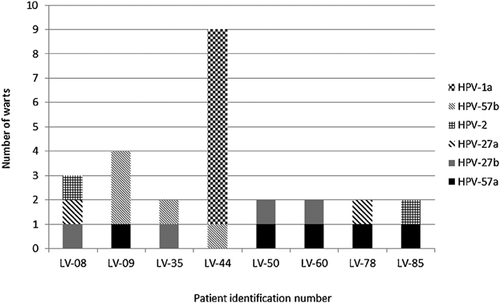
Individuals with multiple warts were 22.2% (16/72), being alpha genotype the most prevalent (n = 14). Among all individuals with multiple warts, half of them (n = 8) had different HPV genotypes (Fig. 1). However, HPV types for each patient belonged to the same genus (alpha), except for one of the patients, who presented several warts from two different genera (alpha and mu) (Fig. 1).

Location of Plantar Warts and HPV Genotypes
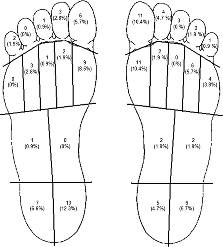
Figure 2 shows the location of all plantar warts. There were no significant differences between the number of warts in toes, midfoot and heel. Taking into account both feet, the most affected areas were the internal ones: medial (inside) zone of the heel (17.1%; 18/105), the first metatarsal head (19.0%; 20/105) and the great toe (16.2%; 17/105).

The most prevalent genotypes detected in all areas belonged to the alpha genus. The most frequently identified genotype in the heel was HPV-57 (51.6%; 16/31), while HPV-2 was more prevalent in barefoot warts (28.9%; 11/38), and both HPV-27 and HPV-57 genotypes in toes (70.9% in total; 22/31) (Table IV).
| Genera | |||||||
|---|---|---|---|---|---|---|---|
| Alfa | Gamma | Mu | |||||
| Location | 2 | 27a | 27b | 57a | 57b | 65 | 1a |
| Both feet | |||||||
| Heel | |||||||
| Medial zone | 2 | 0 | 3 | 7 | 1 | 0 | 5 |
| Lateral zone | 0 | 0 | 0 | 2 | 6 | 0 | 5 |
| Total warts in heel (n = 31) | 2 | 0 | 3 | 9 | 7 | 0 | 10 |
| Midfoot | |||||||
| Medial zone | 0 | 0 | 0 | 1 | 0 | 0 | 1 |
| Lateral zone | 0 | 1 | 1 | 1 | 0 | 0 | 0 |
| Total warts in midfoot (n = 5) | 0 | 1 | 1 | 2 | 0 | 0 | 1 |
| Barefoot | |||||||
| I ray | 8 | 2 | 4 | 2 | 3 | 0 | 1 |
| II ray | 0 | 0 | 0 | 0 | 2 | 1 | 1 |
| III ray | 0 | 0 | 0 | 0 | 0 | 0 | 1 |
| IV ray | 2 | 0 | 2 | 2 | 0 | 0 | 3 |
| V ray | 1 | 1 | 0 | 0 | 1 | 1 | 0 |
| Total warts in barefoot (n = 38) | 11 | 3 | 6 | 4 | 6 | 2 | 6 |
| Toes | |||||||
| 1st | 2 | 4 | 2 | 4 | 2 | 1 | 2 |
| 2nd | 1 | 2 | 1 | 2 | 0 | 0 | 2 |
| 3rd | 0 | 0 | 0 | 0 | 0 | 0 | 1 |
| 4th | 0 | 0 | 1 | 1 | 0 | 0 | 0 |
| 5th | 0 | 0 | 1 | 1 | 1 | 0 | 0 |
| Total warts in toes (n = 31) | 3 | 6 | 5 | 8 | 3 | 1 | 5 |
DISCUSSION
In the present study, we analyzed 105 plantar warts from 72 patients. Laminar sections of inner hyperkeratosis of warts were obtained and DNA was extracted using a standard procedure [Green and Sambrook, 2012]. We were able to obtain PCR amplicons for each of the warts and determine their sequences, by using the method described by Sasagawa and Mitsuishi [2012] to identify the HPV genotypes.
The sample collection method used is a rapid and simple procedure that allows good performance, since all 105 samples were identified. Several other methods have been described in the literature. The study by de Koning et al. [2011], for instance, aimed to validate the use of surface swabs for reliable detection of HPV, and to compare the results with the ones obtained when using scabs and deeper wart portions. They found that the use of swabs was a sensitive method, although it failed to identify two out of 25 samples. In our work, the total number of analyzed samples was much higher and all of them were positively identified. Among studies based on biopsy procedures, Giannaki et al. [2013] collected cutaneous wart biopsies and obtained reliable identification of HPV in 73.5% of cases. In contrast, the study by Sasagawa and Mitsuishi [2012] successfully identified all warts, which were resected using a carbon dioxide laser. Although the sensitivity of their method is very high, the collection procedure based on the extraction of the inner hyperkeratosis of warts is equally sensitive, and in addition it is a simpler and least invasive technique. These results confirm the suitability of the method used in this study.
HPV-57 was the most prevalent genotype detected in women and HPV-1a in men, both genotypes belonging to different genera: alpha and mu, respectively. Bruggink et al. [2012], in their study on the prevalence of wart-associated HPV types in a sample of individuals with cutaneous warts showed similar results.
In this study, warts with HPV-2, -27, and -57 mainly occurred in adults older than 21 years, and were absent or minor in childhood, whereas HPV-1-induced warts were detected in all ages. These findings are not in accordance to those obtained by Bruggink et al. [2012], who found that HPV-2, -27, and -57 types occurred in all ages and HPV-1 warts were mainly detected in childhood. This study included not only plantar warts but also common warts that may account for the differences observed. Moreover, it should be noted that in our study the number of patients younger than 20 years was low (26.4%; 19/72), and the conclusions obtained from these results should be further examined to better determine the age-specific HPV prevalence.
Most of the studies on HPV types of warts that have been published so far are focused not only on plantar warts but also in cutaneous warts, except for the Sasagawa study [Sasagawa and Mitsuishi, 2012]. Our results are similar to those obtained by Sasagawa and Mitsuishi. In our study, all plantar warts were infected with a single HPV type and the most prevalent genotypes were HPV-57 (37.1%) and HPV-27 (23.8%), followed by HPV-1a (20.9%), HPV-2 (15.2%), and HPV-65 (2.8%). Sasagawa and Mitsuishi analyzed 50 plantar warts, identifying HPV-27 as the most prevalent type, and HPV-57 and HPV-2 as relatively common types. However, significant differences between studies are found when comparing the less prevalent HPV types. They rarely found HPV-1a, -4, and -63, whereas in our study HPV-1a was almost as prevalent as HPV-27. Although the median age of the patients involved in both studies was similar, the samples in that study originated from a different source population.
Rübben et al. [1997] describe the most frequent HPV genotypes associated with common warts in palmoplantar location: HPV-2, -27, and -57. These results are in accordance with our data, although they did not detect the HPV-1a genotype in any of the analyzed warts. It is important to highlight that the methodology they used for the identification of HPV genotypes relies on restriction enzyme digestion patterns, and not on sequencing techniques. On the other hand, the study does not differentiate between plantar warts and warts extracted from other anatomical locations.
Other investigations and data available in the literature show that HPV-1 genotype is the virus type with the highest frequency in palmoplantar warts [Burns, 1992; Odom et al., 2004; Weston et al., 2008; Androphy and Lowy, 2009; Handisurya et al., 2009; Conejo-Mir et al., 2010; James et al., 2011], whereas HPV-27 and HPV-57 types are rarely detected [Androphy and Lowy, 2009; Handisurya et al., 2009]. Data extracted from the literature on HPV-2 and HPV-4 frequency are also variable [Burns, 1992; Odom et al., 2004; Weston et al., 2008; Androphy and Lowy, 2009; Handisurya et al., 2009; James et al., 2011], emphasizing the need for greater uniformity in the methodologies used to detect and identify HPV genotypes, as well as to characterize study populations.
Half of patients presenting multiple warts had the same HPV genotype, suggesting auto (self) inoculation from one site to another. Several authors have reported that autoinoculation is one of the causes of newly acquired infections [Androphy, 1989; Burns, 1992; Kirnbauer et al., 1992; Kopera, 2003; Salk and Douglas, 2006; Handisurya et al., 2009; Conejo-Mir et al., 2010]. HPV-2, -27, or -57 genotypes, which are closely related viruses belonging to the alpha genus, were present in all other cases, except for one. The presence of different HPV genotypes from the alfa genus in patients with multiple warts has been previously described by Rübben et al. [1997], although samples in their study were extracted from common warts.
Surprisingly, one of the participants with multiple warts presented two HPV types (HPV-1a and HPV-57b) from two different genera (mu and alpha genus, respectively), indicating that the warts were spread by self-inoculation and by de novo infection. So far, no other studies have reported the presence of different warts from different genera in the same patient.
It is important to highlight differences observed in the time to development of warts based on their HPV genotype. Warts belonging to the mu genus mainly developed in less than 1 year and warts of the alpha genus were primarily detected over a prolonged period of time. Nevertheless, a significant percentage of patients (31.9%) were not able to remember when their warts appeared. Warts can be present but not often recognized clinically, unless they cause harm or are detected by the individual.
Considering the location of plantar warts, the most affected areas were the medial (inside) zone of the heel, the first metatarsal head and the great toe. The observed distribution of warts reflects the usual gait pattern. Plantar warts frequently appear beneath pressure points, which are vulnerable sites on the skin of feet [Perry and Burnfield, 2010]. The most prevalent genotypes found in these locations were HPV-2, -27, and -57 (HPV-57 in the heel, HPV-2 in barefoot warts, and HPV-27 and HPV-57 in toes). These types were also predominantly found in plantar warts in the study by Rübben et al. [1997].
In conclusion, this study provides new insight on plantar warts and their associated HPV genotypes, and evidences the usefulness of the PCR method used. The data presented also suggest that obtaining laminar sections of inner hyperkeratosis of plantar warts is an accurate and reliable collection procedure for further detection of HPV genotypes.


